Tag: Cancer

Three Brothers Are Making Research a Family Affair

0 comments
From left to right: Caleb, Paul, and Adam Worsley sitting on stools in a chemistry lab.
Caleb, Paul, and Adam Worsley. Credit: Pittsburg State University.

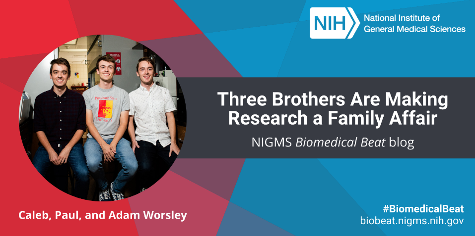
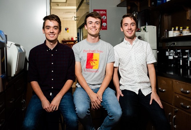
“You’re doing something really important with people who are important to you,” Paul Worsley remarks when asked about having his younger brothers Caleb and Adam as lab mates. The trio are undergraduate students working in the lab of Santimukul Santra, Ph.D., at Pittsburg State University in Pittsburg, Kansas.

Paul seated at his chemistry fume hood. Credit: Pittsburg State University.

All three brothers are part of the Kansas IDeA Networks of Biomedical Research Excellence (K-INBRE). Paul is currently a junior majoring in biology and history. He plans to go to medical school when he graduates, but his time in the lab has given him a love for research—and has even led him to toy with the idea of going to graduate school instead. His twin brothers Caleb and Adam are only freshmen, but they both think they want to pursue scientific research when they graduate.

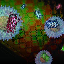
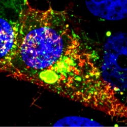
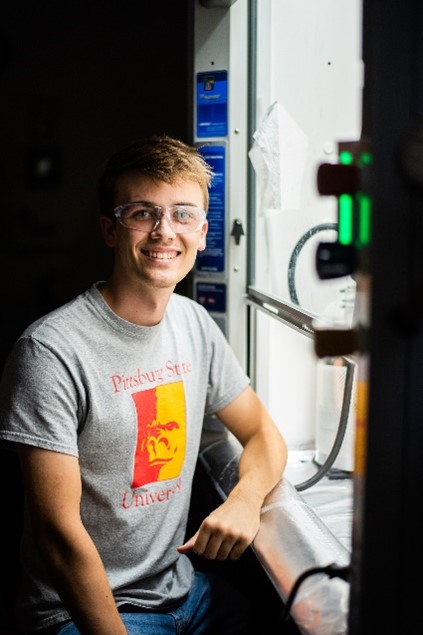
When Paul was a sophomore, he applied for a K-INBRE research spot in Dr. Santra’s lab and was immediately accepted. He quickly realized that organic chemistry in the lab was much different—and more exciting—than anything he’d seen in the classroom. “I like organic synthesis because it really tests your knowledge,” he says. “Answering exam questions is way different than actually doing it in a lab.” Despite the challenges that came with research, Paul was clearly doing great work because one day Dr. Santra joked, “Hey, you got any brothers?” Paul responded, “Actually, yes.”

Continue reading “Three Brothers Are Making Research a Family Affair”

Accelerating the Development of Tests for Endometriosis and Cancer

0 comments

NIGMS’ Small Business Technology Transfer (STTR) program works toward more effective methods for patient screening, diagnosis, and treatment.

Translating lab discoveries into health care products requires large investments of time and resources. Through STTR funding, NIGMS supports researchers interested in transitioning their discoveries and/or inventions into products. Here are the stories of three researchers working with the XLerator Hub, one of the funded programs that supports six southeastern IDeA states and Puerto Rico.

Ending Diagnostic Delays for Endometriosis

A headshot of Dr. Idhaliz Flores-Caldera. Dr. Idhaliz Flores-Caldera.
Credit: Courtesy of Dr. Flores-Caldera.

Idhaliz Flores-Caldera, Ph.D., a professor of basic sciences and OB-GYN at Ponce Health Sciences University in Puerto Rico, has studied endometriosis for nearly 20 years. Endometriosis occurs when endometrial tissue, which typically lines the uterus, grows elsewhere in the body. Dr. Flores-Caldera first had the idea for a noninvasive diagnostic test for the disorder about 10 years ago. But it was only when she learned about funding opportunities from the XLerator Hub that she saw a path to validating her preliminary research findings and eventually commercializing her test.

Dr. Flores-Caldera applied for and was accepted into the hub’s proof-of-concept program, Ideas to Products, which funds researchers to flesh out ideas they want to commercialize. “I am very appreciative of how the program has provided me with tools and knowledge about commercializing a product and the process of patenting a product,” she says. “In general, scientists aren’t educated on this important topic.”

Continue reading “Accelerating the Development of Tests for Endometriosis and Cancer”

Interview With a Scientist – Rommie Amaro: Computational and Theoretical Model Builder

0 comments

Many researchers who search for anti-cancer drugs have labs filled with chemicals and tissue samples. Not Rommie Amaro. Her work uses computers to analyze the shape and behavior of a protein called p53. Defective versions of p53 are associated with more human cancers than any other malfunctioning protein.

Continue reading “Interview With a Scientist – Rommie Amaro: Computational and Theoretical Model Builder”

Our Complicated Relationship With Viruses

9 comments
Illustration of Influenza Virus H1N1. Swine Flu.
Nearly 10 percent of the human genome is derived from the genes of viruses. Credit: Stock image.

When viruses infect us, they can embed small chunks of their genetic material in our DNA. Although infrequent, the incorporation of this material into the human genome has been occurring for millions of years. As a result of this ongoing process, viral genetic material comprises nearly 10 percent of the modern human genome. Over time, the vast majority of viral invaders populating our genome have mutated to the point that they no longer lead to active infections. But they are not entirely dormant.

Sometimes, these stowaway sequences of viral genes, called “endogenous retroviruses” (ERVs), can contribute to the onset of diseases such as cancer. They can also make their hosts susceptible to infections from other viruses. However, scientists have identified numerous cases of viral hitchhikers bestowing crucial benefits to their human hosts—from protection against disease to shaping important aspects of human evolution, such as the ability to digest starch.

Protecting Against Disease

Geneticists Cedric Feschotte, Edward Chuong and Nels Elde Exit icon at the University of Utah have discovered that ERVs lodged in the human genome can jump start the immune system.

For a virus to successfully make copies of itself inside a host cell, it needs molecular tools similar to the ones its host normally uses to translate genes into proteins. As a result, viruses have tools meticulously shaped by evolution to commandeer the protein-producing machinery of human cells.

Continue reading “Our Complicated Relationship With Viruses”

A Bright New Method for Rapidly Screening Cancer Drugs

0 comments
Illustration of red, green and blue fluorescent proteins.
Chemists have devised a new approach to screening cancer drugs that uses gold nanoparticles with red, green and blue outputs provided by fluorescent proteins. Credit: University of Massachusetts Amherst.

Scientists may screen billions of chemical compounds before uncovering the few that effectively treat a disease. But identifying compounds that work is just the first step toward developing a new therapy. Scientists then have to determine exactly how those compounds function.

Different cancer therapies attack cancer cells in distinct ways. For example, some drugs kill cancer cells by causing their outer membranes to rapidly rupture in a process known as necrosis. Others cause more subtle changes to cell membranes, which result in a type of programmed cell death known as apoptosis.

If researchers could distinguish the membrane alterations of chemically treated cancer cells, they could quickly determine how that chemical compound brings about the cells’ death. A new sensor developed by a research team led by Vincent Rotello of the University of Massachusetts Amherst can make these distinctions in minutes.

Continue reading “A Bright New Method for Rapidly Screening Cancer Drugs”

Illuminating Biology

0 comments

This time of year, lights brighten our homes and add sparkle to our holidays. Year-round, scientists funded by the National Institutes of Health use light to illuminate important biological processes, from the inner workings of cells to the complex activity of the brain. Here’s a look at just a few of the ways new light-based tools have deepened our understanding of living systems and set the stage for future medical advances.

RSV infected cell
A new fluorescent probe shows viral RNA (red) in an RSV-infected cell. Credit: Eric Alonas and Philip Santangelo, Georgia Institute of Technology and Emory University.

Visualizing Viral Activity

What looks like a colorful pattern produced as light enters a kaleidoscope is an image of a cell infected with respiratory syncytial virus (RSV) lit up by a new fluorescent probe called MTRIPS (multiply labeled tetravalent RNA imaging probes).

Although relatively harmless in most children, RSV can lead to bronchitis and pneumonia in others. Philip Santangelo of the Georgia Institute of Technology and Emory University, along with colleagues nationwide, used MTRIPS to gain a closer look at the life cycle of this virus.

Once introduced into RSV-infected cells, MTRIPS latched onto the genetic material of individual viral particles (in the image, red), making them glow. This enabled the researchers to follow the entry, assembly and replication of RSV inside the living cells. Continue reading “Illuminating Biology”

Cool Video: How a Microtubule Builds and Deconstructs

0 comments

A microtubule, part of the cell’s skeleton, builds and deconstructs. Credit: Eva Nogales lab, University of California, Berkeley.

In this animation, tubulin proteins snap into place like Lego blocks to build a microtubule, part of the cell’s skeleton. When construction ends, this long hollow cylinder falls to pieces from its top end. The breakdown is critical for many basic biological processes, including cell division, when rapidly shortening microtubules pull chromosomes into each daughter cell.

Until recently, scientists didn’t know exactly what drove microtubules to fall apart. A research team led by Eva Nogales of the Lawrence Berkeley National Laboratory and the University of California, Berkeley, now has an explanation.

Using high-powered microscopy, the scientists peered into the structure of a microtubule and found how a chemical reaction puts the stacking tubulin proteins under intense strain. The only thing keeping the proteins from springing apart is the pressure from the addition of more tubulin. So when assembly stops, the microtubule deconstructs.

The team also learned that Taxol, a common cancer drug, relieves this tension and allows microtubules to remain intact indefinitely. With microtubules frozen in place, a cancer cell cannot divide and eventually dies.

Because of this research, scientists now better understand both the success behind a common cancer drug and the molecular basis underlying the workings of microtubules.

Learn more:
University of California, Berkeley News Release Exit icon
Nogales Lab

Revealing a Key Player in Cancer Metastasis

0 comments
Invadopodia
A newly designed fluorescent biosensor shows where Rac1, a molecule involved in cancer metastasis, is active in this cell. Warmer colors show greater Rac1 activity. Credit: Yasmin Moshfegh, Albert Einstein College of Medicine.

Most of the more than half-a-million deaths caused by cancer each year in the United States result not from the original tumor but from the spread of cancer to new parts of the body, or metastasis. Cancer cells travel from a primary tumor using invadopodia, foot-like protrusions that break through surrounding connective tissue. Invadopodia are driven by protein filaments that repeatedly grow and disassemble. Exactly what guides this cycle was unclear, but scientists suspected a molecule called Rac1 might be involved. A new tool now sheds light on the details.

Researchers led by Louis Hodgson of Albert Einstein College of Medicine developed a fluorescent biosensor that glows wherever Rac1 is active in a cell, and they used it to study highly invasive breast cancer cells taken from rodents and humans. The scientists observed invadopodia form when Rac1 activity was low and disappear when it was high. They then confirmed their findings when they shut down the gene that encodes Rac1 and saw the invadopodia remain intact indefinitely.

This discovery suggests that targeting Rac1 activity with drugs could stop the spread of cancer cells. But a major hurdle remains: Healthy cells, including those that make up our immune system, also rely on the molecule for normal activity. Researchers must find a way to turn off Rac1 in cancer cells without disrupting its function in the rest of the body.

This work also was funded by NIH’s National Cancer Institute.

Learn more:
Albert Einstein College of Medicine News Release Exit icon

Two Proteins That Regulate Energy Use Play Key Role in Stem Cell Development

0 comments
Stem cells. Credit: Julie Mathieu, University of Washington.
The protein HIF1 alpha is beneficial for creating induced pluripotent stem cells (green) from adult human cells. Credit: Julie Mathieu, University of Washington.

Hannele Ruohola-Baker and a team of researchers at the University of Washington recently discovered that two proteins responsible for regulating how cells break down glucose are also essential for stem cell development. The scientists showed that the proteins HIF1 alpha and HIF2 alpha are both required to reprogram adult human cells into pluripotent stem cells, which have the ability to mature into any cell type in the body. Taking a closer look at what each protein does on its own, the researchers found that HIF1 alpha was beneficial for reprogramming throughout the process, whereas HIF2 alpha was required at early stages but was detrimental at later stages of reprogramming. Because the two proteins also play a role in transforming normal cells into cancer cells, the findings could lead to future advances in cancer research.

Learn more:
University of Washington News Release Exit icon
Ruohola-Baker Lab Exit icon
Once Upon a Stem Cell Article from Inside Life Science
Learning About Cancer by Studying Stem Cells Article from Inside Life Science
Sticky Stem Cells Article from Inside Life Science

One Mutation Leads to Another—At Least in Yeast

0 comments
DNA mutation. Credit: Stock image.
Newly discovered genetic effect in yeast could shed light on carcinogenesis. Credit: Stock image.

Cancer cells typically include many gene mutations, extra or missing genes, or even the wrong number of chromosomes. Scientists know that certain genetic changes lead to ones elsewhere. But they’ve had a chicken-and-egg problem trying to figure out which changes trigger which others—or whether mutations accumulate randomly in tumors.

New research led by J. Marie Hardwick Exit icon of Johns Hopkins University sheds light on the issue. She found that incapacitating a single gene in yeast cells—regardless of which gene it was—spurred mutations in one or two other genes. The process was anything but random: If, say, gene X was knocked out, yeast cells almost always developed a secondary mutation in gene Y. It’s as if knocking out one gene disrupts the genomic balance enough that the cell must alter a different gene to compensate.

Significantly, the secondary mutations—but not the original ones—caused altered yeast cell characteristics, including traits linked to cancer. Also, many of the secondary mutations occurred in genes associated with cancer in humans, further suggesting that these secondary changes might play a role in carcinogenesis.

This new information will help researchers better understand the chain of genetic events that lead to cancer. It might also prompt scientists to reevaluate years of research that attributed changes in cell behavior or appearance to a given gene knockout.

This work also was funded by NIH’s National Institute of Neurological Disorders and Stroke.

Learn more:
Johns Hopkins University News Release Exit icon