Tag: Wound Healing

A Tale of Tails: How Reptile Regeneration Could Help Humans

1 comment
A profile picture of Dr. Lozito wearing a white lab coat.
Dr. Thomas Lozito. Credit: Chris Shinn for USC Health Advancement Communications.

“I’ve always been interested in science and in lizards. I got my first pet lizard when I was around 4 years old, and it was love at first sight,” says Thomas Lozito, Ph.D., who now studies the creatures as an assistant professor of orthopaedic surgery, stem cell biology, and regenerative medicine at the University of Southern California (USC) in Los Angeles.

During his childhood, Dr. Lozito turned his parents’ house into a “little zoo” of lizards and amphibians. He sneaked lizards into his dorm room as a college student at Johns Hopkins University in Baltimore, Maryland, where he earned his bachelor’s degree in biomedical engineering. While pursuing his Ph.D. in stem cell biology through a joint program between the National Institutes of Health and Cambridge University in England, he bred lizards and frogs and sold them to earn extra money.

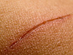
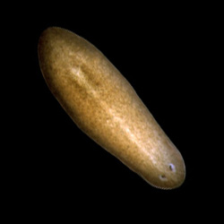
Continue reading “A Tale of Tails: How Reptile Regeneration Could Help Humans”

Reusable Disinfectant Developed from Mussel “Glue”

0 comments
A pile of ocean mussels with shiny black shells.
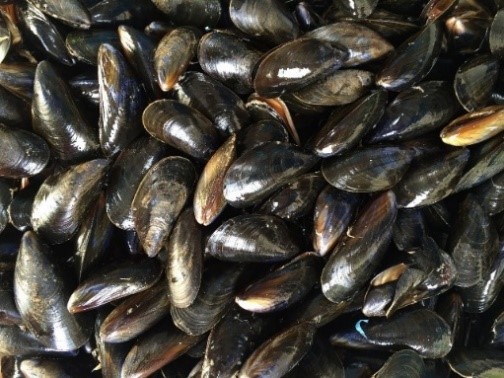
Mimicking mussels’ natural “glue” could have multiple benefits.

Many species have developed unique adaptations to help them thrive in their environments, and scientists in a field called biomimicry use these examples as the basis for tools to help humans. Biomimicry researchers have made a wide range of products, from climbing pads modeled after gecko feet to a faster, sharp-nosed bullet train based on the beak of the kingfisher bird. The animal kingdom also provides inspiration for biomedical products. For instance, scientists at Michigan Technological University in Houghton discovered that a natural “glue” produced by mussels has antimicrobial properties and are developing a way to put these properties to use.

Continue reading “Reusable Disinfectant Developed from Mussel “Glue””

Amazing Organisms and the Lessons They Can Teach Us

1 comment

What do you have in common with rodents, birds, and reptiles? A lot more than you might think. These creatures have organs and body systems very similar to our own: a skeleton, digestive tract, brain, nervous system, heart, network of blood vessels, and more. Even so-called “simple” organisms such as insects and worms use essentially the same genetic and molecular pathways we do. Studying these organisms provides a deeper understanding of human biology in health and disease, and makes possible new ways to prevent, diagnose, and treat a wide range of conditions.

Historically, scientists have relied on a few key organisms, including bacteria, fruit flies, rats, and mice, to study the basic life processes that run bodily functions. In recent years, scientists have begun to add other organisms to their toolkits. Many of these newer research organisms are particularly well suited for a specific type of investigation. For example, the small, freshwater zebrafish grows quickly and has transparent embryos and see-through eggs, making it ideal for examining how organs develop. Organisms such as flatworms, salamanders, and sea urchins can regrow whole limbs, suggesting they hold clues about how to improve wound healing and tissue regeneration in humans.

Continue reading “Amazing Organisms and the Lessons They Can Teach Us”

How Three Physician Scientists Are Taking Strides to Improve Our Health

0 comments

Brain injuries, cancer, infections, and wound healing are some of the complex and pressing health concerns we face today. Understanding the basic science behind these diseases and biological processes is the key to developing new treatments and improving patient outcomes. Physician scientists—medical doctors who also conduct laboratory research—are essential to turning knowledge gained in the lab into innovative treatments, surgical advances, and new diagnostic tools.

In this blog, we highlight the work and impact of three surgeon scientists funded by NIGMS at different stages in their careers: Dr. Nicole Gibran (current grantee), Dr. Rebecca Minter (former grantee), and Dr. Carrie Sims (former grantee). Their work, despite the historical underrepresentation of women in the physician scientist training community, has led to revolutionary surgical treatments, new therapeutics, better screening, and improved quality of life for patients.

Continue reading “How Three Physician Scientists Are Taking Strides to Improve Our Health”

The ECM: A Dynamic System for Moving Our Cells

2 comments

In part I of this series, we mentioned that the extracellular matrix (ECM) makes our tissues stiff or squishy, solid or see-through. Here, we reveal how the ECM helps body cells move around, a process vital for wounds to heal and a fetus to grow.

Sealing and Healing Wounds

MMPs are essential for closing wounds. Credit: Stock image.

When we get injured, the first thing our body does is to form a blood clot to stop the bleeding. Skin cells then start migrating into the wound to close the cut. The ECM is essential for this step, creating a physical support structure—like a road or train track—over which skin cells travel to seal the injured spot.

The ECM is made up of a host of proteins produced before and after injury. Some other proteins called matrix metalloproteinases (MMPs) also crowd into wounds. Because humans have so many different MMPs—a full 24 of them!—it’s been difficult for scientists to figure out what roles, if any, the proteins play in healing scrapes and cuts.

Continue reading “The ECM: A Dynamic System for Moving Our Cells”

Interview With a Scientist: Janet Iwasa, Molecular Animator

0 comments

The world beneath our skin is full of movement. Hemoglobin in our blood grabs oxygen and delivers it throughout the body. Molecular motors in cells chug along tiny tubes, hauling cargo with them. Biological invaders like viruses enter our bodies, hijack our cells and reproduce wildly before bursting out to infect other cells.

To make sense of the subcutaneous world, Janet Iwasa, a molecular animator at the University of Utah, creates “visual hypotheses”—detailed animations that convey the latest thinking of how biological molecules interact.

“It’s really building the animated model that brings insights,” Iwasa told Biomedical Beat in 2014. “When you’re creating an animation, you’re really grappling with a lot of issues that don’t necessarily come up by any other means. In some cases, it might raise more questions, and make people go back and do some more experiments when they realize there might be something missing.”

Iwasa has collaborated with numerous scientists to develop animations of a range of biological processes and structures Exit icon. Recently, she’s undertaken an ambitious, multi-year project to animate HIV reproduction Exit icon.

Interview With a Slime Mold: Racing for New Knowledge

1 comment
Dictyostelium discoideum
Credit: Wikimedia Commons, Usman Bashir.
Dictyostelium discoideum
Natural habitat: Deciduous forest soil and moist leaf litter
Favorite food: Bacteria
Top speed: 8 micrometers per minute

Like the athletes in Rio, the world’s most highly advanced microbial runners recently gathered in Charlestown, Massachusetts, to find out which ones could use chemical cues to most quickly navigate a maze-like microfluidic racecourse. The winners’ prize: credit for helping scientists learn more about how immune system cells navigate through the human body on their way to fight disease.

The finalists were a group of soil-dwelling slime molds called Dictyostelium that were genetically engineered by a pair of Dutch biochemists to detect minuscule chemical changes in the environment. The racers used their enhanced sense of “smell” to avoid getting lost on their way to the finish line.

While researchers have been racing the genetically souped-up microbes at annual events for a few years—another competition is scheduled for October 26—scientists have been studying conventional Dictyostelium for decades to investigate other important basic life processes including early development, gene function, self/non-self recognition, cell-type regulation, chemical signaling and programmed cell death. Continue reading “Interview With a Slime Mold: Racing for New Knowledge”

Visualizing Skin Regeneration in Real Time

2 comments
Top: Colorful skin cells on a zebrafish . Bottom: Cells from the outer surface of the scale.
More than 70 Skinbow colors distinguish hundreds of live cells from a tiny bit (0.0003348 square inches) of skin on the tail fin of an adult zebrafish. The bottom image shows the cells on the outer surface of a scale. Credit: Chen-Hui Chen, Duke University.

Zebrafish, blue-and-white-striped fish that are about 1.5 inches long, can regrow injured or lost fins. This feature makes the small fish a useful model organism for scientists who study tissue regeneration.

To better understand how zebrafish skin recovers after a scrape or amputation, researchers led by Kenneth Poss of Duke University tracked thousands of skin cells in real time. They found that lifespans of individual skin cells on the surface were 8 to 9 days on average and that the entire skin surface turned over in 20 days.

The scientists used an imaging technique they developed called “Skinbow,” which essentially shows the fish’s outer layer of skin cells in a spectrum of colors when viewed under a microscope. Skinbow is based on a technique created to study nerve cells in mice, another model organism.

The research team’s color-coded experiments revealed several unexpected cellular responses during tissue repair and replacement. The scientists plan to incorporate additional imaging techniques to generate an even more detailed picture of the tissue regeneration process.

The NIH director showcased the Skinbow technique and these images on his blog, writing: “You can see more than 70 detectable Skinbow colors that make individual cells as visually distinct from one another as jellybeans in a jar.”

This work was funded in part by NIH under grant R01GM074057.

Interview With a Worm: We’re Not So Different

2 comments
Planarian
Credit: Alejandro Sánchez Alvarado, Stowers Institute for Medical Research.
Schmidtea mediterranea
Home: Freshwater habitats along the Mediterranean
Party trick: Regenerating its head
Most charismatic feature: Eyespots
Work site: Science labs worldwide

The planarian has a power few creatures can match. Remove its head, its tail or nearly any of its body parts, and this aquatic flatworm will simply grow it back. Humans can’t do that, of course. And yet many of the genes that help the planarian regenerate are also found in us. To learn more about this tiny marvel, we “interviewed” a representative. Continue reading “Interview With a Worm: We’re Not So Different”