
In a statement on World AIDS Day 2013, NIH leaders wrote, “In the 25 years that have passed since the first annual commemoration of World AIDS Day, extraordinary scientific progress has been made in the fight against HIV/AIDS. That progress has turned an HIV diagnosis from an almost-certain death sentence to what is now for many, a manageable medical condition and nearly normal lifespan. We have come far, yet not far enough.”
One area of progress is in understanding the structural biology of HIV, the virus that causes AIDS. Capturing details about the virus’ shape has helped scientists better understand how HIV operates and pinpoint its Achilles’ heels. Recently, scientists got a closer look at two key pieces of the virus.
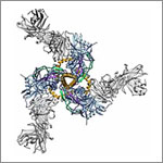
In one study, researchers developed the most detailed picture yet of Env, a three-segment protein on the HIV surface that allows the virus to infect cells. The work illuminated the complex process by which the protein assembles, undergoes radical shape changes during infection and interacts with neutralizing antibodies, which can block many strains of HIV from infecting human cells. The findings also may guide the development of HIV vaccines.
In another study, researchers created the best image yet of the cocoon-like container, or capsid, that carries HIV’s genome. Capsids have been difficult to study because individual imaging techniques had not produced high enough detail. By combining several cutting-edge imaging methods, the scientists pieced together the individual polygonal units of the capsid like a jigsaw puzzle to determine its structure in detail. Now that they know how HIV’s inner vessel looks, the research team is searching for its cracks—potential targets for drug development.
Learn more:
Imaging HIV’S Inner Shell Article from Inside Life Science
Key HIV Protein Structure Revealed Article from NIH Research Matters
Interactive HIV Structural Model
Both of these advances contribute to the fundamental scientific knowledge we have of HIV structure, laying the foundation for understanding structure-function relationships and developing therapeutics against this deadly disease. These advances have another thing in common—they were enabled by NIGMS’ Biomedical Technology Research Resources (BTRRs), which conduct research and development on technologies and instruments and make them available to biomedical researchers around the country:
For more information about the BTRR program and how it’s advancing other areas of basic research, see the Biomedical Technology Research and Resources News Web page.