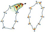
Messenger proteins help the cell make large projections (left). When these proteins aren’t activated, the cell doesn’t move (right). Credit: Devreotes Lab, Johns Hopkins University School of Medicine. View larger image
A new study from Peter Devreotes, Pablo Iglesias and other scientists at Johns Hopkins University sheds light on the way in which cells get around the body to promote embryo development, wound healing and even cancer metastasis. Here’s how they describe cell movement and their findings:
Think of the cell as a rowboat. Sensor proteins on the outside pass on directional signals to messenger proteins that serve as the cell’s coxswain. The coxswain then commands other members of the molecular crew to stay in sync, propelling the cell forward. If there are no sensor signals, the coxswain can still coordinate the cell’s movement, just not in any specific direction—it’s like a boat without a rudder.
Scientists previously thought that the messenger proteins needed the sensor ones to produce both directional and random movements. Because defects in the messenger proteins have been linked to many types of cancer, the new work suggests these molecules could serve as a drug target for immobilizing tumor cells.
Learn more:
New insight into the genes that affect drug responses may help doctors prescribe the medications and doses best suited for each individual. Credit: Jane Ades, NIH’s National Human Genome Research Institute.
Scientists know that variations in certain genes can affect the way a person responds to medications. New research by Wolfgang Sadee at Ohio State University shows that drug responses also depend on previously overlooked parts of DNA—sections that regulate genes, but are not considered genes themselves. This study focused on an important enzyme abbreviated CYP2D6 that processes about one-fourth of all prescription drugs. Differences in the enzyme’s performance, which range from zilch to ultra-rapid, can dramatically alter the effectiveness and safety of certain medications. Researchers discovered two new genetic variants that impact CYP2D6 performance. One of these, located in a non-gene, regulatory region of DNA, doubles or even quadruples enzyme activity. Coupling these findings with genetic tests could help doctors better identify each patient’s CYP2D6 activity level, enabling more precise prescriptions. The findings also open up a whole new area of investigation into genetic factors that impact drug response.
This work also was funded by NIH’s Eunice Kennedy Shriver National Institute of Child Health and Human Development.
Learn more:
Ohio State University News Release (no longer available)
Treating yeast cells with the NAB compound reverses the toxic effects of elevated levels of alpha synuclein protein. Credit: Daniel Tardiff, Whitehead Institute. View larger image
These eye-catching images and the NIGMS-funded research that yielded them were recently featured by NIH Director Francis Collins on his blog. Scientists led by a team at the Whitehead Institute for Biomedical Research engineered yeast to produce too much of a protein, alpha synuclein. In Parkinson’s disease, elevated levels or mutated forms of this protein wreak havoc on the cell. Using the model system, the researchers tested tens of thousands of compounds to identify any that reversed the toxic effects. One did. The compound, abbreviated NAB, worked similarly in an animal model and in rat neurons grown in a lab dish. Collins described the approach as “an innovative strategy for drug hunting that will likely be extended to other conditions.”
Emily Scott Field: Biochemistry Works at: University of Kansas in Lawrence Favorite hobby: Scuba diving Likes watching: “Law & Order” Likes reading: True-life survival stories
Credit: Chuck France, University of Kansas
With an air tank strapped to her back, college student Emily Scott dove to the bottom of the Gulf of Mexico to examine life in an oxygen-starved area called the Dead Zone. The bottom waters had once teemed with red snapper, croaker and shrimp, but to Scott, the region appeared virtually devoid of life. Then, from out of the mud, appeared the long, undulating arms of a brittle star.
As Scott learned, that particular species of brittle star survived in the Dead Zone because it has something many other marine creatures don’t: an oxygen-carrying protein called hemoglobin. This same protein makes our blood red. Key to hemoglobin’s special oxygen-related properties is a small molecular disk called a heme (pronounced HEEM).
Once she saw what it meant to brittle stars, Scott was hooked on heme and proteins that contain it.
Scott’s Findings
Now an associate professor, Scott studies a family of heme proteins called cytochromes P450 (CYP450s). These proteins are enzymes that facilitate many important reactions: They break down cholesterol, help process vitamins and play an important role in flushing foreign chemicals out of our systems.
To better understand CYP450s, Scott uses a combination of two techniques–X-ray crystallography and nuclear magnetic resonance spectroscopy—for capturing the enzymes’ structural and reactive properties.
She hopes to apply her work to design drugs that block certain CYP450 reactions linked with cancer. One target reaction, carried out by CYP2A13, converts a substance in tobacco into two cancer-causing molecules. Another target reaction is carried out by CYP17A1, an enzyme that helps the body produce steroid sex hormones but that, later in life, can fuel the uncontrolled growth of prostate or breast cancer cells.
“I’m fascinated by these proteins and figuring out how they work,” Scott says. “It’s like trying to put together a puzzle—a very addictive puzzle.” Her drive to uncover the unknown and her willingness to apply new techniques have inspired the students in her lab to do the same.
Content adapted from“Hooked on Heme,” an article in the September 2013 issue of Findings magazine.
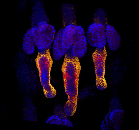
Credit: Yaron Fuchs and Samara Brown in the lab of Hermann Steller, Rockefeller University.
Whether injured by a scrape, minor burn or knife wound, skin goes through the same steps to heal itself. Regrowing hair over new skin is one of the final steps. All the hair you can see on your body is non-living, made up of “dead” cells and protein. It sprouts from living cells in the skin called hair follicle stem cells, shown here in red and orange. For more pictures of hair follicle stem cells—and many other stunning scientific images and videos—go to the NIGMS Image and Video Gallery.
Influenza virus proteins in the act of self-replication. Credit: Wilson, Carragher and Potter labs, Scripps Research Institute.
Flu viruses evolve rapidly, often staying one step ahead of efforts to vaccinate against infections or treat them with antiviral drugs. Work led by Jesse Bloom of the Fred Hutchinson Cancer Research Center has uncovered a surprising new flu mutation that allows influenza to infect cells in a novel way. Normally, a protein called hemagglutinin lets flu viruses attach to cells, and a protein called neuraminidase lets newly formed viruses escape from infected cells. Bloom’s lab has characterized a mutant flu virus where neuraminidase can enable the virus to attach to host cells even when hemagglutinin’s binding is blocked. Although the researchers generated the neuraminidase mutant studied in these experiments in their lab, the same mutation occurs naturally in strains from several recent flu outbreaks. There’s a possibility that flu viruses with such mutations may be able to escape antibodies that block the binding of hemagglutinin.
This work also was funded by NIH’s National Institute of Allergy and Infectious Diseases.
Cancer tumors are like snowflakes—no two ever share the same genetic mutations. Their unique characteristics make them difficult to categorize and treat. A new approach proposed by Trey Ideker and his team at the University of California, San Diego, might offer a solution. Their approach, called network-based stratification (NBS), identifies cancer subtypes by how different mutations in different cancer patients affect the same biological networks, such as genetic pathways. As proof of principle, they applied the method to ovarian, uterine and lung cancer data to obtain biological and clinical information about mutation profiles. Such cancer subtyping shows promise in helping to develop more effective, personalized treatments.
Learn more:
University of California, San Diego News Release Ideker Lab
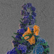
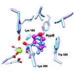
The image shows a comparison of the predicted binding of the substrate to the active site of HpbD (blue) with the binding sites determined experimentally by crystallography (magenta). Credit: Matt Jacobson, University of California, San Francisco; Steve Almo, Albert Einstein College of Medicine.
Sequencing the genomes of almost 7,000 organisms has identified more than 40 million proteins. But how do we figure out what all these proteins do? New results from an initiative led by John Gerlt of the University of Illinois suggest a possible method for identifying the functions of unknown enzymes, proteins that speed chemical reactions within cells. Using high-powered computing, the research team modeled how the structure of a mystery bacterial enzyme, HpbD, might fit like a puzzle piece into thousands of proteins in known metabolic pathways. Since an enzyme acts on other molecules, finding its target or substrate can shed light on its function. The new method narrowed HpbD’s candidate substrate down from more than 87,000 to only four. Follow-up lab work led to the actual substrate, tHypB, and determined the enzyme’s biological role. This combination of computational and experimental methods shows promise for uncovering the functions of many more proteins.
Learn more:
University of Illinois at Urbana-Champaign News Release
Gerlt Lab
Enzyme Function Initiative
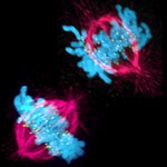
This image shows a cell in two stages of division: prometaphase (top) and metaphase (bottom). To form identical daughter cells, chromosome pairs (blue) separate via the attachment of microtubules made up of tubulin proteins (pink) to specialized structures on centromeres (green). Credit: Lilian Kabeche, Dartmouth.
Chromosome segregation during cell division is like speed dating, according to Geisel School of Medicine at Dartmouth researcher Duane Compton. He and postdoctoral fellow Lilian Kabeche learned that protein cyclin A plays moderator, helping to properly separate chromosomes via the attachment of microtubule fibers to kinetochore structures. Here’s how Compton described the process:
“The chromosomes are testing the microtubules for compatibility—that is, looking for the right match—to make sure there are correct attachments and no errors. The old view of this process held that chromosomes and microtubules pair up individually to find the correct attachment, like conventional dating. However, our results show that the system is more like speed dating. All the chromosomes have to try many connections with microtubules in a short amount of time. Then they all make their final choices at the same time. Cyclin A acts like a timekeeper or referee to make sure no one makes bad connections prematurely.”
Such bad connections can cause chromosome segregation errors that lead to cells with an abnormal number of chromosomes, a hallmark of cancer cells. So in addition to aiding our understanding of a fundamental biological process, the new insights may point to potential ways to correct such errors.
The winners of the 2013 Nobel Prize in physiology or medicine discovered that cells import and export materials using fluid-filled sacs called vesicles. Credit: Judith Stoffer.
Every October, a few scientists receive a call from Sweden that changes their lives. From that day forward, they will be known as Nobel laureates. This year, five of the new Nobelists have received funding from NIGMS.
In physiology or medicine, NIGMS grantees James Rothman (Yale University) and Randy Schekman (University of California, Berkeley) were honored “for their discoveries of machinery regulating vesicle traffic, a major transport system in our cells.” They share the prize with Thomas C. Südhof of Stanford University.
Rothman and Schekman started out working separately and in different systems—Schekman in yeast and Rothman in reconstituted mammalian cells—and their conclusions validated each others’. It’s yet another example of the power of investigator-initiated research and the value of model systems.
Highly accurate molecular models like this one are based on computational techniques developed by the winners of the 2013 Nobel Prize in chemistry. Credit: Rommie E. Amaro, University of California, San Diego.
The three Nobelists in chemistry are NIGMS grantees Martin Karplus (Harvard University), Michael Levitt (Stanford University) and Arieh Warshel (University of Southern California), who developed “multiscale models for complex chemical systems.” They used computational techniques to obtain, for the first time, detailed structural information about proteins and other large molecules. Because of that work, scientists around the world are now able to access, with a few keystrokes, highly accurate models of nearly 100,000 molecular structures. Studying these structures has advanced our understanding of countless diseases, pharmaceuticals and basic biological processes.