Year: 2017

Beauty is in the Eye

2 comments

Our eyes are the gateway to countless brilliant sights. However, as evidenced by the images on this page, the eye itself can be breathtakingly exquisite as well. This May, as we celebrate Healthy Vision Month with the National Eye Institute, we hope sharing the beauty hidden in your eyes will inspire you to take the necessary steps to protect your vision, prevent vision loss and make the most of the vision you have remaining.

Visit NEI to learn more about caring for your eyes.

Happy Healthy Vision Month!

Mammalian eye

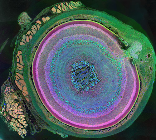
Eyes are beautiful, and they take on a whole new look in this agate-like image, which highlights just how complex mammalian eyes really are. Researchers used staining and imaging techniques to turn each of the 70-plus cell types in this mouse eye a different color. The image won first place in the 2011 International Science and Engineering Visualization Challenge. Credit: Bryan William Jones, University of Utah Moran Eye Center.
Mouse eye

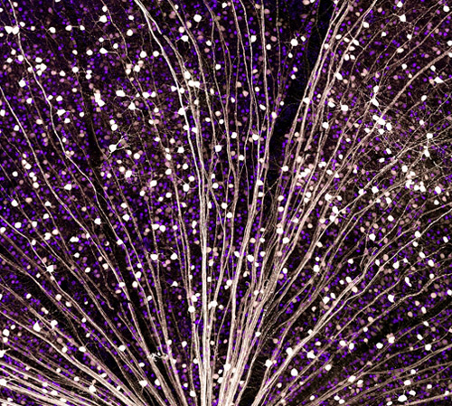
This burst of starry points is actually part of the retina from a mouse eye. The image comes from a research project investigating the promise of gene therapy for glaucoma. Untreated glaucoma is a leading cause of blindness. The disease is characterized by the death of cells called retinal ganglion cells. Scientists are hoping to deliver gene therapy to these cells as a treatment for glaucoma. In this photo, a fluorescent protein (GFP) lights up to show the location of retinal ganglion cells—and to reveal how well the proposed gene therapy technique might work. Credit: Kenyoung Kim, Wonkyu Ju and Mark Ellisman, National Center for Microscopy and Imaging Research, University of California, San Diego.
Mouse eye

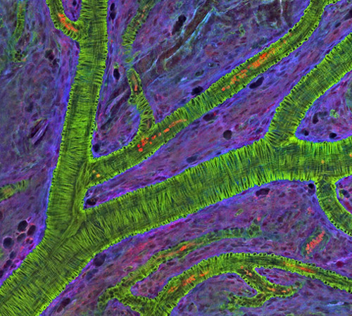
What appears as a tree branch painstakingly wrapped in green wire is a microscopic blood vessel from the retina at the back of a mouse eye. These vessels can help diagnose conditions such as glaucoma and diabetic eye disease. The vessels also have a characteristic appearance in people with high blood pressure. This detailed image was created to help scientists understand what happens in a genetic disease called neurofibromatosis, in which tumors begin to form on nerve tissue. Credit: National Center for Microscopy and Imaging Research, University of California, San Diego.
Mouse eye

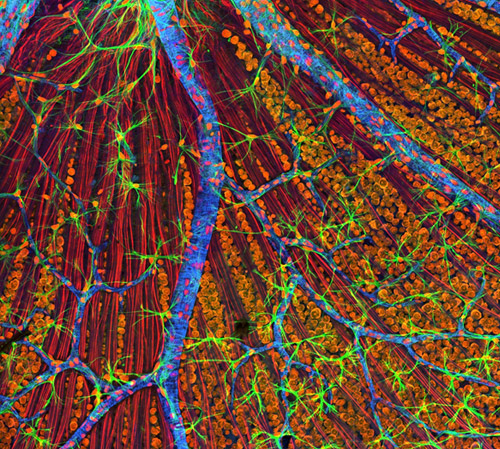
Like a colorful fiber-optic network, this microscopic layer from a mouse’s eye relays information from the retina to the brain. Retinal ganglion neurons (orange) and their associated optic nerve fibers (red) are overlaid with blood vessels (blue) and spidery glial cells (green). By comparing detailed images of healthy eye tissues with similar images of a diseased eye, researchers can learn about changes in biology that occur as eye diseases develop. Credit: National Center for Microscopy and Imaging Research, University of California, San Diego.

The Endoplasmic Reticulum: Networking Inside the Cell

5 comments

Like a successful business networker, a cell’s endoplasmic reticulum (ER) is the structure that reaches out—quite literally—to form connections with many different parts of a cell. In several important ways, the ER enables those other parts, or organelles, to do their jobs. Exciting new images of this key member of the cellular workforce may clarify how it performs its roles. Such knowledge will also help studies of motor neuron and other disorders, such as amyotrophic lateral sclerosis (ALS), that are associated with abnormalities in ER functioning.

Structure Follows Function

Illustration of some of the jobs that the ER performs in the cell.

An illustration of some of the jobs that the endoplasmic reticulum (ER) performs in the cell. Some ER membranes (purple) host ribosomes on their surface. Other ER membranes (blue) extending into the cytoplasm are the site of lipid synthesis and protein folding. The ER passes on newly created lipids and proteins to the Golgi apparatus (green), which packages them into vesicles for distribution throughout the cell. Credit: Judith Stoffer.

Initiated in 1965, the Postdoctoral Research Associate Program (PRAT) is a competitive postdoctoral fellowship program to pursue research in one of the laboratories of the National Institutes of Health. PRAT is a 3-year program providing outstanding laboratory experiences, access to NIH’s extensive resources, mentorship, career development activities and networking. The program places special emphasis on training fellows in all areas supported by NIGMS, including cell biology, biophysics, genetics, developmental biology, pharmacology, physiology, biological chemistry, computational biology, immunology, neuroscience, technology development and bioinformatics

The ER is a continuous membrane that extends like a net from the envelope of the nucleus outward to the cell membrane. Tiny RNA- and protein-laden particles called ribosomes sit on its surface in some places, translating genetic code from the nucleus into amino acid chains. The chains then get folded inside the ER into their three-dimensional protein structures and delivered to the ER membrane or to other organelles to start their work. The ER is also the site where lipids—essential elements of the membranes within and surrounding a cell—are made. The ER interacts with the cytoskeleton—a network of protein fibers that gives the cell its shape—when a cell divides, moves or changes shape. Further, the ER stores calcium ions in cells, which are vital for signaling and other work.

To do so many jobs, the ER needs a flexible structure that can adapt quickly in response to changing situations. It also needs a lot of surface area where lipids and proteins can be made and stored. Scientists have thought that ER structure combined nets of tubules, or small tubes, with areas of membrane sheets. However, recent NIGMS PRAT (Postdoctoral Research Associate; see side bar) fellow Aubrey Weigel, working with her mentor and former PRAT fellow Jennifer Lippincott-Schwartz of the Eunice Kennedy Shriver National Institute of Child Health and Human Development (currently at the Howard Hughes Medical Institute in Virginia) and colleagues, including Nobel laureate Eric Betzig, wondered whether limitations in existing imaging technologies were hiding a better answer to how the ER meets its surface-area structural needs in the periphery, the portion of the cell not immediately surrounding the nucleus. Continue reading “The Endoplasmic Reticulum: Networking Inside the Cell”

New Technology May Help Reduce Serious and Costly Post-Surgical Infections—Using Nothing but Air

7 comments

According to a recent estimate, implant infections following hip and knee replacement surgeries in the U.S. may number 65,000 by 2020, with the associated healthcare costs exceeding $1 billion. A new small, high-tech device could have a significant impact on improving health outcomes and reducing cost for these types of surgeries. The device, Air Barrier System (ABS), attaches on top of the surgical drape and gently emits HEPA-filtered air over the incision site. By creating a “cocoon” of clean air, the device prevents airborne particles—including the bacteria that can cause healthcare-associated infections—from entering the wound.

Air Barrier System
The Air Barrier System creates a “cocoon” of clean air (gray area with size indicated) over a surgical site to remove airborne contaminants and reduce the risk of infection in patients who are receiving an artificial hip, a blood vessel graft, a titanium plate in the spine or other implants.

Scientists recently analyzed the effectiveness of the ABS device in a clinical study—funded by NIGMS—involving nearly 300 patients. Each patient needed an implant, such as an artificial hip, a blood vessel graft in the leg or a titanium plate in the spine. Because implant operations involve inserting foreign materials permanently into the body, they present an even higher risk of infection than many other surgeries, and implant infections can cause life-long problems.

The researchers focused on one of the most common causes of implant infections—the air in the operating room. Although operating rooms are much cleaner than almost any other non-hospital setting, it’s nearly impossible to sterilize the entire room. Instead, the scientists focused on reducing contaminants directly over the surgical site. They theorized that if the air around the wound was cleaner, the number of implant infections might go down. Continue reading “New Technology May Help Reduce Serious and Costly Post-Surgical Infections—Using Nothing but Air”

Six Things to Know About DNA and DNA Repair

3 comments

Deoxyribonucleic acid, better known as DNA, was first identified on a discarded surgical bandage almost 150 years ago. Increasingly sophisticated tools and techniques have allowed scientists to learn more about this chemical compound that includes all the instructions necessary for building a living organism. From among the dozens of fascinating things known about DNA, here are six items touching on the make up of DNA’s double helix, the vast amounts of DNA packed into every human’s cells, common DNA errors and a few ways DNA can repair itself.

1. DNA is in every living thing.

Nucleotide
DNA consists of two long, twisted chains made of nucleotides. Each nucleotide contains one base, one phosphate molecule and the sugar molecule deoxyribose. The bases in DNA nucleotides are adenine, cytosine, guanine and thymine. Credit: NIGMS.

The chemical instructions for building a person—and every other creature on Earth—are contained in DNA. DNA is shaped like a corkscrew-twisted ladder, called a double helix. The two ladder rails are referred to as backbones, made of alternating groups of sugar and phosphate. The ladder’s rungs are made from four different building blocks called bases, arranged in pairs: adenine (A) paired with thymine (T), and cytosine (C) paired with guanine (G). Humans have about 3 billion base pairs in each cell. The order of the base pairs determines the exact instructions encoded in that part of the DNA molecule. Also, the sequence of DNA base pairs in one person is about 99.9 percent identical to that of everyone else.

2. Humans have a lot of DNA.

Humans begin as a single fertilized cell containing (with some rare exceptions) the full complement of DNA—the genome—arranged into 46 discrete chromosomes (23 pairs, with mom and dad each contributing half of each pair) in the cell’s nucleus. There are 6 feet of DNA coiled up tightly in that first cell. All the information in the DNA is replicated each time the cell divides. The amount of DNA packed into all of an adult’s cells is on the order of 100 trillion feet (about 19 billion miles)—so that if the DNA chain was stretched out, it would be long enough to reach back and forth between the Earth and the Sun more than 200 times. Continue reading “Six Things to Know About DNA and DNA Repair”

Birthdays, Nobel Prizes and Basic Research

1 comment
James D. Watson
James D. Watson. Credit: Wikimedia Commons, Cold Spring Harbor Laboratory.

April 6 is the birthday of two Nobel Prize winners in physiology or medicine—James Watson and Edmond H. Fischer. They have also both been NIGMS-supported researchers.

Double helix model
In 1953, Watson and Crick created their historic model of the shape of DNA: the double helix. Credit: Cold Spring Harbor Laboratory archives.

James D. Watson, born on this day in 1928, was honored with the Nobel Prize in 1962. He shared it with Francis H. Compton Crick and Maurice Wilkins “for their discoveries concerning the molecular structure of nucleic acids and its significance for information transfer in living material.” This laid the groundwork for future discoveries. In the early 1950s, Wilkins and another scientist, Rosalind Franklin, worked to determine DNA’s structure. In 1953, Watson and Crick discovered its shape as a double helix. This twisted ladder structure enabled other researchers to unlock the secret of how genetic information is stored, transferred and copied. Franklin is widely recognized as having played a significant role in revealing the physical structure of DNA; due to her death at age 37 in 1958, Franklin did not earn a share of the prize. Read more about DNA.

Continue reading “Birthdays, Nobel Prizes and Basic Research”

Bit by the Research Bug: Priscilla’s Growth as a Scientist

2 comments

This is the third post in a new series highlighting NIGMS’ efforts toward developing a robust, diverse and well-trained scientific workforce.

Priscilla Del Valle
Credit: Christa Reynolds.
Priscilla Del Valle
Academic Institution: The University of Texas at El Paso
Major: Microbiology
Minors: Sociology and Biomedical Engineering
Mentor: Charles Spencer
Favorite Book: The Immortal Life of Henrietta Lacks, by Rebecca Skloot
Favorite Food: Tacos
Favorite music: Pop
Hobbies: Reading and drinking coffee

It’s not every day that you’ll hear someone say, “I learned more about parasites, and I thought, ‘This is so cool!’” But it’s also not every day that you’ll meet an undergraduate researcher like 21-year-old Priscilla Del Valle.

BUILD and the Diversity Program Consortium

The Diversity Program Consortium (DPC) aims to enhance diversity in the biomedical research workforce through improved recruitment, training and mentoring nationwide. It comprises three integrated programs—Building Infrastructure Leading to Diversity (BUILD), which implements activities at student, faculty and institutional levels; the National Research Mentoring Network (NRMN), which provides mentoring and career development opportunities for scientists at all levels; and the Coordination and Evaluation Center (CEC), which is responsible for evaluating and coordinating DPC activities.

Ten undergraduate institutions across the United States have received BUILD grants, and together, they serve a diverse population. Each BUILD site has developed a unique program intended to engage and prepare students for success in the biomedical sciences and maximize opportunities for research training and faculty development. BUILD programs include everything from curricular redesign, lab renovations, faculty training and research grants, to student career development, mentoring and research-intensive summer programs.

Del Valle’s interest in studying infectious diseases and parasites is motivating her to pursue an M.D./Ph.D. focusing on immunology and pathogenic microorganisms. Currently, Del Valle is a junior at The University of Texas at El Paso (UTEP)’s BUILDing SCHOLARS Center. BUILDing SCHOLARS, which stands for “Building Infrastructure Leading to Diversity Southwest Consortium of Health-Oriented Education Leaders and Research Scholars,” focuses on providing undergraduate students interested in the biomedical sciences with academic, financial and professional development opportunities. Del Valle is one of the first cohort of students selected to take part in this training opportunity.

BUILD scholars receive individual support through this training model, and Del Valle says she likes “the way that they [BUILDing SCHOLARS] take care of us and the workshops and opportunities that we have.”

Born in El Paso, Texas, Del Valle moved to Saltillo, Mexico, where she spent most of her childhood. Shortly after graduating from high school, she returned to El Paso to start undergraduate courses at El Paso Community College (EPCC), to pursue an M.D. Del Valle explains that in Mexico, unlike in the United States, careers in medical research are not really emphasized in the student community or in society, so she did not have firsthand experience with research.

Del Valle discovered her passion for research when she was assigned a project on malaria as part of an EPCC course. She was fascinated by the parasite that causes malaria. “It impressed me how something so little could infect a person so harshly,” she says. Continue reading “Bit by the Research Bug: Priscilla’s Growth as a Scientist”

Actin’s Many Roles

2 comments
Skin cancer cells

Skin cancer cells from a mouse. Credit: Catherine and James Galbraith, Oregon Health and Science University, Center for Spatial Systems Biomedicine, Knight Cancer Institute.

This heart-shaped image shows two mouse skin cancer cells connected to each other with actin, a protein that is part of the cellular skeleton. Researchers use mouse cells like these to tease out the molecular methods that cancer uses to invade new tissues in the body. It turns out that actin plays an essential role.

Cells can move as a collective, or independently. Movement of an individual cell requires a series of carefully controlled steps. Among them, a cell must break contacts with its neighbor cells and change its connections to the proteins and fibers around it. In addition, it must sense and follow a chemical path through the tissue it lies in. To do this, a cell changes shape, molding its membrane into flaps or feet called protrusions reaching in the direction it is traveling. Actin, among a variety of other molecules, is involved in all of these steps, but especially the shape change, when it gathers inside the cell membrane to help form the protrusions. Continue reading “Actin’s Many Roles”

On Pi Day, Computational Biologists Share What They Love About Math

0 comments
Another cool fact about Pi: The mirror reflection of the numbers 3 1 4 spells out P I E.

Why do math lovers around the world call March 14 “Pi Day”? Because Pi, the ratio of a circle’s circumference to its diameter, is 3.14. Pi is a Greek letter (π) that represents a constant in math: All circles have the same Pi, regardless of their size. Pi has been calculated out to as many as 1 trillion digits past the decimal, and it can continue forever without repetition or pattern.

In honor of Pi Day, we asked several biomedical researchers in the field of computational biology to tell us why they love math and how they use it in their research. Continue reading “On Pi Day, Computational Biologists Share What They Love About Math”

Online Virus Tracking Tool Nextstrain Wins Inaugural Open Science Prize

0 comments
Nextstrain’s analysis of the genomes from Zika virus obtained in 25 countries over the past few years. 
Credit: Trevor Bedford and Richard Neher, nextstrain.org.

Over the past decade, scientists and clinicians have eagerly deposited their burgeoning biomedical data into publicly accessible databases. However, a lack of computational tools for sharing and synthesizing the data has prevented this wealth of information from being fully utilized.

In an attempt to unleash the power of open-access data, the National Institutes of Health, in collaboration with the Howard Hughes Medical Institute and Britain’s Wellcome Trust, launched the Open Science Prize. Last week, after a multi-stage public voting process, the inaugural award was announced. The winner of the grand prize—and $230,000—is a prototype computational tool called nextstrain Exit icon that tracks the spread of emerging viruses such as Ebola and Zika. This tool could be especially valuable in revealing the transmission patterns and geographic spread of new outbreaks before vaccines are available, such as during the 2013-2016 Ebola epidemic and the current Zika epidemic.

An international team of scientists—led by NIGMS grantee Trevor Bedford of the Fred Hutchinson Cancer Research Center, Seattle, and Richard Neher Exit icon of Biozentrum at the University of Basel, Switzerland—developed nextstrain as an open-access system capable of sharing and analyzing viral genomes. The system mines viral genome sequence data that researchers have made publicly available online. nextstrain then rapidly determines the evolutionary relationships among all the viruses in its database and displays the results of its analyses on an interactive public website.

The image here shows nextstrain’s analysis of the genomes from Zika virus obtained in 25 countries over the past few years. Plotting the relatedness of these viral strains on a timeline provides investigators a sense of how the virus has spread and evolved, and which strains are genetically similar. Researchers can upload genome sequences of newly discovered viral strains—in this case Zika—and find out in short order how their new strain relates to previously discovered strains, which could potentially impact treatment decisions.

Nearly 100 interdisciplinary teams comprising 450 innovators from 45 nations competed for the Open Science Prize. More than 3,500 people from six continents voted online for the winner. Other finalists for the prize focused on brain maps, gene discovery, air-quality monitoring, neuroimaging and drug discovery.

nextstrain was funded in part by NIH under grant U54GM111274.

Field Focus: High-Quality Genome Sequences Inform the Study of Human Evolution

1 comment

Leafing through my favorite biology textbook from a handful of years ago, I was struck by the relative brevity of the chapter on human evolution. While other fields of biological research have enjoyed a steady gallop of productivity over the last few decades due in part to advances in computing power, imaging technology and experimental methods, the study of human evolution can be seen as having lagged behind until recently due to an almost complete dependence on fossil evidence.

Fortunately, contemporary biology textbook chapters on human evolution are being primed for a serious upgrade thanks to the recent availability of high-quality genome sequences from diverse modern human populations as well as from ancient humans and other non-human hominids, including the Neanderthals and Denisovans (but, for purposes of this story, not the Great Apes).

Modern human skull (left) and Neanderthal skull (right), shown to scale. There are not enough Denisovan bone fragments to reconstruct its skull. Credit: Wikimedia Commons, hairymuseummatt.

What are the new resources for studying human evolution?

The cost of DNA sequencing has dropped precipitously in the last decade. As a result, more complete human genome sequences become available for analysis with each passing year.

For example, the 1000 Genomes Project Exit icon includes more than 1,000 full human genome sequences of individuals from European, Asian, American and Sub-Saharan African populations. Earlier this year, the Simons Genome Diversity Project Exit icon further increased the number of available human genomes by adding 300 individuals representing 142 populations around the globe.

Continue reading “Field Focus: High-Quality Genome Sequences Inform the Study of Human Evolution”