Year: 2017

Student Researcher Finds New Clues About Flu with Old Data

1 comment

Do you like to find new uses for old things? Like weaving old shirts into a rug, repurposing bottles into candle holders or turning packing crates into tables? Katie Gostic, a University of California, Los Angeles (UCLA) graduate student, likes finding new uses for old data. She channeled this interest when she analyzed existing data to study whether childhood exposure to flu affects a person’s future immunity to the disease.
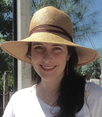
Katie Gostic
Gostic conducted research for the flu project during the summer of 2015 when she was visiting her boyfriend, a tropical biologist, in Alamos, Sonora, Mexico. Credit: Charlie de la Rosa.

As an undergraduate student at Princeton University, Gostic was originally pursuing a degree in engineering. Her focus shifted to biology after taking an infectious disease modeling class. Gostic’s background in math and programming allows her to take large, complex pre-existing data sets and reanalyze them using new tools and methods. The result: Information that wasn’t accessible when the data were first collected.

Now a graduate researcher in the ecology and evolutionary biology lab of James Lloyd-Smith Exit icon, Gostic studies infectious diseases. The lab builds mathematical models to investigate zoonotic diseases—diseases that animals can transmit to humans but that humans don’t frequently spread between each other. Examples include diseases caused by Leptospira, a type of bacteria that infects household pets and many other animals, and monkeypox, a virus whose transmission to humans is increasing since the eradication of smallpox. The lab also studies bird flus, a category of flu viruses that infect birds and other animals and only occasionally jump to people. A very small number of cases of human-to-human transmission of bird flus have been recorded. However, if a bird flu virus mutated in a way that allowed it to spread among humans, it could cause a pandemic. Continue reading “Student Researcher Finds New Clues About Flu with Old Data”

On this Darwin Day, Evolutionary Geneticist Dr. Dan Janes Discusses the Scientific Contributions of Charles Darwin

3 comments

This Sunday, February 12, is Darwin Day—an occasion to recognize the scientific contributions of 19th-century naturalist Charles Darwin. In this video (originally posted on Darwin Day 2016), our own evolutionary geneticist, Dan Janes, answers questions about Darwin and the role of evolution in health and biomedicine.

Continue reading “On this Darwin Day, Evolutionary Geneticist Dr. Dan Janes Discusses the Scientific Contributions of Charles Darwin”

Not Discounting His Future: Austin Works Toward His Goals

0 comments

This is the second post in a new series highlighting NIGMS’ efforts toward developing a robust, diverse and well-trained scientific workforce.

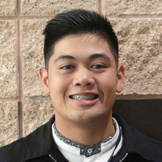
Austin Phanouvong
Credit: Christa Reynolds.
Austin Phanouvong
Academic Institution: Portland State University
Major: Biology
Minor: Chemistry
Mentor: Suzanne Mitchell
Favorite Book: The Alchemist, by Paulo Coelho
Favorite Food: Sushi
Hobbies: Hiking, cooking and traveling

BUILD and the Diversity Program Consortium

The Diversity Program Consortium (DPC) aims to enhance diversity in the biomedical research workforce through improved recruitment, training and mentoring nationwide. It comprises three integrated programs—Building Infrastructure Leading to Diversity (BUILD), which implements activities at student, faculty and institutional levels; the National Research Mentoring Network (NRMN), which provides mentoring and career development opportunities for scientists at all levels; and the Coordination and Evaluation Center (CEC), which is responsible for evaluating and coordinating DPC activities.

Ten undergraduate institutions across the United States have received BUILD grants, and together, they serve a diverse population. Each BUILD site has developed a unique program intended to engage and prepare students for success in the biomedical sciences and maximize opportunities for research training and faculty development. BUILD programs include everything from curricular redesign, lab renovations, faculty training and research grants, to student career development, mentoring and research-intensive summer programs.

Austin Phanouvong, 21, likes biology because it teaches him about life.

“Even the way we walk, the way we breathe – there’s all these little components to it that we don’t even think about but they’re very helpful, and one hiccup in that system can lead to many diseases and sicknesses,” Phanouvong says.

Born and raised in Portland, Oregon, Phanouvong is a senior at Portland State University (PSU), where he is in the Building Infrastructure Leading to Diversity (BUILD) EXITO program. As a student in BUILD EXITO Exit icon (which stands for “Enhancing Cross-Disciplinary Infrastructure and Training at Oregon”), Phanouvong takes biomedical science courses, conducts research and participates in professional development workshops and seminars. He has learned about preparing a CV and writing personal statements. In spring 2017, Phanouvong will graduate with a major in biology and a minor in chemistry. He will continue to pursue research opportunities as he applies for medical school, and he hasn’t ruled out the possibility of pursuing a Ph.D. in the future.

Since high school, Phanouvong’s desire has been to help people through a healthcare career. Originally, he was interested in nursing, but he decided he could push his career plans and education farther by going to medical school. Because of his interest in hands-on research, last year Phanouvong’s honors thesis advisor recommended he apply for the BUILD program, and Phanouvong realized BUILD EXITO would be a great opportunity to engage in research as an undergraduate student. Continue reading “Not Discounting His Future: Austin Works Toward His Goals”

Cool Image: Inside a Biofilm Build-up

1 comment
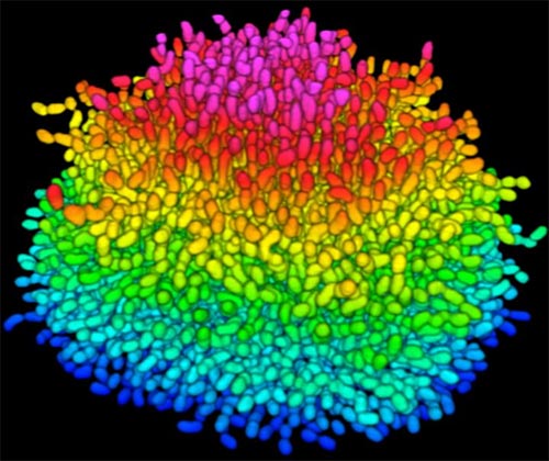
A growing Vibrio cholerae biofilm.

A growing Vibrio cholerae biofilm. Each slightly curved comma shape represents an individual bacterium from assembled confocal microscopy images. Different colors show each bacterium’s position in the biofilm in relation to the surface on which the film is growing. Credit: Jing Yan, Ph.D., and Bonnie Bassler, Ph.D., Department of Molecular Biology, Princeton University, Princeton, NJ.

Bacteria use many methods to overcome threats in their environment. One of these ways is forming colonies called biofilms on surfaces of objects. Often appearing like the bubble-shaped fortress represented in this image, biofilms enable bacteria to withstand attacks, compete for space and survive fluctuations in nutrient supply. Bacteria aggregated within biofilms inside our bodies, for example, resist antibiotic therapy more effectively than free swimming cells, making infections difficult to treat. On the other hand, biofilms are also useful for making microbial fuel cells and for waste-water treatment. Learning how biofilms work, therefore, could provide essential tools in our ongoing battle against disease-causing agents and in our efforts to harness beneficial bacterial behaviors. Researchers are now using new imaging techniques to watch how biofilms grow, cell by cell, and to identify more effective ways of disrupting or fostering them.

Until now, poor imaging resolution meant that scientists could not see what individual bacteria in the films are up to as the biofilms grow. The issue is that bacteria are tiny, making imaging each cell, as well as the ability to distinguish each cell in the biofilm community, problematic. Continue reading “Cool Image: Inside a Biofilm Build-up”

Cool Tools: Pushing the Limits of High-Resolution Microscopy

1 comment

Cell biologists would love to shrink themselves down and actually see, touch and hear the inner workings of cells. Because that’s impossible, they have developed an ever-growing collection of microscopes to study cellular innards from the outside. Using these powerful tools, researchers can exhaustively inventory the molecular bits and pieces that make up cells, eavesdrop on cellular communication and spy on cells as they adapt to changing environments.

In recent years, scientists have developed new cellular imaging techniques that allow them to visualize samples in ways and at levels of detail never before possible. Many of these techniques build upon the power of electron microscopy (EM) to see ever smaller details.

Unlike traditional light microscopy, EM uses electrons, not light, to create an image. To do so, EM accelerates electrons in a vacuum, shoots them out of an electron gun and focuses them with doughnut-shaped magnets onto a sample. When electrons bombard the sample, some pass though without being absorbed while others are scattered. The transmitted electrons land on a detector and produce an image, just as light strikes a detector (or film) in a camera to create a photograph.

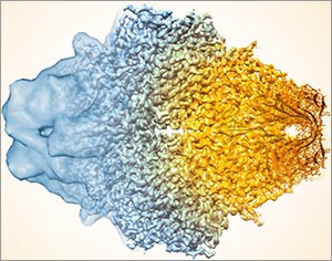
This image, showing a single protein molecule, is a montage. It was created to demonstrate how dramatically cryo-EM has improved in recent years. In the past, cryo-EM was only able to obtain a blobby approximation of a molecule’s shape, like that shown on the far left. Now, the technique yields exquisitely detailed images in which individual atoms are nearly visible (far right). Color is artificially applied. Credit: Veronica Falconieri, Subramaniam Lab, National Cancer Institute.

Transmission electron microscopes can magnify objects more than 10 million times, enabling scientists to see the outline and some details of cells, viruses and even some large molecules. A relatively new form of transmission electron microscopy called cryo-EM enables scientists to view specimens in their natural or near-natural state without the need for dyes or stains.

In cryo-EM—the prefix cry- means “cold” or “freezing”—scientists freeze a biological sample so rapidly that water molecules do not have time to form ice crystals, which could shove cellular materials out of their normal place. Cold samples are more stable and can be imaged many times over, allowing researchers to iteratively refine the image, remove artifacts and produce even sharper images than ever before. Continue reading “Cool Tools: Pushing the Limits of High-Resolution Microscopy”

Interview With a Scientist: Thomas O’Halloran, Metal Maestro

2 comments

Inside our bodies is a surprising amount of metal. Not enough to set off the scanners at the airport or make us rich, but enough to fill each of our cells with billions of metal ions, including calcium, iron, copper and zinc. These ions facilitate critical biological functions.

However, too much of any metal can be toxic, while too little can cause disease. Our cells carefully monitor and control their metal content using a whole series of proteins that bind, sense and transport metal ions.

Keeping tabs on why and how metals flow into and out of our cells is a passion of Thomas O’Halloran Exit icon, professor of chemistry and molecular biosciences at Northwestern University in Illinois. For the past three decades, O’Halloran has investigated how fluctuations in the amount of metal ions inside cells influence gene expression, cell growth and other vital functions. Using a variety of approaches, he has uncovered new types of proteins that bind metal ions and investigated the role that imbalances in these ions play in a number of disease-related physiological processes.

One recent focus of his studies has been how zinc regulates oocyte (egg cell) maturation and fertilization. Ultimately, his work could help us better understand infertility, cancer and certain bacterial infections.

Getting It Done: Chyann’s Path to Graduate School and Research

2 comments

This is the first post in a new series highlighting NIGMS’ efforts toward developing a robust, diverse and well-trained scientific workforce.

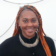
Chyann Richard
Credit: Christa Reynolds.
Chyann Richard
Academic Institution: California State University, Long Beach
Major: Psychology
Mentor: Michelle Barrack
Favorite Book: Outliers, by Malcolm Gladwell
Favorite sports team: Los Angeles Lakers
Favorite music: R&B

“A lot of people would never guess that I’m in research and I take pride in that. I want to be able to represent people that don’t even go this far,” Chyann Richard, 20, says.

BUILD and the Diversity Program Consortium

The Diversity Program Consortium (DPC) aims to enhance diversity in the biomedical research workforce through improved recruitment, training and mentoring nationwide. It comprises three integrated programs—Building Infrastructure Leading to Diversity (BUILD), which implements activities at student, faculty and institutional levels; the National Research Mentoring Network (NRMN), which provides mentoring and career development opportunities for scientists at all levels; and the Coordination and Evaluation Center (CEC), which is responsible for evaluating and coordinating DPC activities.

Ten undergraduate institutions across the United States have received BUILD grants, and together, they serve a diverse population. Each BUILD site has developed a unique program intended to engage and prepare students for success in the biomedical sciences and maximize opportunities for research training and faculty development. BUILD programs include everything from curricular redesign, lab renovations, faculty training and research grants, to student career development, mentoring and research-intensive summer programs.

Currently a junior at California State University, Long Beach (CSULB), Richard is majoring in psychology. After she graduates with a bachelor’s degree in 2018, she plans to continue to a Ph.D. program and do research in behavioral neuroscience.

Richard is among a select group of undergraduate college students participating in the Building Infrastructure Leading to Diversity (BUILD) program. The BUILD programs focus on finding innovative approaches to increase student engagement in the biomedical sciences, through interventions at student, faculty and institutional levels. As a BUILD scholar, Richard is conducting laboratory research and preparing for graduate school through career development seminars, presentations and other activities.

Richard loves how research introduces her to new ideas and allows her to share these concepts with others, including her parents.

“Because they’ve been teaching me my whole life … now I’ve got a one-up because I know about research and they don’t. That’s really fun,” she says.

Richard’s interest in behavioral neuroscience is both personal and scientific. During Richard’s junior year of high school, her mother was diagnosed with generalized anxiety disorder. This sparked Richard to take an Advanced Placement (AP) psychology course, where she began learning about the prevalence of and treatments for such disorders.

“[The class] started bringing [my mom’s condition] into perspective – that it wasn’t just some random thing,” Richard says. Continue reading “Getting It Done: Chyann’s Path to Graduate School and Research”