Author: Abbey Bigler-Coyne

Headshot of Abbey Bigler

Abbey is a science writer who enjoys making important biological science and public health information accessible to everyone.

Posts by Abbey Bigler-Coyne

Make Like a Cell and Split: Comparing Mitosis and Meiosis

12 comments

Your body is made up of trillions of cells that all originate from just one—a fertilized egg. The massive multiplication of cells after conception is possible thanks to cell division, which occurs when one cell splits into two. Cell division not only enables growth but also replaces damaged or dead cells and makes reproduction possible. There are two kinds of cell division: mitosis and meiosis.

On the left, a cell goes through the stages of mitosis to split into two cells that each have two sets of chromosomes. On the right, a cell goes through the phases of meiosis to divide into four cells that each have a single set of chromosomes. Mitosis is shown on the left, and meiosis is shown on the right. Credit: Judith Stoffer. Click to enlarge
Continue reading “Make Like a Cell and Split: Comparing Mitosis and Meiosis”

Researcher Shares Science en Español and Builds a Community

0 comments
A headshot of Dr. Ramos-Benítez.
Dr. Marcos Ramos-Benítez. Credit: Courtesy of Dr. Ramos-Benítez.

“For me, science is the perfect way to harmonize creative thinking and critical thinking,” says Marcos Ramos-Benítez, Ph.D., a fellow in the NIGMS Postdoctoral Research Associate Training (PRAT) program.

Dr. Ramos-Benítez researches interactions between pathogens—such as the viruses that cause Ebola and COVID-19—and their hosts. He’s also the founder and president of Ciencia en tus Manos (“Science in Your Hands”), a nonprofit organization that presents scientific information in Spanish and aims to provide a community to support the next generation of scientists.

Continue reading “Researcher Shares Science en Español and Builds a Community”

Navajo Students Engage With Public Health Research Through NARCH

0 comments

Navajo students are contributing to public health efforts in diabetes, COVID-19, domestic violence, and maternal and child health through the Navajo Native American Research Center for Health (NARCH) Partnership. “Our goal is to really enhance the educational pathways available to Navajo students from high school to graduate school and beyond,” says Mark Bauer, Ph.D., a co-director of the Navajo NARCH Partnership and professor at Diné College—a tribal college on the Navajo Nation. (Diné means “the people” and is how Navajo people refer to themselves in their native language.)

Continue reading “Navajo Students Engage With Public Health Research Through NARCH”

Cool Images: Beautiful Bits of Blue

1 comment

Most cells are naturally colorless, which is why scientists often use fluorescent tags and other tools to color cell structures and make them easier to study. (Check out the Pathways imaging issue for more on scientific imaging techniques). Here, we’re showcasing cell images that feature shades of blue. Visit our Image and Video Gallery for additional images of cells in all the colors of the rainbow, as well as other scientific photos, illustrations, and videos.

Cool Images
Many blue circles, each surrounded by yellow dots. All the structures are encased in gray webs.
Credit: Torsten Wittmann, University of California, San Francisco.
A large circle made up of light blue dots, with darker blue spots underneath and outside of it.
Credit: California Institute for Regenerative Medicine.
Two blue circles encapsulated in red threads and surrounded by other scattered blue circles.
Credit: Deepak Srivastava, Gladstone Institutes, via CIRM.

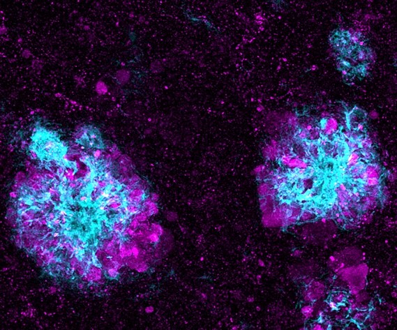
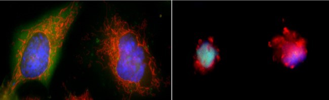
This image shows lysosomes (purple) within nerve cells that surround amyloid plaques (blue) in a research model of Alzheimer’s disease. Lysosomes help the body dispose of proteins and other molecules that have become damaged or worn out. Scientists have linked the accumulation of lysosomes around amyloid plaques to impaired waste disposal in nerve cells. This impairment ultimately causes nerve cell death, a hallmark of Alzheimer’s disease.

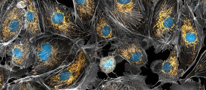
Mitochondria appear in yellow and cell nuclei in blue in this photo of cow cells. The gray webs are the cells’ cytoskeletons. Mitochondria generate energy, nuclei store DNA, and the cytoskeleton gives cells shape and support.

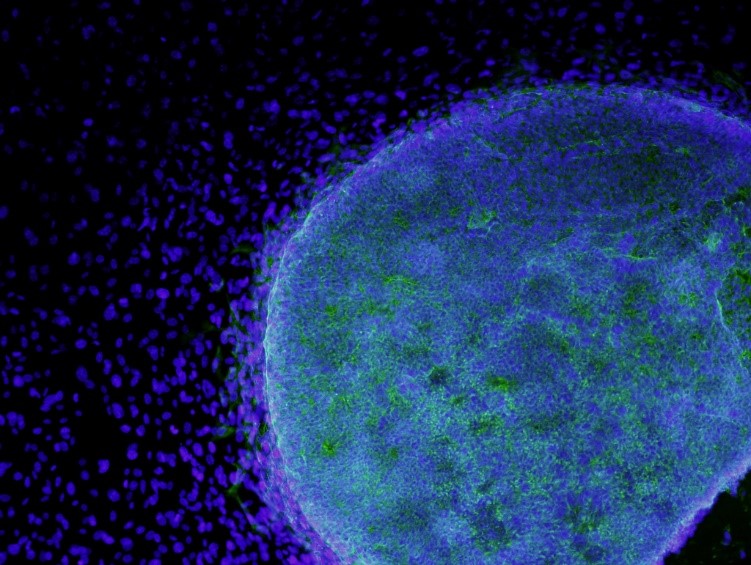
Here, stem cells (light blue) are growing on fibroblasts (dark blue). Stem cells are of great interest to researchers because they can develop into many different cell types. Fibroblasts are the most common cell type in connective tissue. They secrete collagen proteins that help build structural frameworks, and they play an important role in wound healing.

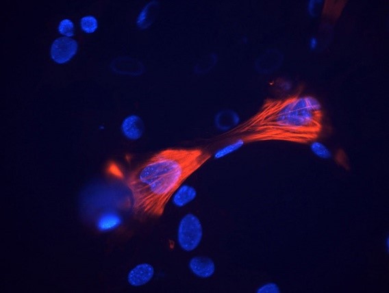
These smooth muscle cells were grown from stem cells. Smooth muscle cells are found in the walls of certain organs, such as the stomach, and can’t be controlled voluntarily. Red indicates smooth muscle proteins, and blue indicates nuclei.
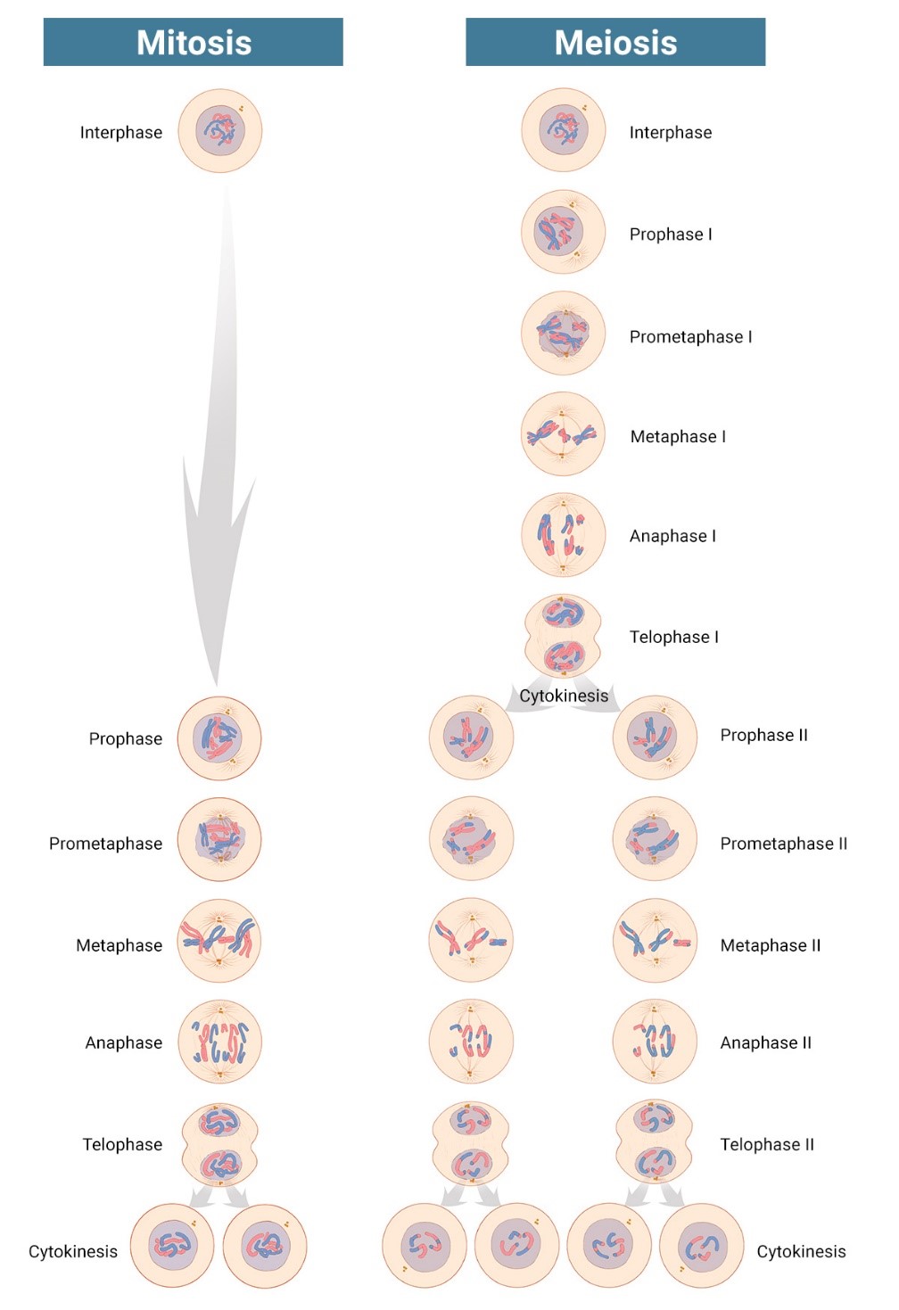
Biology Beyond the Lab: Using Computers to Study Life

1 comment
A headshot of Dr. Melissa Wilson.
Learn more about Dr. Melissa Wilson’s computational biology research in another Biomedical Beat blog post. Credit: Jacob Sahertian, ASU.

“You’re not going to be able to do biology without understanding programming in the future,” Melissa Wilson, Ph.D., an associate professor of genomics, evolution, and bioinformatics at Arizona State University, said in her 2019 NIGMS Early Career Investigator Lecture. “You don’t have to be an expert programmer. But without understanding programming, I can assert you won’t be able to do biology in the next 20 years.”

A growing number of researchers, like Dr. Wilson, are studying biology using computers and mathematical methods. Some of them started in traditional biology or other life science labs, while others studied computer science or math first. Here, we’re featuring two researchers who took different paths to computational biology.

Continue reading “Biology Beyond the Lab: Using Computers to Study Life”

In Other Words: Translation Isn’t Only for Languages

0 comments

In everyday use, most people understand translation to mean converting words from one language to another. But when biologists talk about translation, they mean the process of making proteins based on the genetic information encoded in messenger RNA (mRNA). Proteins are essential for virtually every process in our bodies, from transporting oxygen to defending against infection, so translation is vital for keeping us alive and healthy.

Below the title “Translation: In Other Words,” two images are separated by a jagged line. On the left, is a large speech bubble with the word “hello” surrounded by smaller speech bubbles with greetings in other languages, and on the right is a ribosome producing a protein. Under the images, text reads, “Did you know? In biomedical science, translation refers to the process of making proteins based on genetic information encoded in messenger RNA.”
Credit: NIGMS.
Continue reading “In Other Words: Translation Isn’t Only for Languages”

Science Snippet: Apoptosis Explained

5 comments

Apoptosis is the process by which cells in the body die in a controlled and predictable way because they have DNA damage or are no longer needed. The term comes from a Greek word meaning “falling off,” as in leaves falling from a tree.

When a cell undergoes apoptosis, it shrinks and pulls away from its neighbors. As the cytoskeleton that gives it shape and structure collapses, the envelope around the cell’s nucleus breaks down, and its DNA breaks into pieces. Its surface changes, signaling its death to other cells and leading a healthy cell to engulf the dying one and recycle its components.

On the left, two large cells with clear, smooth edges. On the right, two smaller cells with ragged edges.
Two cells in a healthy state (left) and entering apoptosis (right). Credit: Hogan Tang of the Denise Montell Lab, Johns Hopkins School of Medicine.
Continue reading “Science Snippet: Apoptosis Explained”

Cool Video: A Biological Lava Lamp

1 comment
Several spheres contorting and lighting up inside a cone-shaped structure.
Credit: Jasmin Imran Alsous and Jonathan Jackson, Martin Lab, Massachusetts Institute of Technology.

What looks like a bubbling lava lamp is actually part of an egg cell’s maturation process. In many animals, the egg cell develops alongside sister cells. These sister cells are called nurse cells in the fruit fly (Drosophila melanogaster), and their job is to “nurse” an immature egg cell, or oocyte. Toward the end of oocyte development, the nurse cells transfer all their contents into the oocyte in a process called nurse cell dumping. This video captures this transfer, showing significant shape changes on the part of the nurse cells (blue), which are powered by wavelike activity of the protein myosin (red).

Researchers created the video using a confocal laser scanning microscope. Learn about this type of microscope and other scientific imaging tools by stepping into our virtual imaging lab, and check out more basic science videos and photos in the NIGMS Image and Video Gallery.

This post is a great supplement to Pathways: The Imaging Issue.

The video was taken using a confocal laser scanning microscopy (sometimes shortened to just “confocal microscopy”), one of the techniques mentioned in the Pathways timeline (1970s).

Learn more in our Educator’s Corner.

More Than 25 Years of Competition and Collaboration Advance the Prediction of Protein Shapes

0 comments

Proteins (such as hemoglobin, actin, and amylase) are workhorse molecules that contribute to virtually every activity in the body. Some of proteins’ many jobs include carrying oxygen from your lungs to the rest of your body (hemoglobin), allowing your muscles to move (actin and myosin), and digesting your food (amylase, pepsin, and lactase). All proteins are made up of chains of amino acids that fold into specific 3D structures, and each protein’s structure allows it to perform its distinct job. Proteins that are misfolded or misshapen can cause diseases such as Parkinson’s or cataracts.

While it’s straightforward to use the genetic code to predict amino acid sequences of proteins from gene sequences, the vast diversity of protein shapes and many factors that influence a protein’s 3D structure make it much more complicated to create simple folding rules that could be used to predict proteins’ structures from these sequences. Scientists have worked on this problem for nearly 50 years, and NIGMS has supported many of their efforts, including the Critical Assessment of Structure Prediction (CASP) program.

Continue reading “More Than 25 Years of Competition and Collaboration Advance the Prediction of Protein Shapes”

Career Conversations: Q&A with Molecular Biologist Hong Liu

0 comments
A scientist wearing a lab coat and holding a pipette in front of a workbench with scientific instruments.
Dr. Hong Liu in the lab.
Credit: Courtesy of Dr. Hong Liu.

“A scientific career is really worth it,” says Hong Liu, Ph.D., an assistant professor of biochemistry and molecular biology at Tulane University School of Medicine in New Orleans, Louisiana. Check out the highlights of our interview with Dr. Liu below to learn about his journey as a scientist and his advice for students.

Q: What makes a career in science exciting?

A: I think there are at least two things that make a science career very exciting. The first is that doing science means you have freedom to explore a lot of new ideas. The second thing is it’s rewarding. The “rewarding” I’m talking about here is not like how much money you can make. It’s rewarding in the answers you find and the new knowledge you reveal.

Continue reading “Career Conversations: Q&A with Molecular Biologist Hong Liu”