You’ve probably heard news stories and other talk about CRISPR. If you’re not a scientist—well, even if you are—it can seem a bit complex. Here’s a brief recap of what it’s all about.
In 1987, scientists noticed weird, repeating sequences of DNA in bacteria. In 2002, the abbreviation CRISPR was coined to describe the genetic oddity. By 2006, it was clear that bacteria use CRISPR to defend themselves against viruses. By 2012, scientists realized that they could modify the bacterial strategy to create a gene-editing tool. Since then, CRISPR has been used in countless laboratory studies to understand basic biology and to study whether it’s possible to correct faulty genes that cause disease. Here’s an illustration of how the technique works.
Six NIGMS grantees are among this year’s winners of the Presidential Award for Excellence in Science, Mathematics and Engineering Mentoring (PAESMEM). The award was established by the White House in 1995. This year, it went to 27 individuals and 14 organizations.
PAESMEM recipients were honored during a 3-day event in Washington, D.C. The event featured a gala presentation ceremony and a White House tour. In addition, each winner received a $10,000 grant from the National Science Foundation, which manages PAESMEM on behalf of the White House Office of Science and Technology Policy.
The event also included the first-ever White House State-Federal STEM Education Summit. During the summit, awardees joined leaders in education and workforce development from across the nation, including U.S. territories and several Native American tribes, to discuss trends and future priorities in STEM education. The discussions will inform the development of the next Federal STEM Education 5-Year Strategic Plan, which must be updated every 5 years according to the America COMPETES Reauthorization Act of 2010.
Many researchers who search for anti-cancer drugs have labs filled with chemicals and tissue samples. Not Rommie Amaro. Her work uses computers to analyze the shape and behavior of a protein called p53. Defective versions of p53 are associated with more human cancers than any other malfunctioning protein.
Our sense of touch provides us with bits of information about our surroundings that inform the decisions we make. When we touch something, our nervous system transmits signals through nerve endings that feed information to our brain. This enables us to sense the stimulus and take the appropriate action, like drawing back quickly when we touch a hot stovetop.
Bacteria are single cells and lack a nervous system. But like us, they rely on their “sense” of touch to make decisions—or at least change their behavior. For example, bacteria’s sense of touch is believed to trigger the cells to form colonies, called biofilms, on surfaces they make contact with. Bacteria may form biofilms as a way to defend themselves, share limited nutrients, or simply to prevent being washed away in a flowing liquid.
Humans can be harmed by biofilms because these colonies serve as a reservoir of disease-causing cells that are responsible for high rates of human infection. Biofilms can protect at least some cells from being affected by antibiotics. The surviving reservoir of bacteria then have more time to evolve resistance to antibiotics.
At the same time, some biofilms can be valuable; for example, they help to break down waste in water treatment plants and to drive electrical current as part of microbial fuel cells.
Until recently, scientists thought that bacteria formed biofilms and caused infections in response to chemical signals they received from their environments. But research in 2014 showed that the bacterium Pseudomonas aeruginosa could infect a variety of living tissues—from plants to many kinds of animals—simply by making contact with them. In the past year, multiple groups of investigators have learned more about how bacteria sense that they have touched a surface and how that sense translates to changes in their behavior. This understanding could lead to new ways of preventing infections or harmful biofilm formation.
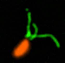
Making Contact
Pili (green) on cells from the bacterium Caulobacter crescentus (orange). Scientists used a fluorescent dye to stain pili so they could watch the structures extend and retract. Credit: Courtney Ellison, Indiana University.
When they first make contact with a surface, bacteria change from free-ranging, swimming cells to stationary ones that secrete a sticky substance, tethering them in one place. To form a biofilm, they begin replicating, creating an organized mass stable enough to resist shaking and to repel potential invaders (see https://biobeat.nigms.nih.gov/2017/01/cool-image-inside-a-biofilm-build-up/).
How do swimming bacteria sense that they have found a potential surface to colonize? Working with the bacterium Caulobacter crescentus, Indiana University Ph.D. student Courtney Ellison and her colleagues, under the direction of professor of biology and NIGMS grantee Yves Brun, recently showed that hair-like structures on the cell’s surface, called pili, play a role here. The researchers found that as a bacterial cell swims in a fluid, its pili are constantly stretching out and retracting. When the cell makes contact with a surface, the pili stop moving, start producing a sticky substance and use it to hold onto the surface. Continue reading “Feeling Out Bacteria’s Sense of Touch”
How “membrane-less” organelles help with key cellular functions
Scientists have long known that animal and plant cells have specialized subdivisions called organelles. These organelles are surrounded by a semi-permeable barrier, called a membrane, that both organizes the organelles and insulates them from the rest of the cell’s mix of proteins, salt, and water. This set-up helps to make cells efficient and productive, aiding in energy production and other specialized functions. But, because of their semi-permeable membranes, organelles can’t regroup and reform in response to stress or other outside changes. Cells need a rapid response team working alongside the membrane-bound organelles to meet these fluctuating needs. Until recently, who those rapid responders were and how they worked has been a mystery.
Recent research has led biologists to learn that the inside of a cell or an organelle is not just a lot of different molecules dissolved in water. Instead, we now know that cells contain many pockets of liquid droplets (one type of liquid surrounded by a liquid of different density) with specialized composition and function that are not surrounded by membranes. Because these “membrane-less organelles” are not confined, they can rapidly come together in response to chemical signals, such as those that indicate stress, and equally rapidly fall apart when they are no longer needed, or when the cell is about to divide. This enables membrane-less organelles to be “rapid responders.” They can have complex, multilayered structures that help them to perform many critical cell functions with multiple steps, just like membrane-bound organelles. Scientists even suspect that the way these organelles form as droplets may shed light on how life on Earth first took shape (see sidebar “Could This Be How Life First Took Shape?” at bottom of page).
The Many Membrane-less Organelles
Scientists have identified more than a dozen membrane-less organelles at work in mammalian cells. Several kinds found inside the nucleus—including nuclear speckles, paraspeckles, and Cajal bodies—help with cell growth, stress response, the metabolizing (breaking down) of RNA, and the control of gene expression—the process by which information in a gene is used in the synthesis of a protein. Out in the cytoplasm, P-bodies, germ granules, and stress granules are membrane-less organelles that are involved in metabolizing or protecting messenger RNA (mRNA), controlling which mRNAs are made into proteins, and in maintaining balance, or homeostasis, of the cell’s overall health.
The nucleolus, located inside the nucleus, is probably the largest of the membrane-less organelles. It acts as a factory to assemble ribosomes, the giant molecular machines that “translate” messenger RNAs to make all cellular proteins.
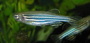
Name: Danio rerio Hometown: Freshwater ponds and rivers of India, Nepal, and neighboring countries Occupation: Research Long-term goal: Solving the basic mysteries of life Work site: More than 600 science labs worldwide
That’s me and some other zebrafish, swimming in a tank in one of the more than 600 labs around the world that use us to study embryo development, genetics, and all kinds of human diseases. Credit: Wikimedia Commons, Azul.
Apart from the tell-tale stripes that give me my nickname, zebrafish, I look a lot like your standard minnow swimming in the shallows of any pond, lake, or river. But I like to think I’m more important than that. In fact, researchers around the world have turned to me and my extended family to understand some of the most basic mysteries of life. From studying us, they’re learning about how embryos develop, how cancer works, and whether someday humans might be able to rebuild a heart, repair a spinal cord injury, or regrow a severed limb.
Why us? Because zebrafish are pretty special and researchers think we’re easy to work with. First, unlike your standard lab mouse or rat, we lay lots of eggs, producing baby fish that grow up fast. We develop outside our mothers and go from egg to embryo to free-swimming larva in just 3 days (check out this video of how we grow, cell by cell, during the first 24 hours). Within 3 months, we’re fully mature.
Not only do zebrafish moms have many babies at the same time, and not only do these babies grow up quickly, but our eggs and embryos are see-through, so scientists can literally watch us grow one cell at a time. We stay mostly transparent for a few weeks after hatching. That makes it super easy for scientists to monitor us for both normal and abnormal development. In fact, scientists have learned how to turn off the genes that give our skin its color. These zebrafish, named casper, after the “friendly ghost” of cartoon fame, stay semi-transparent, or translucent, through adulthood.
And last, but certainly not least, did I mention that we can regenerate? If parts of my body are damaged, even to a significant degree, they can regrow. This holds true for my heart, fins, spinal cord, and even brain tissue. Our regenerative capacity is seemingly unlimited; my caudal fin, for example, can grow back dozens of times. Continue reading “Zebrafish Scrapbook”
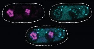
These glowing images of yeast (Schizosaccharomyces kambucha) reproductive cells show an example of a selfish gene at work. Here, the selfish gene boosts its chances of being passed to the next generation by producing both a toxin (stained cyan) and an antitoxin (stained magenta). Cells with a copy of the selfish gene are protected by the antitoxin, left and bottom ovals. Those without the selfish gene are destroyed by the toxin. Scientists suspect that selfish genes could be operating throughout many organisms’ genomes, possibly having a major impact on how genetic material is inherited over generations. Credit: Image courtesy of María Angélica Bravo Núñez and Nicole Nuckolls.
There’s an old saying that rules are meant to be broken. In the 1860s, Gregor Mendel found that each copy of a gene in an organism has an equal chance of being passed to the next generation. According to this simple rule, each version of a gene gets passed to offspring with the same frequency. The natural selection process can then operate efficiently, favoring the genes that produce an advantage for an organism’s survival or reproductive success and, over successive generations, eliminating genes from the gene pool that bring a disadvantage.
Of course, the way organisms inherit genes is not as straightforward as Mendel’s work predicted. In natural systems, inheritance often fails to follow the rules. One culprit scientists are identifying again and again are what are called “selfish genes”: one or more genes that have evolved a method of skewing inheritance in their favor.
Scientists refer to these selfish genes—which are often complexes of multiple genes working together—as “selfish” because they enhance their own transmission to the next generation, sometimes by killing off any of the organism’s reproductive cells that lack copies of those genes. Using a variety of techniques, the genes are effective at passing themselves on to future generations. However, their methods set up a conflict within the organism by damaging its fertility; overall, fewer reproductive cells or offspring survive to produce a new generation.
Selfish genes are a challenge for scientists to identify, but researchers say that knowing more about these genes could help explain a range of genetic mysteries, from causes of infertility to details on how species evolve. The methods these genes use could also be harnessed to help control the reproduction of certain populations such as mosquitos that spread disease.
One recently described selfish gene system is found in the yeast cells pictured above. Sarah Zanders and her colleagues at the Stowers Institute for Medical Research in Kansas City, Missouri, and the Fred Hutchinson Cancer Research Center in Seattle, Washington, study selfish gene systems in yeast to understand how common they are and how they affect a species’ fertility and evolution. “Usually we think about infertility stemming from the good guys failing. For example, a gene that normally promotes fertility could be mutated so that it can no longer do its job,” says Zanders. “But selfish genes are another potential source of infertility. Learning general principles about selfish genes in simple models will guide future searches for selfish genes that could be contributing to human infertility.”
Recently, the scientists discovered a single selfish gene, wtf4, that encodes both a toxin and an antitoxin protein. When yeast produce their reproductive cells, called spores, the wtf4 toxin protein is released into the immediate vicinity, but the antitoxin remains inside spores that contain a copy of wtf4. The toxin destroys all the spores that don’t have the antitoxin protein. Although the yeast has fewer spores—and therefore reduced fertility—each spore carries wtf4, ensuring that the gene will be passed to the next generation of yeast.
This is the fourth post in a new series highlighting NIGMS’ efforts toward developing a robust, diverse and well-trained scientific workforce.
Marina Z. Nakhla Hometown: West Los Angeles, California Blogs For:Ottobock “Life in Motion,” a forum for the amputee community, where she’s covered topics ranging from medical insurance to dating. Influential Book: The Catcher in the Rye by J.D. Salinger Favorite TV Show: Grey’s Anatomy Languages: English and Arabic Unusual Fact: Gets a new pair of legs every year or two
Nakhla at her graduation from California State University, Northridge, where she graduated with a B.A. in psychology with honors. She is currently a second-year master’s student there studying clinical psychology. Credit: Christina Nakhla.
When Marina Z. Nakhla was just a toddler, she lost both of her legs. Now 22 and a graduate student at California State University, Northridge (CSUN), she has hurdled obstacles most of us never face.
Nakhla conducts research to better understand the decrease in mental abilities experienced by people with brain diseases. She is a scholar in CSUN’s Research Initiative for Scientific Enhancement (RISE) Program. This training program aims to enrich and diversify the pool of future biomedical researchers. Her long-term goal is to earn a Ph.D., to work as a clinical psychologist and to continue conducting research in neuropsychology. Along the way, she aspires to be a leader to her peers and an advocate for underrepresented people, particularly those with disabilities.
I first learned about Nakhla from an email message titled “CSUN RISE Student.” The acronym, pronounced “see [the] sun rise,” is an apt motto for a program that prepares students for a bright future in science. I believe it also encapsulates Nakhla’s positive, forward-looking mindset, despite the obstacles she has faced. Here’s her story:
Q: What got you interested in science?
A: Growing up, I was always drawn to science. I enjoyed learning how things work. I first became interested in psychology after reading The Catcher in the Rye in high school. I was so intrigued by Holden Caulfield’s thought processes and experiences of alienation and depression, despite the fact that he came from a wealthy family and went to a good school.
Why are some people more prone to experiencing depression? Why are some peoples’ thought processes so different than others? What factors contribute to resiliency? How can we help these people? These questions also made me think about the significant adversities that I had personally experienced. My desire to know more about the brain, as well as my personal experiences, instilled my passion to make a difference in others’ lives through science. Continue reading “RISE-ing Above: Embracing Physical Disability in the Lab”
Medications are designed to treat diseases and make us healthier. But our bodies don’t know that. To them, medications are merely foreign molecules that need to be removed.
Before our bodies can get rid of these drug molecules, enzymes in the liver do the chemical work of preparing the molecules for removal. There are hundreds of different versions of these drug-processing enzymes. Some versions work quickly, others work slowly. In some cases, the versions you have determine how well a medication works for you, and whether you experience side effects from it.
Namandjé Bumpus, a researcher at Johns Hopkins University School of Medicine, is interested in how human bodies respond to HIV medications. She studies the enzymes that process these drugs. Her research team discovered that a genetic variant of a liver enzyme impacts the way some people handle a particular HIV drug. This variant is found in around 80 percent of people of European descent. She describes her work in this video.