Author: Emily Carlson

Headshot of emily Carlson

Posts by Emily Carlson

Field Focus: Asking Our Expert About Modeling Ebola

0 comments
Irene Eckstrand
NIGMS’ Irene Eckstrand answers questions about modeling Ebola. Credit: National Institute of General Medical Sciences.

Ebola is the focus of many NIH-supported research efforts, from analyzing the genetics of virus samples to evaluating the safety and effectiveness of treatments and vaccines. Researchers involved in our Models of Infectious Disease Agent Study, or MIDAS, have been using computational methods to forecast the potential course of the outbreak and the impact of various intervention strategies.

Wondering how their work is going, I recently asked our modeling expert Irene Eckstrand a few questions.

How useful are the forecasts?

Forecasts give us a range of possible outcomes. In addition to being a useful public health tool to prepare for an outbreak, they’re an important research tool to test assumptions about how a disease may spread. When we compare the predicted and actual outcomes, we can confirm assumptions, such as the groups of individuals who are more likely to spread the infection to others. Continually doing this helps refine the models and ensure that their forecasts are as accurate as possible.

What are some of the challenges the modelers face?
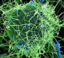
Ebola virus
Ebola virus particles (green) attached to and budding from a cell (blue). Credit: National Institute of Allergy and Infectious Diseases.

We need data to build and test models. The data available from this outbreak have been more limited than in most previous outbreaks of Ebola simply because the public health systems are overwhelmed with sick people, and recording information is a secondary priority.

Another issue with forecasting future trends is incorporating information about the deployment of resources and the implementation of interventions that actually slowed the outbreak. We also need to incorporate changes in people’s behavior. If people think an outbreak is leveling off, they may relax the precautions they’ve been taking—and that could lead to another spike in the disease.

What other Ebola-related projects are the MIDAS modelers working on?

The MIDAS researchers are:

  • Modeling logistical factors such as the number and placement of treatment beds and staffing needs.
  • Tracking potential transmission within and between communities and at hospitals and funerals.
  • Developing a method to estimate the amount of underreporting of case data.
  • Applying models of “tipping points” to look for evidence that the disease curve is slowing.
  • Estimating the number of people who are infected but not symptomatic.
  • Creating new resources for Ebola modelers, including standards for using infectious disease data.
  • Calculating the risk of importation of cases for a wide variety of countries based on travel networks.

How are the modelers working together?

The MIDAS modelers conference call 1-2 times a week to discuss results, modeling strategies, data sources and questions amenable to modeling. They also participate in discussions with government and other academic groups, so there’s a sizable number of modelers working on a wide variety of public health, logistical and basic research questions.

If you’re interested in learning more about Ebola, Irene recommends a video overview of the 2014 outbreak from Penn State University Exit icon and a slide presentation on the myths and realities of the disease from Nigeria’s Kaduna State University Exit icon.

4 Timely Facts About Our Biological Clocks

0 comments
Illustration of circadian rhythm.
Genes and proteins run biological clocks that help keep daily rhythms in synch. Credit: Wikimedia Commons.

After you roll your clocks back by an hour this Sunday, you may feel tired. That’s because our bodies—more specifically, our circadian rhythms—need a little time to adjust. These daily cycles are run by a network of tiny, coordinated biological clocks.

NIGMS’ Mike Sesma tracks circadian rhythm research being conducted in labs across the country, and he shares a few timely details about our internal clocks:

1. They’re incredibly intricate.

Biological clocks are composed of genes and proteins that operate in a feedback loop. Clock genes contain instructions for making clock proteins, whose levels rise and fall in a regular cyclic pattern. This pattern in turn regulates the activity of the genes. Many of the results from circadian rhythm research this year have uncovered more parts of the molecular machinery that fine-tune the clock. Earlier in the month, we blogged about an RNA molecule that cues the internal clock.

2. Every organism has them—from algae to zebras.

Many of the clock genes and proteins are similar across species, allowing researchers to make important findings about human circadian processes by studying the clock components of organisms like fruit flies, bread mold and plants.

3. Whether we’re awake or asleep, our clocks keep ticking.

While they might get temporarily thrown off by changes in light or temperature, our clocks usually can reset themselves.

4. Nearly everything about how our body works is tied to biological clocks.

Our clocks influence alertness, hunger, metabolism, fertility, mood and other physiological conditions. For this reason, clock dysfunction is associated with various disorders, including insomnia, diabetes and depression. Even drug efficacy has been linked to our clocks: Studies have shown that some drugs might be more effective if given earlier in the day.

Learn more:
Circadian Rhythms Fact Sheet

The Sweet Side of Chemistry

0 comments
Glycans
Simple sugars connect in long, branched structures to create glycans. Credit: Stock image.

We’re in the middle of National Chemistry Week Exit icon, which this year focuses on “The Sweet Side of Chemistry—Candy.” Studying sugar chemistry is also relevant to our health.

The sugar in chocolate, taffy and other confections is a type of simple sugar called sucrose. In our bodies, sugars can exist in many forms, ranging from individual units like glucose to long, branched chains known as glycans containing thousands of individual sugar units linked together.

Glycans are involved in just about every aspect of how our cells work. They help make sure proteins are folded into the proper shape so they function correctly. They act as ZIP codes that direct newly made proteins to the right cellular locations. Some divert white blood cells to infection sites, and others serve as anchors for viruses to latch onto.

Because of the diverse and critical roles that glycans play in our bodies, chemists want to learn more about these molecules, with a long-term goal of harnessing them to treat or prevent disease. Read about some of their discoveries in the Why Sugars Might Surprise You article from Inside Life Science and the Life is Sweet article from Findings magazine. The You Are What You Eat chapter from ChemHealthWeb offers more details about the chemistry of sugar.

Mighty Mitochondria

0 comments
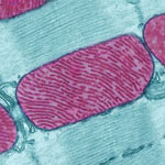
Mitochondria from the heart muscle cell of a rat.
Mitochondria (red) from the heart muscle cell of a rat. Nearly all our cells have these structures. Credit: Thomas Deerinck, National Center for Microscopy and Imaging Research Exit icon.

Meet mitochondria: cellular compartments, or organelles, that are best known as the powerhouses that convert energy from the food we eat into energy that runs a range of biological processes.

As you can see in this close-up of mitochondria from a rat’s heart muscle cell, the organelles have an inner membrane that folds in many places (and that appears here as striations). This folding vastly increases the surface area for energy production. Nearly all our cells have mitochondria, but cells with higher energy demands have more. For instance, a skin cell has just a few hundred, while the cell pictured here has about 5,000.

Scientists are discovering there’s more to mitochondria than meets the eye, especially when it comes to understanding and treating disease.

Read more about mitochondria in this Inside Life Science article.

A Crisper View of the CRISPR Gene-Editing Mechanism

0 comments
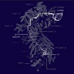
Structural model of the Cascade surveillance machine
Structural model of the Cascade surveillance machine. Credit: Ryan Jackson, Montana State University. Click for larger image

To dismantle the viruses that infect them, bacteria have evolved an immune system that identifies invading viral DNA and signals for its destruction. This gene-editing system is called CRISPR, and it’s being harnessed as a tool for modifying human genes associated with disease.

Taking another important step toward this potential application, researchers now know the structure of a key CRISPR component: a multi-subunit surveillance machine called Cascade that identifies the viral DNA. Shaped like a sea horse, Cascade is composed of 11 proteins and CRISPR-related RNA. A research team led by Blake Wiedenheft of Montana State University used X-ray crystallography and computational analysis to determine Cascade’s configuration. In a complementary study, Scott Bailey of Johns Hopkins University and his colleagues determined the structure of the complex bound to a viral DNA target.

Like blueprints, these structural models help scientists understand how Cascade assembles into an efficient surveillance machine and, more broadly, how the CRISPR system functions and how to adapt it as a tool for basic and clinical research.

Learn more:
Montana State University News Release Exit icon
Johns Hopkins University News Release Exit icon

Intercepting Amyloid-Forming Proteins

0 comments
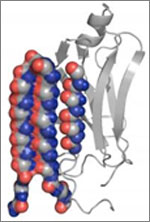
Structure of a protein involved in disease-associated amyloid fibrils.
A molecule targets the intermediary structure of a protein involved in disease-associated amyloid fibrils. Credit: University of Washington.

Alzheimer’s disease, type 2 diabetes and many other illnesses are linked to the buildup of proteins whose structures have changed into shapes that enable the formation of cell-entangling threads called amyloid fibrils. About 10 years ago, researchers led by Valerie Daggett of the University of Washington used computer simulations to suggest that such proteins, on their way to creating fibrils, form an intermediary structure called an alpha sheet that’s even more toxic to cells than fibrils. Now Daggett’s team has experimentally investigated this possibility. The scientists made alpha sheet molecules expected to bind to amyloid-forming proteins in the computationally predicted intermediate state. When they tested the molecules on two amyloid disease-related proteins, they observed a substantial reduction in fibril formation. The work is still very preliminary, but it highlights a potential new avenue for treating a range of amyloid-related diseases.

This work also was funded by NIH’s National Institute of Allergy and Infectious Diseases.

Learn more:
University of Washington News Release Exit icon
Daggett Lab Exit icon
Monster Mash: Protein Folding Gone Wrong Article from Inside Life Science

The “Virtuous Cycle” of Technology and Science

0 comments
A scientist looking through a  microscope. Credit: Stock image.
Whether it’s a microscope, computer program or lab technique, technology is at the heart of biomedical research. Credit: Stock image.

Whether it’s a microscope, computer program or lab technique, technology is at the heart of biomedical research. Its central role is particularly clear from this month’s posts.

Some show how different tools led to basic discoveries with important health applications. For instance, a supercomputer unlocked the secrets of a drug-making enzyme, a software tool identified disease-causing variations among family members and high-powered microscopy revealed a mechanism allowing microtubules—and a cancer drug that targets them—to work.

Another theme featured in several posts is novel uses for established technologies. The scientists behind the cool image put a new spin on a long-standing imaging technology to gain surprising insights into how some brain cells dispose of old parts. Similarly, the finding related to sepsis demonstrates yet another application of a standard lab technique called polymerase chain reaction: assessing the immune state of people with this serious medical condition.

“We need tools to answer questions,” says NIGMS’ Doug Sheeley, who oversees biomedical technology research resource grants. “When we find the answers, we ask new questions that then require new or improved tools. It’s a virtuous cycle that keeps science moving forward.”

Raking the Family Tree for Disease-Causing Variations

1 comment
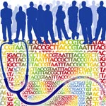
Silhouettes of people with nucleic acid sequences and a stethoscope.
A new software tool analyzes disease-causing genetic variations within a family. Credit: NIH’s National Human Genome Research Institute.

Changes in your DNA sequence occur randomly and rarely. But when they do happen, they can increase your risk of developing common, complex diseases, such as cancer. One way to identify disease-causing variations is to study the genomes of family members, since the changes typically are passed down to subsequent generations.

To rake through a family tree for genetic variations with the highest probabilities of causing a disease, researchers combined several commonly-used statistical methods into a new software tool called pVAAST. The scientific team, which included Mark Yandell and Lynn Jorde of the University of Utah and Chad Huff of the University of Texas MD Anderson Cancer Center, used the tool to identify the genetic causes of a chronic intestinal inflammation disease and of developmental defects affecting the heart, face and limbs.

The results confirmed previously identified genetic variations for the developmental diseases and pinpointed a previously unknown variation for the intestinal inflammation. Together, the findings confirm the ability of the tool to detect disease-causing genetic changes within a family. Another research team has already used the software tool to discover rare genetic changes associated with family cases of breast cancer. These studies are likely just the beginning for studying genetic patterns of diseases than run in a family.

This work also was funded by NIH’s National Institute of Diabetes and Digestive and Kidney Diseases; National Cancer Institute; National Human Genome Research Institute; National Heart, Lung, and Blood Institute; and National Institute of Mental Health.

Learn more:
University of Utah News Release (no longer available)
Yandell Exit icon, Jorde and Huff Exit icon Labs

New Compound Improves Insulin Levels in Preliminary Studies

1 comment
compound
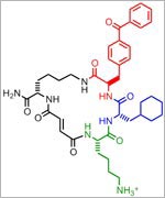
A new compound (chemical structure shown here) blocks the activity of an enzyme involved in glucose regulation.

The discovery of a compound that slows the natural degradation of insulin in mice opens up a new area of investigation in the search for drugs to treat diabetes. The research team, which included David Liu Exit icon and Alan Saghatelian Exit icon of Harvard University, Markus Seeliger of Stony Brook University School of Medicine, and Wei-Jen Tang Exit icon of the University of Chicago focused on insulin-degrading enzyme, or IDE. Using a method called DNA-templated synthesis, the scientists made 14,000 small molecules and found one that bound to the enzyme, suggesting it might modulate the enzyme’s activity. Work in test tubes and in animal models confirmed this—and showed that blocking IDE activity improved insulin levels and glucose tolerance. The researchers also learned that the enzyme is misnamed: In addition to insulin, it degrades two other hormones involved in glucose regulation.

NIGMS’ Peter Preusch says, “This is a very interesting fusion of chemical methods and biology that has uncovered new basic science findings about insulin processing with potential clinical impact.”

This work also was funded by NIH’s National Cancer Institute and the Office of the Director.

Learn more:
Harvard University News Article Exit icon
Chemistry of Health Booklet

Revealing the Human Proteome

0 comments
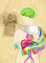
An artistic interpretation of the human proteome. Credit: Corinne Sandone and Jennifer Fairman, Johns Hopkins University.

Genes control the most basic functions of the cell, including what proteins to make and when. In 2003, the Human Genome Project created a draft map of our genes, and now researchers have completed a draft map of the human proteome—the set of all our proteins. The map, which includes proteins encoded by more than 17,000 genes as well as ones from regions of the genome previously thought to be non-coding, will help advance a broad range of research into human health and disease.

Read more about the proteome map in this NIH Research Matters article.