Category: Cells

There’s an “Ome” for That

1 comment
In the 13 years since the sequencing of the human genome, the list of “omes” has proliferated. Drop us a comment with your favorite ome—we may feature it in a follow-up post next month.

Have you ever collected coins, cards, toy trains, stuffed animals? Did you feel the need to complete the set? If so, then you may be a completist. A completist will go to great lengths to acquire a complete set of something.

Scientists can also be completists who are inspired to identify and catalog every object in a particular field to further our understanding of it. For example, a comprehensive parts list of the human body—and of other organisms that are important in biomedical research—could aid in the development of novel treatments for diseases in the same way that a parts list for a car enables auto mechanics to build or repair a vehicle.

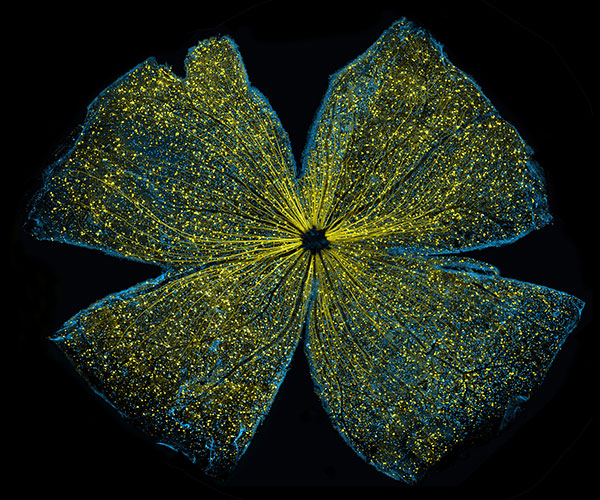
More than 15 years ago, scientists figured out how to catalog every gene in the human body. In the years since, rapid advances in technology and computational tools have allowed researchers to begin to categorize numerous aspects of the biological world. There’s actually a special way to name these collections: Add “ome” to the end of the class of objects being compiled. So, the complete set of genes in the body is called the “genome,” and the complete set of proteins is called the “proteome.”

Below are three -omes that NIH-funded scientists work with to understand human health.

Genome

Illustration of the entire outer shell of the bacteriophage MS2. Credit: Wikimedia Commons, Naranson.

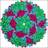
The genome is the original -ome. In 1976, Belgium scientists identified all 3,569 DNA bases—the As, Cs, Gs and Ts that make up DNA’s code—in the genes of bacteriophage MS2, immortalizing this bacteria-infecting virus as possessing the first fully sequenced genome.

Over the next two decades, a small handful of additional genomes from other microorganisms followed. The first animal genome was completed in 1998. Just 5 years later, scientists identified all 3.2 billion DNA bases in the human genome, representing the work of more than 1,000 researchers from six countries over a period of 13 years. Continue reading “There’s an “Ome” for That”

The Irresistible Resistome: How Infant Diapers Might Help Combat Antibiotic Resistance (sort of)

5 comments
Gautam Dantas
Credit: Pablo Tsukayama, Ph.D.,
Washington University School of Medicine
Gautam Dantas
Born: Mumbai, India
Most proud of: His family, which brings him joy and pride
Favorite lab tradition: OOFF! Official Optional Formal Fridays, when members of his lab can dress up, eat bread—made in the lab’s own bread machine—and drink beer and wine together at the end of the day
When not in the lab, he: Enjoys home brewing, pickling and canning, and spending time with his wife and children. He also attends musical performances, including those of his wife, who sings in the St. Louis Symphony Chorus
Advice to aspiring scientists: Pursue hobbies, take risks, explore beyond your comfort zone. “You can do a Ph.D., but also have other experiences.” He says his own outside activities refine his focus in the lab, keep him grounded and help him be an empathetic mentor to his students. Plus, he met his wife while singing in the chorus of Macalester College in St. Paul, Minnesota

When I Grow Up…

Gautam Dantas remembers the day in 10th grade when he first wanted to be a scientist. It was the day he had a new biology teacher, a visiting researcher from the U.S. The teacher passionately described his own biochemical studies of how organisms live together in communities. By the end of the class, Dantas had resolved to earn a Ph.D. in biochemistry.

He ended up doing much more—gaining expertise in computational biology, protein design and synthetic biology. He now combines his skills and knowledge in multifaceted research that spans four departments at Washington University in St. Louis. His goal: to better understand and help combat a vital public health threat—drug-resistant bacteria.

“Our motivation is that we are living in the antibiotic era, and antibiotic resistance is getting out of control,” Dantas says. “We have very few new antibiotics we can use, so we’re kind of scrambling [to find new ways to treat bacterial diseases].”

His research focuses on one of the groups most vulnerable to bacterial infections—newborn babies.

According to his lab’s website Exit icon, the research is “at the interface of microbial genomics, ecology, synthetic biology, and systems biology,” and it aims “to understand, harness, and engineer the biochemical processing potential of microbial communities.” They do it by scrounging around in infant diapers.

Antibiotic Angst

Since their introduction in the 1940s, antibiotic drugs have saved countless lives. Simultaneously, they weeded out strains of bacteria easily killed by the drugs, allowing drug-resistant strains to thrive. Every year, at least 2 million people in the U.S. become infected and at least 23,000 die from drug-resistant bacteria, according to the Center for Disease Control and Prevention. Continue reading “The Irresistible Resistome: How Infant Diapers Might Help Combat Antibiotic Resistance (sort of)”

You’ve Got Questions, We’ve Got Answers: Cell Day 2016

0 comments

Students from Connecticut to Washington State and points in between peppered our experts with questions during the recent live Cell Day web chat. They fielded questions about cell structures, microscopes and other tools, life as a scientist, and whether there are still discoveries to be made in cell biology. One of the Cell Day moderators, Jessica Faupel-Badger, even gave a shout-out to the Biomedical Beat blog as a great way to keep up with new and exciting discoveries being made every day. Thanks!

The full transcript with all the questions and answers is now available. We’ve recapped some of the highlights below.

[Check out our Facebook Live post-chat video Exit icon for a bonus answer to the question “If you put lizard DNA into human cells, could humans regrow their limbs?”]


Being a Scientist

Patrick Brown
Prior to joining NIGMS in 2016 as a program director, Patrick Brown, was a high school chemistry teacher in Maryland.

What do you think is the best thing about being a biologist? Why do you love your job so much? (Assuming you do!)

Patrick Brown answered: I love that question! And, I love being a scientist. There are so many things that I like about my career choice. The answer is simple—I like learning! I like learning about different living organisms and how they may be the same or different. I also really enjoy the multi-cultural aspect of science. I get to interact with so many different people from different parts of the world who are all studying different aspects of science that are just as interesting as my own, and we are all interested in knowing more about life.

Gram stained cerebrospinal fluid with gram-positive anthrax bacilli. Credit: Wikimedia Commons, Yuval Madar.

How did you know that biology was the career for you? In other words, what motivated you to become a biologist?

Amy Kullas answered: I remember being in my high school biology class, gazing through a microscope, and seeing the mixture of beautiful purple and pink cocci after performing my first Gram stain Exit icon. It was at that moment that I got hooked on science. I majored in microbiology in college and then went on to graduate school.

What does a typical day at work look like?

Amicia Elliott answered: The truth is that every day at work is an adventure. A typical day includes some of the following things: reading scientific papers, thinking about and designing experiments (my favorite part!), carrying out those experiments, data analysis and discussing results. Scientists work long hours to accomplish all of these things, but it is mostly a labor of love!

In a 2014 Molecular Cell Exit icon paper, NIGMS Director Jon Lorsch and colleagues determined the structure of initiation complexes.

What was the most interesting experiment you have conducted?

Jon Lorsch answered: In my lab, we study how proteins are synthesized by the eukaryotic ribosome. We have learned a great deal about how the ribosome and the proteins that help it (called translation factors) find the start codon in the messenger RNA. Recently, in collaboration with a group in the UK, we used cryo-electron microscopy to determine the three-dimensional structure of various ‘initiation complexes’ – the small subunit of the ribosome bound to mRNA, tRNA and initiation factors. Being able to see how this process works in three dimensions is amazing!

Continue reading “You’ve Got Questions, We’ve Got Answers: Cell Day 2016”

NIGMS Is on Instagram!

0 comments

Science is beautiful.

For several years, we’ve used this blog to highlight pictures we think are cool, scientifically relevant and visually striking. The images were created by NIGMS-funded researchers in the process of doing their research. Many come from our Life: Magnified collection, which features dozens of stunning photos of life, close-up. We’ll continue to bring you interesting images and information here on Biomedical Beat, but if you can’t get enough of them, we have a new way to share our visual content with you: Instagram.

We’re pleased to announce the launch of our NIGMS Instagram account. We’ll highlight gorgeous images, and bring you the science behind them—straight from our mobile device to yours. Instagram lets us label our images with subject-specific hashtags. You can find our pictures by going on Instagram and searching for #NIGMS. If you’re already an Instagram user, you can follow us @NIGMS_NIH. You can even see our page using a web browser at https://www.instagram.com/nigms_nih/ Exit icon. Let us know what you think!

Also, if you have any stunning images or videos that relate to scientific areas supported by NIGMS, please send them to us. They might end up on our Instagram feed!

See those finger-like projections? They are called villi. This image shows the small intestine, where most of the nutrients from the food we eat are absorbed into the bloodstream. The villi increase the organ’s surface area, making nutrient absorption more efficient. Credit: National Center for Microscopy and Imaging Research.
#science Exit icon #biology Exit icon #research Exit icon #cells Exit icon #cellbiology Exit icon #microscopy Exit icon #nigms Exit icon #scienceisbeautiful Exit icon #sciart Exit icon #nigms Exit icon #nih Exit icon #NCMIR Exit icon #UCSD Exit icon #smallbowel Exit icon  #nutrients Exit icon #nutrient Exit icon #anatomyphysiology Exit icon

We need zinc. It’s an essential nutrient for growth and development, fending off invading microbes, healing injuries, and all sorts of cellular processes. We get the mineral through our diet, but people in certain parts of the world don’t get enough. Researchers study how plants acquire and process zinc, hoping to find ways to increase the nutrient in food crops. Using synchrotron X-ray fluorescence technology, scientists created this heat map of zinc in a leaf from a plant called Arabidopsis thaliana (zinc levels from lowest to highest: blue, green, red, white). Credit: Suzana Car and Mary Lou Guerinot, Dartmouth College.
#science Exit icon #biology Exit icon #research Exit icon #botany Exit icon #plant Exit icon #arabidopsis Exit icon #zinc Exit icon #microscopy Exit icon #synchrotron Exit icon #x-ray Exit icon #modelorganism Exit icon #leaf Exit icon #heatmap Exit icon #scienceisbeautiful Exit icon #nigms Exit icon #nih Exit icon

Beautiful brain! This image shows the cerebellum, which is the brain’s locomotion control center. Every time you shoot a basketball, tie your shoe or chop an onion, your cerebellum fires into action. Found at the base of your brain, the cerebellum is a single layer of tissue with deep folds like an accordion. People with damage to this region of the brain often have difficulty with balance, coordination and fine motor skills. Credit: Tom Deerinck and Mark Ellisman, NCMIR
#science Exit icon #biology Exit icon #research Exit icon #cerebellum Exit icon #neuroscience Exit icon #neurobiology Exit icon #brain Exit icon #brains Exit icon #brainimages Exit icon #neuroanatomy Exit icon #microscopy Exit icon #scienceisbeautiful Exit icon #sciart Exit icon #nigms Exit icon #nih Exit icon #NCMIR Exit icon #UCSD Exit icon

Scientists can learn a lot by studying pigment cells, which give animals their colorful skins, eyes, hair and scales. We can even gain insight into skin cancers, like melanoma, that originate from pigment cells. Pigment cells can form all sorts of patterns, like these stripes on the fin of a pearl danio, a type of tropical minnow. Credit: David Parichy, University of Washington.
#biology Exit icon #marinebiology Exit icon #marinescience Exit icon #cell Exit icon #pigmentcell Exit icon #xanthophore Exit icon #science Exit icon #research Exit icon #cellbiology Exit icon #microscopy Exit icon #scienceisbeautiful Exit icon #scienceiscool Exit icon #zebrafish Exit icon #universityofwashington Exit icon

Lighting Up the Promise of Gene Therapy for Glaucoma

0 comments
Retinal ganglion cells in the mouse.

Retinal ganglion cells in the mouse retina that do (bright, yellow spots throughout) and do not (blue streaks, mostly along the edges) contain a specific gene that scientists introduced with a virus. Credit: Kenyoung (“Christine”) Kim, Wonkyu Ju and Mark Ellisman, National Center for Microscopy and Imaging Research, University of California, San Diego.

What looks like the gossamer wings of a butterfly is actually the retina of a mouse, delicately snipped to lay flat and sparkling with fluorescent molecules. Researchers captured this image while investigating the promise of gene therapy for glaucoma, a progressive eye disease. It all happened at the National Center for Microscopy and Imaging Research Exit icon (NCMIR) at the University of California, San Diego.

Glaucoma is the leading cause of irreversible blindness. It is characterized by the slow, steady death of certain nerve cells in the retina. If scientists can prevent the death of these cells, which are called retinal ganglion cells, it might be possible to slow the progression of glaucoma. Some researchers are examining the possibility of using gene therapy to do just that.

A major challenge of gene therapy is finding a way to get therapeutic genes into the right cells without damaging the cells in any way. Scientists have had success using a non-disease-causing virus (adeno-associated serotype 2) for this task. Continue reading “Lighting Up the Promise of Gene Therapy for Glaucoma”

Newly Identified Cell Wall Construction Workers: A Novel Antibiotic Target?

0 comments
SEDS

A family of proteins abbreviated SEDS (bright, pink) help build bacterial cell walls, so they are a potential target for new antibiotic drugs. Credit: Rudner lab, Harvard Medical School.

Scientists have identified a new family of proteins that, like the targets of penicillin, help bacteria build their cell walls. The finding might reveal a new strategy for treating a range of bacterial diseases.

The protein family is nicknamed SEDS, because its members help control the shape, elongation, division and spore formation of bacterial cells. Now researchers have proof that SEDS proteins also play a role in constructing cell walls. This image shows the movement of a molecular machine that contains a SEDS protein as it constructs hoops of bacterial cell wall material.

Any molecule involved in building or maintaining cell walls is of immediate interest as a possible target for antibiotic drugs. That’s because animals, including humans, don’t have cell walls—we have cell membranes instead. So disabling cell walls, which bacteria need to survive, is a good way to kill bacteria without harming patients.

This strategy has worked for the first antibiotic drug, penicillin (and its many derivatives), for some 75 years. Now, many strains of bacteria have evolved to resist penicillins—and other antibiotics—making the drugs less effective.

According to the Centers for Disease Control and Prevention, drug-resistant strains of bacteria Exit icon infect at least 2 million people, killing more than 20,000 of them in the U.S. every year. Identifying potential new drug targets, like SEDS proteins, is part of a multi-faceted approach to combating drug-resistant bacteria.

Get Your Cell Biology Questions Ready for Cell Day

0 comments

What do you get when you mix a room full of scientists with a classroom full of students who have questions about cells? Cell Day 2016! During this free web chat, middle and high school students will have the opportunity to ask our scientists at NIGMS about cell biology, biochemistry, research careers and more. Join us on Thursday, November 3 anytime from 10 a.m. to 3 p.m. EDT. Registration (no longer available).

The ECM: A Dynamic System for Moving Our Cells

2 comments

In part I of this series, we mentioned that the extracellular matrix (ECM) makes our tissues stiff or squishy, solid or see-through. Here, we reveal how the ECM helps body cells move around, a process vital for wounds to heal and a fetus to grow.

Sealing and Healing Wounds

MMPs are essential for closing wounds. Credit: Stock image.

When we get injured, the first thing our body does is to form a blood clot to stop the bleeding. Skin cells then start migrating into the wound to close the cut. The ECM is essential for this step, creating a physical support structure—like a road or train track—over which skin cells travel to seal the injured spot.

The ECM is made up of a host of proteins produced before and after injury. Some other proteins called matrix metalloproteinases (MMPs) also crowd into wounds. Because humans have so many different MMPs—a full 24 of them!—it’s been difficult for scientists to figure out what roles, if any, the proteins play in healing scrapes and cuts.

Continue reading “The ECM: A Dynamic System for Moving Our Cells”

The Extracellular Matrix, a Multitasking Marvel

1 comment

In part II of this series, we reveal how the ECM helps body cells move around, a process vital for wounds to heal and a fetus to grow. Here we introduce the extracellular matrix (ECM) and discuss how it makes our tissues stiff or squishy, solid or see-through.

When we think about how our bodies are made and what they do, we usually focus on organs, tissues and cells. These structures have well-known roles. But around, within and between them is a less understood material that also plays an essential part in making us what we are.

This gelatinous filler material is known as the extracellular matrix (ECM). Once thought to be the biological equivalent of bubble wrap, we now know that the ECM is a dynamic, physiologically active component of all our tissues. It guides cell shape, orientation and function.

The ECM is found in all of our body parts. In some tissues, it’s a thin layer separating cells, like mortar between bricks. In other tissues, it’s the major constituent.

The ECM is most prevalent in connective tissue, the material that forms our skeletons, cushions our internal organs and winds between blood vessels and around nerves. In connective tissue, the ECM is more abundant than the cells suspended within it.

The extracellular matrix meets the needs of each body part. In teeth and bones, it’s rock-hard. In corneas, it’s a transparent gel that acts like a camera lens. In tendons, it forms strong fibers that bind muscle to bone. Credit: Stock image.

What makes the ECM truly unique is its variability: Its texture, composition and functions vary by body part. That’s because the ECM’s deceptively simple recipe of water, fibrous proteins and carbohydrates has virtually endless variations.

In general, the fibrous proteins give the ECM its texture and help cells adhere properly. Carbohydrates in the ECM absorb water and swell to form a gel that acts as an excellent shock absorber. Continue reading “The Extracellular Matrix, a Multitasking Marvel”

The Science of Size: Rebecca Heald Explores Size Control in Amphibians

0 comments
Rebecca Heald
Credit: Mark Hanson.
Rebecca Heald
Grew up in: Greenville, Pennsylvania
Studied at: Hamilton College, Rice University, Harvard Medical School
Job site: University of California, Berkeley
Favorite hobby: Cycling

A 50-pound frog isn’t some freak of nature or a creepy Halloween prank. It’s a thought experiment conceived by Rebecca Heald, a cell biologist at the University of California, Berkeley Exit icon, who is studying the factors that control size in animals.

Heald’s “50-pound frog project” speaks to the power of evolution and to scientists’ ability to modify the physical characteristics of an organism by altering its genome. The project also incorporates many of Heald’s fascinating discoveries studying amphibian eggs and embryos.

In amphibians, unlike in mammals, there are striking correlations among the size of the animals’ genomes (an organism’s complete set of genes) and several aspects of the animals’ size. For example, amphibians with large genomes tend to be bigger than those with smaller genomes. Larger genomes also correspond to larger cells and larger organelles (specialized cellular structures such as the nucleus). Heald has also demonstrated that these seemingly fixed parameters can be tweaked in the lab. Continue reading “The Science of Size: Rebecca Heald Explores Size Control in Amphibians”