Category: Genes

Meet Jasmine Johnson and Gabe Vela

0 comments
Jasmine Johnson and Gabriel 'Gabe' Vela
Jasmine Johnson and Gabriel “Gabe” Vela
Field: Genetics of sleep and obesity
Worked as researchers at: The Jackson Laboratory, Bar Harbor, Maine
Graduated from high school at: Rockdale Magnet School for Science and Technology in Conyers, Ga.
Now freshman at: Stanford University in Palo Alto, Calif. (Johnson) and Southern Polytechnic State University in Marietta, Ga. (Vela)
Fascinating fact: Johnson presented her research at the 2013 White House Science Fair
Credit: Joe Piergrossi

Jasmine Johnson and Gabe Vela might still be teenagers, but they are also seasoned scientists. It all started 3 years ago, when, as high school juniors, they took the research course Independent Studies in Computational Biology at The Jackson Laboratory in Bar Harbor, Maine. They were hooked. They continued to do research until they graduated, working part-time for 2 academic years and full-time for 2 summers.

They worked with statistical geneticist Gary Churchill, using computational biology to explore the relationship between sleep and obesity. They focused on finding genes that regulate sleep and understanding how sleep affects the body. One goal of the research is to tease out a genetic explanation for why sleep deprivation increases the risk of obesity.

Working in a lab “completely changed what I thought I was going to do with my life,” said Vela. “Now I’m going to focus more on research than anything else.”

For Johnson, the experience provided the opportunity to present her research at the 2013 White House Science Fair, where she hobnobbed with some political hot shots.

“It was an amazing experience,” she said. Having “important White House officials be interested in my project … inspired me.”

Johnson and Vela visited NIH a few months ago and talked with us about their research experiences, their lives and their future goals. Jasmine Johnson & Gabriel Vela on their experience as high school researchers at The Jackson Laboratory in Bar Harbor, Maine.

Learn More
Article Exit icon about Johnson and Vela and other young researchers, from The Jackson Laboratory’s magazine The Search.
Article about the work of Gary Churchill, from NIH’s Findings magazine.

Neanderthal DNA—Still Among Us

0 comments
Map of Europe and Asia showing the presumed range of where Neanderthals lived.
For tens of thousands of years, Neanderthals lived in Europe and Asia (presumed range shown in blue) and interbred with humans, passing on some DNA to present-day people. Credit: Ryulong, Wikimedia Commons.
View larger image

Many of us have some Neanderthal genes. Before Neanderthals went extinct about 30,000 years ago, they interbred with humans living in Europe and Asia. Today’s descendants of those pairings inherited about 2 percent of their genomes from the big-brained hominids.

A research group led by David Reich at Harvard Medical School recently completed an analysis to determine the extent and identity of Neanderthal DNA in modern-day human populations. The group found that many traits in present-day people—including skin characteristics and susceptibility to various diseases—can be traced to Neanderthal DNA.

It also appears that, genetically speaking, Neanderthals and humans weren’t completely compatible. Based on the uneven distribution of Neanderthal DNA in today’s genomes, the scientists concluded that many of the male offspring of Neanderthal-human unions were infertile. In the animal world, this phenomenon is known as hybrid infertility, where the offspring of a male from one subspecies and a female from another have low or no fertility.

Studying human genes passed down through Neanderthals—as well as regions of the human genome notably devoid of Neanderthal DNA—provides an increasingly complete picture of the genetic landscape that contributed to health, disease and diversity among humans today.

Learn more:
Harvard Medical School News Release
Reich Lab Exit icon

Targeting Toxic RNA Molecules in Muscular Dystrophy

0 comments
Genetic defect that causes myotonic dystrophy type 2 and used that information to design drug candidates to counteract the disease. Credit: Ilyas Yildirim, Northwestern University.
Scientists revealed a detailed image of the genetic defect that causes myotonic dystrophy type 2 and used that information to design drug candidates to counteract the disease. Credit: Ilyas Yildirim, Northwestern University. View larger image

Myotonic dystrophy type 2 (DM2) is a relatively rare, inherited form of adult-onset muscular dystrophy that has no cure. It’s caused by a genetic defect in which a short series of nucleotides—the chemical units that spell out our genetic code—is repeated more times than normal. When the defective gene is transcribed, the resulting RNA repeat forms a hairpin-like structure that binds to and disables a protein called MBNL1.

Now, research led by Matthew Disney of The Scripps Research Institute (TSRI), Florida Campus, has revealed the detailed, three-dimensional structure of the RNA defect in DM2 and used this information to design small molecules that bind to the aberrant RNA. These designer molecules, even in small amounts, significantly improved disease-associated defects in a cellular model of DM2, and thus hold potential for reversing the disorder.

Drugs that target toxic RNA molecules associated with diseases such as DM2 are few and far between, as developing such compounds is technically challenging. The “bottom-up” approach that the scientists used to design potent new drug candidates, by first studying in detail how the RNA structure interacts with small molecules, is unconventional, noted Jessica Childs-Disney of TSRI, who was lead author of the paper with Ilyas Yildirim of Northwestern University. But it may serve as an effective strategy for pioneering the use of small molecules to manipulate disease-causing RNAs—a central focus of the Disney lab.

This work also was funded by NIH’s National Cancer Institute.

Learn more:
The Scripps Research Institute News Release Exit icon
Disney Lab

Meet Dave Cummings

0 comments
Dave Cummings. Credit: Marcus Emerson, PLNU.
Dave Cummings
Field: Environmental microbiology
Works at: Point Loma Nazarene University, San Diego, Calif.
Hobbies: Hiking, backpacking, fly-fishing
Dream home: One that doesn’t need a lot of work
Credit: Marcus Emerson, PLNU

In college, as a pre-med student majoring in biology and chemistry, Dave Cummings grew frustrated with the traditional “cookbook” approach to doing labs in his science classes. Turned off by having to follow step-by-step lab procedures that had little to do with scientific discovery, Cummings changed his major to English literature. Studying literature, he says, “helped me find myself” and taught him to think critically.

Ultimately, Cummings says, “I came to realize that it wasn’t the practice of medicine that got me excited, but the science behind it all.” He decided to pursue a graduate degree in biology and—after “knocking on a lot of doors”—was accepted at the University of Idaho, where he earned his master’s and doctoral degrees and discovered his passion for microbiology.

Today, Cummings applies his critical thinking skills to his work as professor of biology back at his alma mater, Point Loma Nazarene University (PLNU), a small university focused on undergraduate education. There he studies the role of urban storm water in spreading genes for antibiotic resistance in natural environments, and pursues his enthusiasm for training the next generation of scientists. He enlists his students in his research, giving them what he calls “real, live, on-the-ground” research experience that relatively few undergraduate students at larger universities receive.

Cummings’ Findings

Antibiotic resistance, which can transform once-tractable bacterial infections into diseases that are difficult or impossible to treat, is a major public health challenge of the 21st century. The most common way that bacteria become drug resistant is by acquiring genes that confer antibiotic resistance from other bacteria. Often, such drug resistance genes are found on small, circular pieces of DNA called plasmids that are readily passed from one species of bacteria to another.

Urban wetlands provide ideal conditions for bacteria to mingle, swap genes and spread antibiotic resistance, notes Cummings. He focuses on the wetlands around San Diego, which act as a giant mixing bowl for storm runoff, human sewage, animal waste, naturally occurring plant and soil microorganisms, and plasmids indigenous to the wetlands.

Cummings and his students examine sediment samples from these wetlands in search of plasmids that carry resistance genes. They’ve found that during winter rains, the San Diego wetlands receive runoff containing antibiotic-resistant bacteria and plasmids, which can persist at low, but detectable, levels into the dry summer months.

“We know that urban storm water carries with it a lot of antibiotic-resistant bacteria, and along with that the DNA instructions [or genes] that code for the resistance,” says Cummings. “We have solid evidence of genes encoding resistance to clinically important antibiotics washing … into coastal wetlands in San Diego.”

“We’re trying to understand the scope of the problem, and ultimately what threat that poses to human health,” he says. His concern is that the drug-defying bacterial genes will accumulate in the wetlands, and then “[find] their way back to us, where they will augment and amplify the problem of resistance.”

Precisely how resistance genes might move from the environment into people is not yet known. One way this could occur is through direct contact with contaminated water or sediment by anglers, swimmers, surfers and other recreational users. Fish, birds and insects could also transmit resistance genes from contaminated wetlands to humans.

“There is good evidence elsewhere that birds are important vectors of drug-resistant pathogens, and this is my favorite possibility,” says Cummings. “Hopefully someday we can test that hypothesis.”

Cummings’ studies, done in collaboration with Ryan Botts at PLNU and Eva Top at the University of Idaho, could reveal antibiotic resistance genes with the potential to move into new species of disease-causing bacteria and back into human populations. By identifying these genes and raising awareness of the problem, he hopes to aid future efforts to mitigate the spread of antibiotic resistance.

One Mutation Leads to Another—At Least in Yeast

0 comments
DNA mutation. Credit: Stock image.
Newly discovered genetic effect in yeast could shed light on carcinogenesis. Credit: Stock image.

Cancer cells typically include many gene mutations, extra or missing genes, or even the wrong number of chromosomes. Scientists know that certain genetic changes lead to ones elsewhere. But they’ve had a chicken-and-egg problem trying to figure out which changes trigger which others—or whether mutations accumulate randomly in tumors.

New research led by J. Marie Hardwick Exit icon of Johns Hopkins University sheds light on the issue. She found that incapacitating a single gene in yeast cells—regardless of which gene it was—spurred mutations in one or two other genes. The process was anything but random: If, say, gene X was knocked out, yeast cells almost always developed a secondary mutation in gene Y. It’s as if knocking out one gene disrupts the genomic balance enough that the cell must alter a different gene to compensate.

Significantly, the secondary mutations—but not the original ones—caused altered yeast cell characteristics, including traits linked to cancer. Also, many of the secondary mutations occurred in genes associated with cancer in humans, further suggesting that these secondary changes might play a role in carcinogenesis.

This new information will help researchers better understand the chain of genetic events that lead to cancer. It might also prompt scientists to reevaluate years of research that attributed changes in cell behavior or appearance to a given gene knockout.

This work also was funded by NIH’s National Institute of Neurological Disorders and Stroke.

Learn more:
Johns Hopkins University News Release Exit icon

Abnormal Mitochondria Might Cause Resistance to Radiation Therapy

2 comments
Mitochondria. Credit: Judith Stoffer.
Bean-shaped mitochondria are cells’ power plants. The highly folded inner membranes are the site of energy generation. Credit: Judith Stoffer. View larger image

Why some cancers are resistant to radiation therapy has baffled scientists, but research on abnormalities in mitochondria, often described as cells’ power plants, could offer new details. A research team led by Maxim Frolov Exit icon of the University of Illinois at Chicago learned that the E2F gene, which plays a role in the natural process of cell death, contributes to the function of mitochondria. Fruit flies with a mutant version of the E2F gene had misshapen mitochondria that produced less energy than normal ones. Flies with severely damaged mitochondria were more resistant to radiation-induced cell death. Studies using human cells revealed similar effects. The work could help explain why people with cancer respond differently to radiation therapy and might aid the development of drugs that enhance mitochondrial function, thereby improving the effectiveness of radiation therapy.

This work also was funded by NIH’s National Cancer Institute.

Learn more:
University of Illinois at Chicago News Release Exit icon

Stop the (Biological) Clock

0 comments
Molecular structure of the three proteins in blue-green algae’s circadian clock.  Credit: Johnson Lab, Vanderbilt University.
Molecular structure of the three proteins in blue-green algae’s circadian clock. Credit: Johnson Lab, Vanderbilt University.

Many microorganisms can sense whether it’s day or night and adjust their activity accordingly. In tiny blue-green algae, the “quartz-crystal” of the time-keeping circadian clock consists of only three proteins, making it the simplest clock found in nature. Researchers led by Carl Johnson of Vanderbilt University recently found that, by manipulating these clock proteins, they could lock the algae into continuously expressing its daytime genes, even during the nighttime.

Why would one want algae to act like it’s always daytime? The kind used in Johnson’s study is widely harnessed to produce commercial products, from drugs to biofuels. But even when grown in constant light, algae with a normal circadian clock typically decrease production of biomolecules when nighttime genes are expressed. When the researchers grew the algae with the daytime genes locked “on” in constant light, the microorganism’s output increased by as much as 700 percent. This proof of concept experiment may be applicable to improving the commercial production of compounds such as insulin and some anti-cancer drugs.

Learn more:

Vanderbilt University Press Release Exit icon
Johnson Laboratory Exit icon
Circadian Rhythms Fact Sheet

Genetic Discovery Could Enable More Precise Prescriptions

0 comments
Prescription pad with DNA illustration on it. Credit: Jane Ades, NIH’s National Human Genome Research Institute.
New insight into the genes that affect drug responses may help doctors prescribe the medications and doses best suited for each individual. Credit: Jane Ades, NIH’s National Human Genome Research Institute.

Scientists know that variations in certain genes can affect the way a person responds to medications. New research by Wolfgang Sadee Exit icon at Ohio State University shows that drug responses also depend on previously overlooked parts of DNA—sections that regulate genes, but are not considered genes themselves. This study focused on an important enzyme abbreviated CYP2D6 that processes about one-fourth of all prescription drugs. Differences in the enzyme’s performance, which range from zilch to ultra-rapid, can dramatically alter the effectiveness and safety of certain medications. Researchers discovered two new genetic variants that impact CYP2D6 performance. One of these, located in a non-gene, regulatory region of DNA, doubles or even quadruples enzyme activity. Coupling these findings with genetic tests could help doctors better identify each patient’s CYP2D6 activity level, enabling more precise prescriptions. The findings also open up a whole new area of investigation into genetic factors that impact drug response.

This work also was funded by NIH’s Eunice Kennedy Shriver National Institute of Child Health and Human Development.

Learn more:

Ohio State University News Release (no longer available)

NIH Director Blogs About Value of Model Organisms in Drug Discovery Research

0 comments
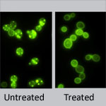
(Left) Untreated yeast cells, (Right) Treated yeast cells. Credit: Daniel Tardiff, Whitehead Institute.
Treating yeast cells with the NAB compound reverses the toxic effects of elevated levels of alpha synuclein protein. Credit: Daniel Tardiff, Whitehead Institute. View larger image

These eye-catching images and the NIGMS-funded research that yielded them were recently featured by NIH Director Francis Collins on his blog. Scientists led by a team at the Whitehead Institute for Biomedical Research engineered yeast to produce too much of a protein, alpha synuclein. In Parkinson’s disease, elevated levels or mutated forms of this protein wreak havoc on the cell. Using the model system, the researchers tested tens of thousands of compounds to identify any that reversed the toxic effects. One did. The compound, abbreviated NAB, worked similarly in an animal model and in rat neurons grown in a lab dish. Collins described the approach as “an innovative strategy for drug hunting that will likely be extended to other conditions.”

Healing Wounds, Growing Hair

1 comment
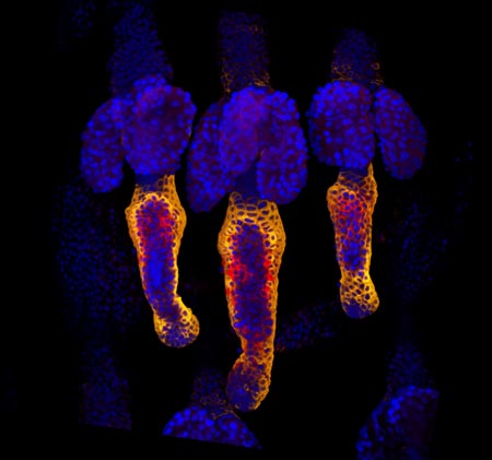
Wound healing in process. Credit: Yaron Fuchs and Samara Brown in the lab of Hermann Steller, Rockefeller University.

Credit: Yaron Fuchs and Samara Brown in the lab of Hermann Steller, Rockefeller University.

Whether injured by a scrape, minor burn or knife wound, skin goes through the same steps to heal itself. Regrowing hair over new skin is one of the final steps. All the hair you can see on your body is non-living, made up of “dead” cells and protein. It sprouts from living cells in the skin called hair follicle stem cells, shown here in red and orange. For more pictures of hair follicle stem cells—and many other stunning scientific images and videos—go to the NIGMS Image and Video Gallery.

Learn more:

Rockefeller University News Release Exit icon
Steller Lab Exit icon