Category: Genes

E. Coli Bacteria as Medical Sensors and Hard Drives?

1 comment
E.Coli
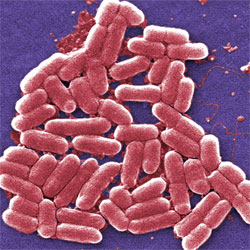
Modified E. coli bacteria can serve as sensors and data storage devices for environmental and medical monitoring. Credit: Centers for Disease Control and Prevention. View larger image

E. coli bacteria help us digest our food, produce vitamin K and have served as a model organism in research for decades. Now, they might one day be harnessed as environmental or medical sensors and long-term data storage devices Exit icon.

MIT researchers Timothy Lu Exit icon and Fahim Farzadfard modified the DNA of E. coli cells so that the cells could be deployed to detect a signal (for example, a small molecule, a drug or the presence of light) in their surroundings. To create the modified E. coli, the scientists inserted into the bacteria a custom-designed genetic tool.

When exposed to the specified signal, the tool triggers a series of biochemical processes that work together to introduce a single mutation at a specific site in the E. coli’s DNA. This genetic change serves to record exposure to the signal, and it’s passed on to subsequent generations of bacteria, providing a continued record of exposure to the signal. In essence, the modified bacteria act like a hard drive, storing biochemical memory for long periods of time. The memory can be retrieved by sequencing the bacteria or through a number of other laboratory techniques. Continue reading “E. Coli Bacteria as Medical Sensors and Hard Drives?”

Correcting a Cellular Routing Error Could Treat Rare Kidney Disease

0 comments
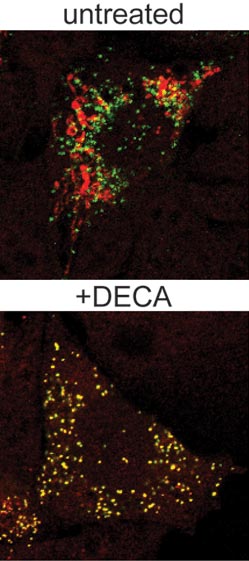
AGT protein and peroxisomes in untreated and treated cells.
The altered AGT protein (red) and peroxisomes (green) appear in different places in untreated cells (top), but they appear together (shown in yellow) in cells treated with DECA (bottom). Credit: Carla Koehler/Reproduced with permission from Proceedings of the National Academy of Sciences USA. View larger image.

Our cells have organized systems to route newly created proteins to the places where they’re needed to do their jobs. For some people born with a rare and potentially fatal genetic kidney disorder called PH1, a genetically altered form of a particular protein mistakenly ends up in mitochondria instead of in another organelle, the peroxisome. This cellular routing error of the AGT protein results in the harmful buildup of oxalate, which leads to kidney failure and other problems at an early age.

In new work led by UCLA biochemist Carla Koehler Exit icon, researchers used a robotic screening system to identify a compound that interferes with the delivery of proteins to mitochondria. Koehler’s team Exit icon showed that adding a small amount of the compound, known as DECA, to cells grown in the laboratory prevented the altered form of the AGT protein from going to the mitochondria and sent it to the peroxisome. The compound also reduced oxalate levels in a cell model of PH1.

The team’s findings suggest that DECA, which is already approved by the Food and Drug Administration for treating certain bacterial infections, could be a promising candidate for treating children affected by PH1. And, Koehler notes, the screening strategy that she and her team used to identify DECA as a potential therapy may help researchers identify other new therapies for the disorder.

This work was funded in part by NIH under grant R01GM061721.

How Instructions for Gene Activity Are Passed Across Generations

0 comments
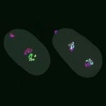
C. elegans embryos
Images of C. elegans embryos show transmission of an epigenetic mark (green) during cell division from a one-cell embryo (left) to a two-cell embryo (right). Credit: Laura J. Gaydos.

Chemical tags that cells attach to DNA or to DNA-packaging proteins across the genome—called epigenetic marks—can alter gene activity, or expression, without changing the underlying DNA code. As a result, these epigenetic changes can influence health and disease. But it’s a matter of debate as to whether and how certain epigenetic changes on DNA-packaging proteins can be passed from parents to their offspring.

In studies with a model organism, the worm C. elegans, researchers led by Susan Strome of the University of California, Santa Cruz, have offered new details that help resolve the debate.

Strome’s team created worms with a genetic change that knocks out the enzyme responsible for making a particular methylation mark, a type of epigenetic mark that can turn off gene expression at certain points of an embryo’s development. Then the scientists bred the knockout worms with normal ones. Looking at the chromosomes from the resulting eggs, sperm and dividing cells of embryos after fertilization, the researchers found that the methylation marks are passed from both parents to offspring. The enzyme, however, is passed to the offspring just by the egg cell. For embryos with the enzyme, the epigenetic marks are passed faithfully through many cell divisions. For those without it, the epigenetic mark can be passed through a few cell divisions.

Because all animals use the same enzyme to create this particular methylation mark, the results have implications for parent-to-child epigenetic inheritance as well as cell-to-cell inheritance in other organisms.

This work was funded in part by NIH under grants R01GM034059, T32GM008646 and P40OD010440.

Learn more:

University of California, Santa Cruz News Release
Dynamic DNA Section from The New Genetics Booklet

Stem Cells Do Geometry

2 comments
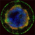
Human embryonic cells
As seen under a microscope, human embryonic cells (colored dots) confined to circles measuring 1 millimeter across start to specialize and form distinct layers similar to those seen in early development. Credit: Aryeh Warmflash, Rockefeller University. View larger image

Each fluorescent point of light making up the multicolored rings in this image is an individual human embryonic cell in the early stages of development. Scientists seeking to understand the molecular cues responsible for early embryonic patterning found that human embryonic cells confined to areas of precisely controlled size and shape begin to specialize, migrate and organize into distinct layers just as they would under natural conditions.

Read the Inside Life Science article to learn more about this research, which has opened a new window for studying early development and could advance efforts aimed at using human stem cells to replace diseased cells and regenerate lost or injured body parts.

4 Timely Facts About Our Biological Clocks

0 comments
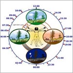
Illustration of circadian rhythm.
Genes and proteins run biological clocks that help keep daily rhythms in synch. Credit: Wikimedia Commons.

After you roll your clocks back by an hour this Sunday, you may feel tired. That’s because our bodies—more specifically, our circadian rhythms—need a little time to adjust. These daily cycles are run by a network of tiny, coordinated biological clocks.

NIGMS’ Mike Sesma tracks circadian rhythm research being conducted in labs across the country, and he shares a few timely details about our internal clocks:

1. They’re incredibly intricate.

Biological clocks are composed of genes and proteins that operate in a feedback loop. Clock genes contain instructions for making clock proteins, whose levels rise and fall in a regular cyclic pattern. This pattern in turn regulates the activity of the genes. Many of the results from circadian rhythm research this year have uncovered more parts of the molecular machinery that fine-tune the clock. Earlier in the month, we blogged about an RNA molecule that cues the internal clock.

2. Every organism has them—from algae to zebras.

Many of the clock genes and proteins are similar across species, allowing researchers to make important findings about human circadian processes by studying the clock components of organisms like fruit flies, bread mold and plants.

3. Whether we’re awake or asleep, our clocks keep ticking.

While they might get temporarily thrown off by changes in light or temperature, our clocks usually can reset themselves.

4. Nearly everything about how our body works is tied to biological clocks.

Our clocks influence alertness, hunger, metabolism, fertility, mood and other physiological conditions. For this reason, clock dysfunction is associated with various disorders, including insomnia, diabetes and depression. Even drug efficacy has been linked to our clocks: Studies have shown that some drugs might be more effective if given earlier in the day.

Learn more:
Circadian Rhythms Fact Sheet

Outwitting Antibiotic Resistance

0 comments
Marine scene with fish and corals
The ocean is a rich source of microbes that could yield infection-fighting natural molecules. Credit: National Oceanic and Atmospheric Administration Exit icon.

Antibiotics save countless lives and are among the most commonly prescribed drugs. But the bacteria and other microbes they’re designed to eradicate can evolve ways to evade the drugs. This antibiotic resistance, which is on the rise due to an array of factors, can make certain infections difficult—and sometimes impossible—to treat.

Read the Inside Life Science article to learn how scientists are working to combat antibiotic resistance, from efforts to discover potential new antibiotics to studies seeking more effective ways of using existing ones.

An RNA Molecule That Cues the Internal Clock

0 comments
Clock
Dysfunction in our internal clocks may lead to insufficient sleep, which has been linked to an increased risk for chronic diseases. Credit: Stock image.

Our internal clocks tell us when to sleep and when to eat. Because they are sensitive to changes in daytime and nighttime cues, they can get thrown off by activities like traveling across time zones or working the late shift. Dysfunction in our internal clocks may lead to insufficient sleep, which has been linked to an increased risk for chronic diseases like high blood pressure, diabetes, depression and cancer.

Researchers led by Yi Liu Exit icon of the University of Texas Southwestern Medical Center have uncovered a previously unknown mechanism by which internal clocks run and are tuned to light cues. Using the model organism Neurospora crassa (a.k.a., bread mold), the scientists identified a type of RNA molecule called long non-coding RNA (lncRNA) that helps wind the internal clock by regulating how genes are expressed. When it’s produced, the lncRNA identified by Liu and his colleagues blocks a gene that makes a specific clock protein.

This inhibition works the other way, too: The production of the clock protein blocks the production of the lncRNA. This rhythmic gene expression helps the body stay tuned to whether it’s day or night.

The researchers suggest that a similar mechanism likely exists in the internal clocks of other organisms, including mammals. They also think that lncRNA-protein pairs may contribute to the regulation of other biologic processes.

Learn more:
University of Texas Southwestern Medical Center News Release Exit icon
Circadian Rhythms Fact Sheet

Meet Scott Poethig

1 comment
Scott Poethig
Fields: Plant biology, cell and developmental biology, genetics
Works at: University of Pennsylvania
Studied at: College of Wooster, Yale University
Favorite musicians: Nick Drake and Bruce Springsteen
High school job: Radio D.J.
Favorite book: “The Little Prince,” by Antoine de Saint-Exupéry

When Scott Poethig signed up for a developmental biology course in his senior year of college, he expected to learn how organisms transition from single cells to juveniles to adults. He did not expect to learn just how much scientists still didn’t know about this process.

“It was the first course I had taken as an undergraduate where I felt that I could ask a question that there wasn’t an answer to already,” he recalls. “I thought, ‘Wow! This is amazing.’”

Poethig already had an interest in plant biology and an independent research project studying corn viruses. He immediately saw the potential in combining his knowledge of plants with his questions about how organisms grow. “There seemed to be a lot of low hanging fruit in plant development,” he says.

Today, Poethig is the head of a plant development lab at the University of Pennsylvania. His work probes the complex molecular mechanisms that drive the transition from a young seedling to an adult plant that hasn’t yet produced seeds.

“The analogous period in human development is the interval between birth and puberty,” he explains. “People think of puberty as the major developmental transition in postnatal human development, but a lot of change happens before that point.”

His Findings

Poethig discovered that for the mustard plant Arabidopsis, a model organism frequently studied by geneticists, change begins early. Before these plants begin to flower—a sign of reproductive maturity—they undergo a process of vegetative maturation. In Arabidopsis, Poethig found that juvenile plants can be distinguished from adult plants by where hairs are produced on a leaf. Juvenile plants only produce hairs on the upper surface of the leaf, whereas adult plants produce leaves with hairs on both the upper and lower surfaces.

By studying mutant Arabidopsis plants where the adult pattern of hair development is either delayed or advanced, Poethig identified microRNAs as key players in this developmental transition.

MicroRNA molecules commonly block the expression of specific genes. Poethig found that in Arabidopsis, a type of microRNA prevents development. Young plants have high levels of this microRNA and cannot fully mature. When those levels drop, plants progress to adulthood.

MicroRNAs similarly control development in the nematode C. elegans. Scientists study the genetics of this tiny worm to better understand related developmental processes in more complex organisms. Because plants also use microRNAs to regulate development, Poethig’s discoveries may contribute to our understanding of how these molecules govern development in animals, including humans.

Poethig now wants to learn what determines the timing of developmental changes. He asks: “Why do microRNA levels drop? What’s the signal that causes that? What is the plant measuring?” His current hypothesis: sugar.

In a recent study, he found that giving plants additional sugar reduced microRNA levels and sped up development. Meanwhile, mutant plants that couldn’t produce enough sugar on their own through photosynthesis had increased microRNA levels and delayed development compared to normal plants.

This research may one day advance our understanding of how nutrition and genetics interact to affect human development. “In essentially all organisms, aging and the timing of developmental processes are strongly affected by nutrition,” Poethig explains. “In humans, childhood obesity is sometimes associated with early puberty, and it is important to understand the molecular basis for this effect.”

Poethig believes that studying microRNAs in plants may also lead to discoveries in human genetics outside of developmental biology. “MicroRNAs control a wide range of gene activity in plants and animals,” Poethig explains. “In humans, these molecules control the activity of as many as 30 percent of our genes. So understanding how microRNAs work in plants could help us understand their function in humans.”

Besides studying the Arabidopsis plants in his lab, Poethig also studies the plants in his kitchen, and uses his fascination with the history, culture and politics of food to excite others about science. Watch video.
Field Focus: Precision Gene Editing with CRISPR

0 comments
Bacterial cells infected by viruses.
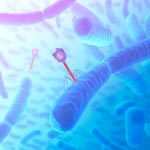
Bacterial cells can be infected by viruses (shown in red and purple) and have evolved ways to defend themselves. Credit: Stock image.

Like humans, bacteria can be infected by viruses and have evolved ways to defend themselves. Researchers are now adapting this bacterial “immune system” to precisely and efficiently edit genes in cells from humans and a wide range of other organisms. Scientists are excited about the tremendous potential of this powerful tool for advancing biomedical research and treating diseases.

The bacterial defense system is called CRISPR, for clustered regularly interspaced short palindromic repeats. A breakthrough in understanding CRISPR came from examining bacteria used by the dairy industry for the production of yogurt and cheese. In a study published in 2007, researchers showed that these bacteria insert viral DNA sequences into their own genomes and use that information to disarm the virus when it attacks again. Subsequent research has shown that the CRISPR system consists of small RNA molecules that target specific viral DNA sequences and proteins that cut the DNA, thus destroying the virus.

Researchers have already adapted CRISPR into a gene-editing tool that’s quicker, cheaper and more precise than existing methods. Researchers can use CRISPR to add, delete, rev up or tone down certain genes as well as create animal models for studying human diseases. The ability to precisely target genes in human cells is expected to speed progress in the development of gene-based therapies.

Although much is known about CRISPR, we still have a lot to learn. For example, how do bacterial cells obtain and insert the viral DNA into their genome? What triggers production of the CRISPR RNA molecules? How are invading viral DNAs targeted for destruction? This last question is answered in part by a pair of findings described in an earlier post, A Crisper View of the CRISPR Gene-Editing Mechanism. We also want to figure out how we can make the CRISPR gene-editing tool even more versatile and precise.

The CRISPR story offers a good example of how studying basic biological processes leads to new—and sometimes unexpected—insights and applications.

Emily Carlson also contributed to this blog post.

Related advances:
CRISPR/Cas9 Protein Complex Can Be Programmed to Recognize and Cleave RNA
CRISPR System Adapted to Reversibly Regulate Gene Expression

A Crisper View of the CRISPR Gene-Editing Mechanism

0 comments
Structural model of the Cascade surveillance machine
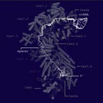
Structural model of the Cascade surveillance machine. Credit: Ryan Jackson, Montana State University. Click for larger image

To dismantle the viruses that infect them, bacteria have evolved an immune system that identifies invading viral DNA and signals for its destruction. This gene-editing system is called CRISPR, and it’s being harnessed as a tool for modifying human genes associated with disease.

Taking another important step toward this potential application, researchers now know the structure of a key CRISPR component: a multi-subunit surveillance machine called Cascade that identifies the viral DNA. Shaped like a sea horse, Cascade is composed of 11 proteins and CRISPR-related RNA. A research team led by Blake Wiedenheft of Montana State University used X-ray crystallography and computational analysis to determine Cascade’s configuration. In a complementary study, Scott Bailey of Johns Hopkins University and his colleagues determined the structure of the complex bound to a viral DNA target.

Like blueprints, these structural models help scientists understand how Cascade assembles into an efficient surveillance machine and, more broadly, how the CRISPR system functions and how to adapt it as a tool for basic and clinical research.

Learn more:
Montana State University News Release Exit icon
Johns Hopkins University News Release Exit icon