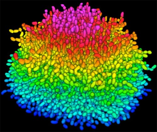
A growing Vibrio cholerae biofilm. Each slightly curved comma shape represents an individual bacterium from assembled confocal microscopy images. Different colors show each bacterium’s position in the biofilm in relation to the surface on which the film is growing. Credit: Jing Yan, Ph.D., and Bonnie Bassler, Ph.D., Department of Molecular Biology, Princeton University, Princeton, NJ.
Bacteria use many methods to overcome threats in their environment. One of these ways is forming colonies called biofilms on surfaces of objects. Often appearing like the bubble-shaped fortress represented in this image, biofilms enable bacteria to withstand attacks, compete for space and survive fluctuations in nutrient supply. Bacteria aggregated within biofilms inside our bodies, for example, resist antibiotic therapy more effectively than free swimming cells, making infections difficult to treat. On the other hand, biofilms are also useful for making microbial fuel cells and for waste-water treatment. Learning how biofilms work, therefore, could provide essential tools in our ongoing battle against disease-causing agents and in our efforts to harness beneficial bacterial behaviors. Researchers are now using new imaging techniques to watch how biofilms grow, cell by cell, and to identify more effective ways of disrupting or fostering them.
Until now, poor imaging resolution meant that scientists could not see what individual bacteria in the films are up to as the biofilms grow. The issue is that bacteria are tiny, making imaging each cell, as well as the ability to distinguish each cell in the biofilm community, problematic. Continue reading “Cool Image: Inside a Biofilm Build-up”
Cell biologists would love to shrink themselves down and actually see, touch and hear the inner workings of cells. Because that’s impossible, they have developed an ever-growing collection of microscopes to study cellular innards from the outside. Using these powerful tools, researchers can exhaustively inventory the molecular bits and pieces that make up cells, eavesdrop on cellular communication and spy on cells as they adapt to changing environments.
In recent years, scientists have developed new cellular imaging techniques that allow them to visualize samples in ways and at levels of detail never before possible. Many of these techniques build upon the power of electron microscopy (EM) to see ever smaller details.
Unlike traditional light microscopy, EM uses electrons, not light, to create an image. To do so, EM accelerates electrons in a vacuum, shoots them out of an electron gun and focuses them with doughnut-shaped magnets onto a sample. When electrons bombard the sample, some pass though without being absorbed while others are scattered. The transmitted electrons land on a detector and produce an image, just as light strikes a detector (or film) in a camera to create a photograph.
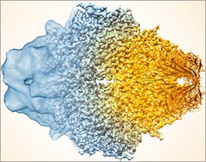
This image, showing a single protein molecule, is a montage. It was created to demonstrate how dramatically cryo-EM has improved in recent years. In the past, cryo-EM was only able to obtain a blobby approximation of a molecule’s shape, like that shown on the far left. Now, the technique yields exquisitely detailed images in which individual atoms are nearly visible (far right). Color is artificially applied. Credit: Veronica Falconieri, Subramaniam Lab, National Cancer Institute.
Transmission electron microscopes can magnify objects more than 10 million times, enabling scientists to see the outline and some details of cells, viruses and even some large molecules. A relatively new form of transmission electron microscopy called cryo-EM enables scientists to view specimens in their natural or near-natural state without the need for dyes or stains.
In cryo-EM—the prefix cry- means “cold” or “freezing”—scientists freeze a biological sample so rapidly that water molecules do not have time to form ice crystals, which could shove cellular materials out of their normal place. Cold samples are more stable and can be imaged many times over, allowing researchers to iteratively refine the image, remove artifacts and produce even sharper images than ever before. Continue reading “Cool Tools: Pushing the Limits of High-Resolution Microscopy”
Inside our bodies is a surprising amount of metal. Not enough to set off the scanners at the airport or make us rich, but enough to fill each of our cells with billions of metal ions, including calcium, iron, copper and zinc. These ions facilitate critical biological functions.
However, too much of any metal can be toxic, while too little can cause disease. Our cells carefully monitor and control their metal content using a whole series of proteins that bind, sense and transport metal ions.
Keeping tabs on why and how metals flow into and out of our cells is a passion of Thomas O’Halloran, professor of chemistry and molecular biosciences at Northwestern University in Illinois. For the past three decades, O’Halloran has investigated how fluctuations in the amount of metal ions inside cells influence gene expression, cell growth and other vital functions. Using a variety of approaches, he has uncovered new types of proteins that bind metal ions and investigated the role that imbalances in these ions play in a number of disease-related physiological processes.
One recent focus of his studies has been how zinc regulates oocyte (egg cell) maturation and fertilization. Ultimately, his work could help us better understand infertility, cancer and certain bacterial infections.
This is the first post in a new series highlighting NIGMS’ efforts toward developing a robust, diverse and well-trained scientific workforce.
Credit: Christa Reynolds.
Chyann Richard Academic Institution: California State University, Long Beach Major: Psychology Mentor: Michelle Barrack Favorite Book: Outliers, by Malcolm Gladwell Favorite sports team: Los Angeles Lakers Favorite music: R&B
“A lot of people would never guess that I’m in research and I take pride in that. I want to be able to represent people that don’t even go this far,” Chyann Richard, 20, says.
BUILD and the Diversity Program Consortium
The Diversity Program Consortium (DPC) aims to enhance diversity in the biomedical research workforce through improved recruitment, training and mentoring nationwide. It comprises three integrated programs—Building Infrastructure Leading to Diversity (BUILD), which implements activities at student, faculty and institutional levels; the National Research Mentoring Network (NRMN), which provides mentoring and career development opportunities for scientists at all levels; and the Coordination and Evaluation Center (CEC), which is responsible for evaluating and coordinating DPC activities.
Ten undergraduate institutions across the United States have received BUILD grants, and together, they serve a diverse population. Each BUILD site has developed a unique program intended to engage and prepare students for success in the biomedical sciences and maximize opportunities for research training and faculty development. BUILD programs include everything from curricular redesign, lab renovations, faculty training and research grants, to student career development, mentoring and research-intensive summer programs.
Currently a junior at California State University, Long Beach (CSULB), Richard is majoring in psychology. After she graduates with a bachelor’s degree in 2018, she plans to continue to a Ph.D. program and do research in behavioral neuroscience.
Richard is among a select group of undergraduate college students participating in the Building Infrastructure Leading to Diversity (BUILD) program. The BUILD programs focus on finding innovative approaches to increase student engagement in the biomedical sciences, through interventions at student, faculty and institutional levels. As a BUILD scholar, Richard is conducting laboratory research and preparing for graduate school through career development seminars, presentations and other activities.
Richard loves how research introduces her to new ideas and allows her to share these concepts with others, including her parents.
“Because they’ve been teaching me my whole life … now I’ve got a one-up because I know about research and they don’t. That’s really fun,” she says.
Richard’s interest in behavioral neuroscience is both personal and scientific. During Richard’s junior year of high school, her mother was diagnosed with generalized anxiety disorder. This sparked Richard to take an Advanced Placement (AP) psychology course, where she began learning about the prevalence of and treatments for such disorders.
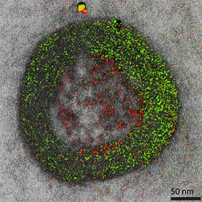
Color electron micrograph of an endosome, a cell organelle. Credit: Ranjan Ramachandra, UCSD
As his Christmas gift to himself each year, the late biochemist Roger Tsien treated himself to two weeks of uninterrupted research in his lab. This image is a product of those annual sojourns. While it may look like a pine wreath dotted with crimson berries, it is in fact one of the world’s first color electron micrographs—and the method used to create it may dramatically advance cell imaging.
Electron microscopy (EM) is a time-honored technique for visualizing cell structures that uses beams of accelerated electrons to magnify objects up to 10 million times their actual size. Standard EM images are in grayscale and any color is added in with computer graphics programs after the image is made. With their new technique, Tsien, who received a Nobel Prize for his development of green fluorescent protein into a tool for visualizing details in living cells using light microscopes, and his colleagues have found a way to incorporate color labeling directly into EM. Continue reading “Cool Image: Adding Color to the Gray World of Electron Microscopy”
In the 13 years since the sequencing of the human genome, the list of “omes” has proliferated. Drop us a comment with your favorite ome—we may feature it in a follow-up post next month.
Have you ever collected coins, cards, toy trains, stuffed animals? Did you feel the need to complete the set? If so, then you may be a completist. A completist will go to great lengths to acquire a complete set of something.
Scientists can also be completists who are inspired to identify and catalog every object in a particular field to further our understanding of it. For example, a comprehensive parts list of the human body—and of other organisms that are important in biomedical research—could aid in the development of novel treatments for diseases in the same way that a parts list for a car enables auto mechanics to build or repair a vehicle.
More than 15 years ago, scientists figured out how to catalog every gene in the human body. In the years since, rapid advances in technology and computational tools have allowed researchers to begin to categorize numerous aspects of the biological world. There’s actually a special way to name these collections: Add “ome” to the end of the class of objects being compiled. So, the complete set of genes in the body is called the “genome,” and the complete set of proteins is called the “proteome.”
Below are three -omes that NIH-funded scientists work with to understand human health.
Genome
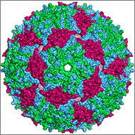
Illustration of the entire outer shell of the bacteriophage MS2. Credit: Wikimedia Commons, Naranson.
The genome is the original -ome. In 1976, Belgium scientists identified all 3,569 DNA bases—the As, Cs, Gs and Ts that make up DNA’s code—in the genes of bacteriophage MS2, immortalizing this bacteria-infecting virus as possessing the first fully sequenced genome.
Over the next two decades, a small handful of additional genomes from other microorganisms followed. The first animal genome was completed in 1998. Just 5 years later, scientists identified all 3.2 billion DNA bases in the human genome, representing the work of more than 1,000 researchers from six countries over a period of 13 years. Continue reading “There’s an “Ome” for That”
Credit: Pablo Tsukayama, Ph.D., Washington University School of Medicine
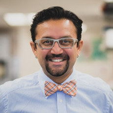
Gautam Dantas Born: Mumbai, India Most proud of: His family, which brings him joy and pride Favorite lab tradition: OOFF! Official Optional Formal Fridays, when members of his lab can dress up, eat bread—made in the lab’s own bread machine—and drink beer and wine together at the end of the day When not in the lab, he: Enjoys home brewing, pickling and canning, and spending time with his wife and children. He also attends musical performances, including those of his wife, who sings in the St. Louis Symphony Chorus Advice to aspiring scientists: Pursue hobbies, take risks, explore beyond your comfort zone. “You can do a Ph.D., but also have other experiences.” He says his own outside activities refine his focus in the lab, keep him grounded and help him be an empathetic mentor to his students. Plus, he met his wife while singing in the chorus of Macalester College in St. Paul, Minnesota
When I Grow Up…
Gautam Dantas remembers the day in 10th grade when he first wanted to be a scientist. It was the day he had a new biology teacher, a visiting researcher from the U.S. The teacher passionately described his own biochemical studies of how organisms live together in communities. By the end of the class, Dantas had resolved to earn a Ph.D. in biochemistry.
He ended up doing much more—gaining expertise in computational biology, protein design and synthetic biology. He now combines his skills and knowledge in multifaceted research that spans four departments at Washington University in St. Louis. His goal: to better understand and help combat a vital public health threat—drug-resistant bacteria.
“Our motivation is that we are living in the antibiotic era, and antibiotic resistance is getting out of control,” Dantas says. “We have very few new antibiotics we can use, so we’re kind of scrambling [to find new ways to treat bacterial diseases].”
His research focuses on one of the groups most vulnerable to bacterial infections—newborn babies.
According to his lab’s website, the research is “at the interface of microbial genomics, ecology, synthetic biology, and systems biology,” and it aims “to understand, harness, and engineer the biochemical processing potential of microbial communities.” They do it by scrounging around in infant diapers.
Antibiotic Angst
Since their introduction in the 1940s, antibiotic drugs have saved countless lives. Simultaneously, they weeded out strains of bacteria easily killed by the drugs, allowing drug-resistant strains to thrive. Every year, at least 2 million people in the U.S. become infected and at least 23,000 die from drug-resistant bacteria, according to the Center for Disease Control and Prevention. Continue reading “The Irresistible Resistome: How Infant Diapers Might Help Combat Antibiotic Resistance (sort of)”
Students from Connecticut to Washington State and points in between peppered our experts with questions during the recent live Cell Day web chat. They fielded questions about cell structures, microscopes and other tools, life as a scientist, and whether there are still discoveries to be made in cell biology. One of the Cell Day moderators, Jessica Faupel-Badger, even gave a shout-out to the Biomedical Beat blog as a great way to keep up with new and exciting discoveries being made every day. Thanks!
The full transcript with all the questions and answers is now available. We’ve recapped some of the highlights below.
[Check out our Facebook Live post-chat video for a bonus answer to the question “If you put lizard DNA into human cells, could humans regrow their limbs?”]
Being a Scientist
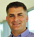
Prior to joining NIGMS in 2016 as a program director, Patrick Brown, was a high school chemistry teacher in Maryland.
What do you think is the best thing about being a biologist? Why do you love your job so much? (Assuming you do!)
Patrick Brown answered: I love that question! And, I love being a scientist. There are so many things that I like about my career choice. The answer is simple—I like learning! I like learning about different living organisms and how they may be the same or different. I also really enjoy the multi-cultural aspect of science. I get to interact with so many different people from different parts of the world who are all studying different aspects of science that are just as interesting as my own, and we are all interested in knowing more about life.
How did you know that biology was the career for you? In other words, what motivated you to become a biologist?
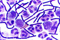
Amy Kullas answered: I remember being in my high school biology class, gazing through a microscope, and seeing the mixture of beautiful purple and pink cocci after performing my first Gram stain. It was at that moment that I got hooked on science. I majored in microbiology in college and then went on to graduate school.
What does a typical day at work look like?
Amicia Elliott answered: The truth is that every day at work is an adventure. A typical day includes some of the following things: reading scientific papers, thinking about and designing experiments (my favorite part!), carrying out those experiments, data analysis and discussing results. Scientists work long hours to accomplish all of these things, but it is mostly a labor of love!
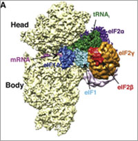
In a 2014 Molecular Cell paper, NIGMS Director Jon Lorsch and colleagues determined the structure of initiation complexes.
What was the most interesting experiment you have conducted?
Jon Lorsch answered: In my lab, we study how proteins are synthesized by the eukaryotic ribosome. We have learned a great deal about how the ribosome and the proteins that help it (called translation factors) find the start codon in the messenger RNA. Recently, in collaboration with a group in the UK, we used cryo-electron microscopy to determine the three-dimensional structure of various ‘initiation complexes’ – the small subunit of the ribosome bound to mRNA, tRNA and initiation factors. Being able to see how this process works in three dimensions is amazing!
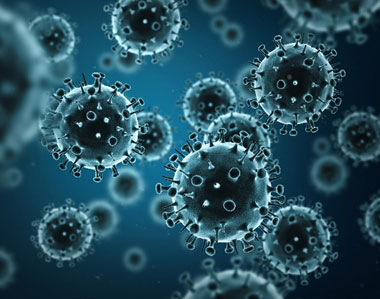
Nearly 10 percent of the human genome is derived from the genes of viruses. Credit: Stock image.
When viruses infect us, they can embed small chunks of their genetic material in our DNA. Although infrequent, the incorporation of this material into the human genome has been occurring for millions of years. As a result of this ongoing process, viral genetic material comprises nearly 10 percent of the modern human genome. Over time, the vast majority of viral invaders populating our genome have mutated to the point that they no longer lead to active infections. But they are not entirely dormant.
Sometimes, these stowaway sequences of viral genes, called “endogenous retroviruses” (ERVs), can contribute to the onset of diseases such as cancer. They can also make their hosts susceptible to infections from other viruses. However, scientists have identified numerous cases of viral hitchhikers bestowing crucial benefits to their human hosts—from protection against disease to shaping important aspects of human evolution, such as the ability to digest starch.
For a virus to successfully make copies of itself inside a host cell, it needs molecular tools similar to the ones its host normally uses to translate genes into proteins. As a result, viruses have tools meticulously shaped by evolution to commandeer the protein-producing machinery of human cells.
An exhibit called “Minerals in Medicine” opened at the NIH Clinical Center last month (see slideshow). The display features a fascinating overview of how dozens of minerals are used to create drugs and medical instruments useful in treating disease and maintaining health. The minerals ranged from commonplace ones like quartz, which is used to make medical instruments, to more exotic ones like hubnerite, a source of the metal tungsten, which is used in radiation shielding.
Inspired by this collection, which is co-sponsored by NIH and the Smithsonian Institution, we highlight here examples of “Metals in Medicine.”
Copper and Fat Metabolism
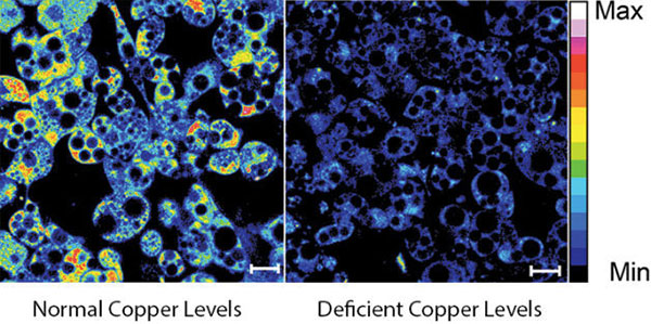
Fluorescent imaging of copper in white fat cells from mice. The left panel shows fat cells with normal levels of copper, and the right panel shows fat cells deficient in copper. Credit: Lakshmi Krishnamoorthy and Joseph Cotruvo Jr., University of California, Berkeley.
What does a metal like copper have to do with our ability to breakdown fat? Researchers explored this question by observing mice with Wilson’s disease—a rare, inherited condition that causes copper to accumulate in the liver, brain and other vital organs. The mice with the condition usually have larger deposits of fat compared to healthy mice. To confirm that fat metabolism is somehow compromised in these mice, the researchers treated them with a drug that induces the breakdown of fat. And indeed they found that less fat was metabolized in mice with the disease.
In an effort to investigate what role copper may be playing in fat metabolism, the researchers examined adipose tissue, or fat, cells under a microscope to track the metal’s interactions with various proteins in the cell. They discovered that copper inhibits an enzyme called PDE3. This enzyme usually prevents another enzyme called cAMP from helping to break down fat. The researchers concluded that copper actually promotes fat metabolism. This work shows that transition metal nutrients can play signaling roles, which has been previously thought to be restricted to alkali and alkaline earth metals like sodium, potassium and calcium.