NIGMS Is on Instagram!

0 comments

Science is beautiful.

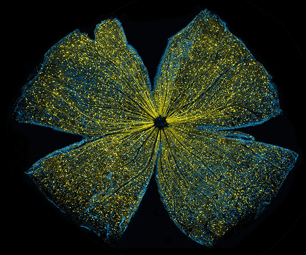
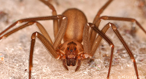
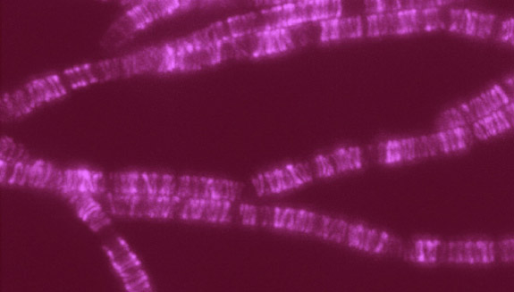
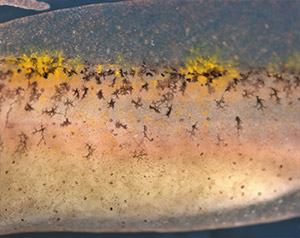
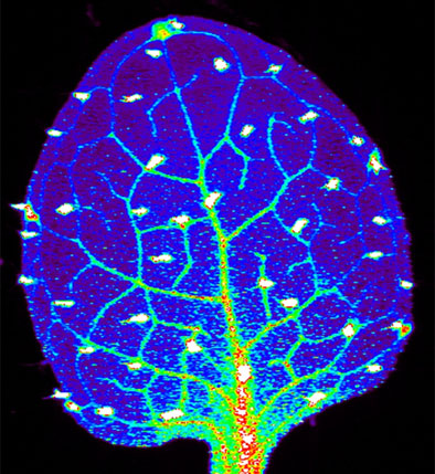
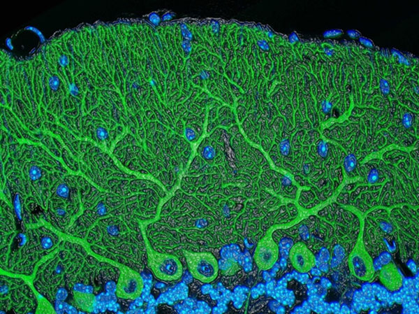
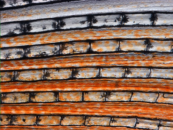
For several years, we’ve used this blog to highlight pictures we think are cool, scientifically relevant and visually striking. The images were created by NIGMS-funded researchers in the process of doing their research. Many come from our Life: Magnified collection, which features dozens of stunning photos of life, close-up. We’ll continue to bring you interesting images and information here on Biomedical Beat, but if you can’t get enough of them, we have a new way to share our visual content with you: Instagram.

We’re pleased to announce the launch of our NIGMS Instagram account. We’ll highlight gorgeous images, and bring you the science behind them—straight from our mobile device to yours. Instagram lets us label our images with subject-specific hashtags. You can find our pictures by going on Instagram and searching for #NIGMS. If you’re already an Instagram user, you can follow us @NIGMS_NIH. You can even see our page using a web browser at https://www.instagram.com/nigms_nih/ Exit icon. Let us know what you think!

Also, if you have any stunning images or videos that relate to scientific areas supported by NIGMS, please send them to us. They might end up on our Instagram feed!

See those finger-like projections? They are called villi. This image shows the small intestine, where most of the nutrients from the food we eat are absorbed into the bloodstream. The villi increase the organ’s surface area, making nutrient absorption more efficient. Credit: National Center for Microscopy and Imaging Research.
#science Exit icon #biology Exit icon #research Exit icon #cells Exit icon #cellbiology Exit icon #microscopy Exit icon #nigms Exit icon #scienceisbeautiful Exit icon #sciart Exit icon #nigms Exit icon #nih Exit icon #NCMIR Exit icon #UCSD Exit icon #smallbowel Exit icon  #nutrients Exit icon #nutrient Exit icon #anatomyphysiology Exit icon

We need zinc. It’s an essential nutrient for growth and development, fending off invading microbes, healing injuries, and all sorts of cellular processes. We get the mineral through our diet, but people in certain parts of the world don’t get enough. Researchers study how plants acquire and process zinc, hoping to find ways to increase the nutrient in food crops. Using synchrotron X-ray fluorescence technology, scientists created this heat map of zinc in a leaf from a plant called Arabidopsis thaliana (zinc levels from lowest to highest: blue, green, red, white). Credit: Suzana Car and Mary Lou Guerinot, Dartmouth College.
#science Exit icon #biology Exit icon #research Exit icon #botany Exit icon #plant Exit icon #arabidopsis Exit icon #zinc Exit icon #microscopy Exit icon #synchrotron Exit icon #x-ray Exit icon #modelorganism Exit icon #leaf Exit icon #heatmap Exit icon #scienceisbeautiful Exit icon #nigms Exit icon #nih Exit icon

Beautiful brain! This image shows the cerebellum, which is the brain’s locomotion control center. Every time you shoot a basketball, tie your shoe or chop an onion, your cerebellum fires into action. Found at the base of your brain, the cerebellum is a single layer of tissue with deep folds like an accordion. People with damage to this region of the brain often have difficulty with balance, coordination and fine motor skills. Credit: Tom Deerinck and Mark Ellisman, NCMIR
#science Exit icon #biology Exit icon #research Exit icon #cerebellum Exit icon #neuroscience Exit icon #neurobiology Exit icon #brain Exit icon #brains Exit icon #brainimages Exit icon #neuroanatomy Exit icon #microscopy Exit icon #scienceisbeautiful Exit icon #sciart Exit icon #nigms Exit icon #nih Exit icon #NCMIR Exit icon #UCSD Exit icon

Scientists can learn a lot by studying pigment cells, which give animals their colorful skins, eyes, hair and scales. We can even gain insight into skin cancers, like melanoma, that originate from pigment cells. Pigment cells can form all sorts of patterns, like these stripes on the fin of a pearl danio, a type of tropical minnow. Credit: David Parichy, University of Washington.
#biology Exit icon #marinebiology Exit icon #marinescience Exit icon #cell Exit icon #pigmentcell Exit icon #xanthophore Exit icon #science Exit icon #research Exit icon #cellbiology Exit icon #microscopy Exit icon #scienceisbeautiful Exit icon #scienceiscool Exit icon #zebrafish Exit icon #universityofwashington Exit icon

Lighting Up the Promise of Gene Therapy for Glaucoma

0 comments
Retinal ganglion cells in the mouse.

Retinal ganglion cells in the mouse retina that do (bright, yellow spots throughout) and do not (blue streaks, mostly along the edges) contain a specific gene that scientists introduced with a virus. Credit: Kenyoung (“Christine”) Kim, Wonkyu Ju and Mark Ellisman, National Center for Microscopy and Imaging Research, University of California, San Diego.

What looks like the gossamer wings of a butterfly is actually the retina of a mouse, delicately snipped to lay flat and sparkling with fluorescent molecules. Researchers captured this image while investigating the promise of gene therapy for glaucoma, a progressive eye disease. It all happened at the National Center for Microscopy and Imaging Research Exit icon (NCMIR) at the University of California, San Diego.

Glaucoma is the leading cause of irreversible blindness. It is characterized by the slow, steady death of certain nerve cells in the retina. If scientists can prevent the death of these cells, which are called retinal ganglion cells, it might be possible to slow the progression of glaucoma. Some researchers are examining the possibility of using gene therapy to do just that.

A major challenge of gene therapy is finding a way to get therapeutic genes into the right cells without damaging the cells in any way. Scientists have had success using a non-disease-causing virus (adeno-associated serotype 2) for this task. Continue reading “Lighting Up the Promise of Gene Therapy for Glaucoma”

Exploring the Evolution of Spider Venom to Improve Human Health

1 comment
Brown recluse

Female brown recluse spider. Credit Matt Bertone, North Carolina State University.

This Halloween, you’re not likely to see many trick-or-treaters dressed as spiders. Google Trends pegs “Spider” as the 87th most searched-for Halloween costume, right between “Hippie” and “The Renaissance.” But don’t let your guard down. Spiders are everywhere.

“I grew up on a farm in Indiana and had the luxury of exploring and turning over rocks and being curious. Any feelings of being grossed out by spiders were rapidly replaced by my feelings of awe for how amazing and diverse these creatures are.”– Greta Binford”

More than 46,000 species of spiders creepy crawl across the globe, on every continent except Antarctica. Each species produces a venom composed of an average of 500 distinct toxins, putting the conservative estimate of unique venom compounds at more than 22 million. This staggering diversity of venoms, collectively referred to as the venome, has only begun to be explored. Continue reading “Exploring the Evolution of Spider Venom to Improve Human Health”

Interview With a Scientist: Laura Kiessling, Carbohydrate Scientist

4 comments

The outside of every cell on Earth—from the cells in your body to single-celled microorganisms—is blanketed with a coat of carbohydrates, or sugar molecules, that extend from the cell surface, branching off and bending as they interface with the extra-cellular space. The specific patterns in which these carbohydrates are arranged serve as an ID code that help cells recognize each other. For example, human liver cells have one pattern, while human red blood cells another. Certain diseases can even alter the pattern of surface carbohydrates, which is one way the body can recognize damaged cells. On foreign cells, including invading bacteria such as Streptococcus pneumoniae, the carbohydrate coat is even more distinct.

Laura Kiessling, a professor of chemistry at the University of Wisconsin, Madison, studies how carbohydrate coats are assembled and how cells use these coats to tell friend from foe. The implications of her research suggest strategies for targeting tumors, fighting diseases of inflammation and, as she discusses in this video, developing new classes of antibiotics.

Newly Identified Cell Wall Construction Workers: A Novel Antibiotic Target?

0 comments
SEDS

A family of proteins abbreviated SEDS (bright, pink) help build bacterial cell walls, so they are a potential target for new antibiotic drugs. Credit: Rudner lab, Harvard Medical School.

Scientists have identified a new family of proteins that, like the targets of penicillin, help bacteria build their cell walls. The finding might reveal a new strategy for treating a range of bacterial diseases.

The protein family is nicknamed SEDS, because its members help control the shape, elongation, division and spore formation of bacterial cells. Now researchers have proof that SEDS proteins also play a role in constructing cell walls. This image shows the movement of a molecular machine that contains a SEDS protein as it constructs hoops of bacterial cell wall material.

Any molecule involved in building or maintaining cell walls is of immediate interest as a possible target for antibiotic drugs. That’s because animals, including humans, don’t have cell walls—we have cell membranes instead. So disabling cell walls, which bacteria need to survive, is a good way to kill bacteria without harming patients.

This strategy has worked for the first antibiotic drug, penicillin (and its many derivatives), for some 75 years. Now, many strains of bacteria have evolved to resist penicillins—and other antibiotics—making the drugs less effective.

According to the Centers for Disease Control and Prevention, drug-resistant strains of bacteria Exit icon infect at least 2 million people, killing more than 20,000 of them in the U.S. every year. Identifying potential new drug targets, like SEDS proteins, is part of a multi-faceted approach to combating drug-resistant bacteria.

Get Your Cell Biology Questions Ready for Cell Day

0 comments

What do you get when you mix a room full of scientists with a classroom full of students who have questions about cells? Cell Day 2016! During this free web chat, middle and high school students will have the opportunity to ask our scientists at NIGMS about cell biology, biochemistry, research careers and more. Join us on Thursday, November 3 anytime from 10 a.m. to 3 p.m. EDT. Registration (no longer available).

The ECM: A Dynamic System for Moving Our Cells

2 comments

In part I of this series, we mentioned that the extracellular matrix (ECM) makes our tissues stiff or squishy, solid or see-through. Here, we reveal how the ECM helps body cells move around, a process vital for wounds to heal and a fetus to grow.

Sealing and Healing Wounds

MMPs are essential for closing wounds. Credit: Stock image.

When we get injured, the first thing our body does is to form a blood clot to stop the bleeding. Skin cells then start migrating into the wound to close the cut. The ECM is essential for this step, creating a physical support structure—like a road or train track—over which skin cells travel to seal the injured spot.

The ECM is made up of a host of proteins produced before and after injury. Some other proteins called matrix metalloproteinases (MMPs) also crowd into wounds. Because humans have so many different MMPs—a full 24 of them!—it’s been difficult for scientists to figure out what roles, if any, the proteins play in healing scrapes and cuts.

Continue reading “The ECM: A Dynamic System for Moving Our Cells”

The Extracellular Matrix, a Multitasking Marvel

1 comment

In part II of this series, we reveal how the ECM helps body cells move around, a process vital for wounds to heal and a fetus to grow. Here we introduce the extracellular matrix (ECM) and discuss how it makes our tissues stiff or squishy, solid or see-through.

When we think about how our bodies are made and what they do, we usually focus on organs, tissues and cells. These structures have well-known roles. But around, within and between them is a less understood material that also plays an essential part in making us what we are.

This gelatinous filler material is known as the extracellular matrix (ECM). Once thought to be the biological equivalent of bubble wrap, we now know that the ECM is a dynamic, physiologically active component of all our tissues. It guides cell shape, orientation and function.

The ECM is found in all of our body parts. In some tissues, it’s a thin layer separating cells, like mortar between bricks. In other tissues, it’s the major constituent.

The ECM is most prevalent in connective tissue, the material that forms our skeletons, cushions our internal organs and winds between blood vessels and around nerves. In connective tissue, the ECM is more abundant than the cells suspended within it.

The extracellular matrix meets the needs of each body part. In teeth and bones, it’s rock-hard. In corneas, it’s a transparent gel that acts like a camera lens. In tendons, it forms strong fibers that bind muscle to bone. Credit: Stock image.

What makes the ECM truly unique is its variability: Its texture, composition and functions vary by body part. That’s because the ECM’s deceptively simple recipe of water, fibrous proteins and carbohydrates has virtually endless variations.

In general, the fibrous proteins give the ECM its texture and help cells adhere properly. Carbohydrates in the ECM absorb water and swell to form a gel that acts as an excellent shock absorber. Continue reading “The Extracellular Matrix, a Multitasking Marvel”

The Science of Size: Rebecca Heald Explores Size Control in Amphibians

0 comments
Rebecca Heald
Credit: Mark Hanson.
Rebecca Heald
Grew up in: Greenville, Pennsylvania
Studied at: Hamilton College, Rice University, Harvard Medical School
Job site: University of California, Berkeley
Favorite hobby: Cycling

A 50-pound frog isn’t some freak of nature or a creepy Halloween prank. It’s a thought experiment conceived by Rebecca Heald, a cell biologist at the University of California, Berkeley Exit icon, who is studying the factors that control size in animals.

Heald’s “50-pound frog project” speaks to the power of evolution and to scientists’ ability to modify the physical characteristics of an organism by altering its genome. The project also incorporates many of Heald’s fascinating discoveries studying amphibian eggs and embryos.

In amphibians, unlike in mammals, there are striking correlations among the size of the animals’ genomes (an organism’s complete set of genes) and several aspects of the animals’ size. For example, amphibians with large genomes tend to be bigger than those with smaller genomes. Larger genomes also correspond to larger cells and larger organelles (specialized cellular structures such as the nucleus). Heald has also demonstrated that these seemingly fixed parameters can be tweaked in the lab. Continue reading “The Science of Size: Rebecca Heald Explores Size Control in Amphibians”

Pigment Cells: Not Just Pretty Colors

2 comments

If you’ve ever visited an aquarium or snorkeled along a coral reef, you’ve witnessed the dazzling colors and patterns on tropical fish. The iridescent stripes and dots come from pigment cells, which also tint skin, hair and eyes in all kinds of animals, including humans. Typically, bright colors help attract mates, while dull ones provide camouflage. In humans, pigment helps protect skin from DNA-damaging UV light.

Researchers study cellular hues not only to decipher how they color our world, but also to understand skin cancers that originate from pigment cells. Some of these researchers work their way back, developmentally speaking, to focus on the type of cell, known as a neural crest cell, that is the precursor of pigment cells.

Present at the earliest stages of development, neural crest cells migrate throughout an embryo and transform into many different types of cells and tissues, including nerve cells, cartilage, bone and skin. The images here, from research on neural crest cells in fish and salamanders, showcase the beauty and versatility of pigment cells in nature’s palette.

Xanthophores
Pigment cells called xanthophores, shown here in the skin of the popular laboratory animal zebrafish, glow brightly under light. Credit: David Parichy, University of Washington.
Melanocytes
Dark pigment cells, called melanocytes, like these in pearl danio, a tropical minnow and relative of zebrafish, assemble in skin patterns that allow the animals to blend into their surroundings or attract mates. Credit: David Parichy, University of Washington.
Fin of pearl danio
Pigment cells can form all sorts of patterns, like these stripes on the fin of pearl danio. Credit: David Parichy, University of Washington.
Salamander skin
Pigment cells arise from neural crest cells. Here, pigment cells can be seen migrating in the skin of a salamander where they will form distinct color patterns. Credit: David Parichy, University of Washington.