A Labor Day-Themed Collection: Hard-Working Cell Structures

2 comments

Hard labor might be the very thing we try to avoid on Labor Day. But our cells and their components don’t have the luxury of taking a day off. Their non-stop work is what keeps us going and healthy.

Scientists often compare cells with small factories. Just like a factory, a cell contains specialized compartments and machines—including organelles and other structures—that each play their own roles in getting the job done. In the vignettes below, we give a shout out to some of these tireless cellular workers.

Energy Generators
Credit: Thomas Deerinck, National Center for Microscopy and Imaging Research
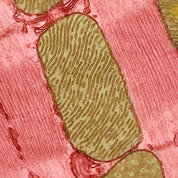
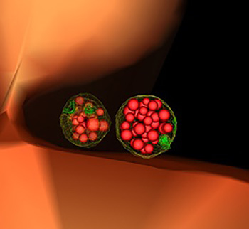
Mitochondria are the cell’s power plants. They convert energy from food into a molecule called ATP that fuels virtually every process in the cell. As shown here, mitochondria (brown) often have distinct, oblong shapes. Like most other organelles, mitochondria are encased in an outer membrane. But they also have an inner membrane that folds many times, increasing the area available for energy production. In addition, mitochondria store calcium ions, help make hemoglobin—the vital iron-containing protein that allows red blood cells to carry oxygen—and even take part in producing some hormones. Defects in mitochondria can lead to a host of rare but often incurable diseases that range from mild to devastating. Researchers are studying mitochondria to better understand their manifold jobs in the cell and to find treatments for mitochondrial diseases.

Continue reading “A Labor Day-Themed Collection: Hard-Working Cell Structures”

Interview With a Scientist: Janet Iwasa, Molecular Animator

0 comments

The world beneath our skin is full of movement. Hemoglobin in our blood grabs oxygen and delivers it throughout the body. Molecular motors in cells chug along tiny tubes, hauling cargo with them. Biological invaders like viruses enter our bodies, hijack our cells and reproduce wildly before bursting out to infect other cells.

To make sense of the subcutaneous world, Janet Iwasa, a molecular animator at the University of Utah, creates “visual hypotheses”—detailed animations that convey the latest thinking of how biological molecules interact.

“It’s really building the animated model that brings insights,” Iwasa told Biomedical Beat in 2014. “When you’re creating an animation, you’re really grappling with a lot of issues that don’t necessarily come up by any other means. In some cases, it might raise more questions, and make people go back and do some more experiments when they realize there might be something missing.”

Iwasa has collaborated with numerous scientists to develop animations of a range of biological processes and structures Exit icon. Recently, she’s undertaken an ambitious, multi-year project to animate HIV reproduction Exit icon.

Interview With a Slime Mold: Racing for New Knowledge

1 comment
Dictyostelium discoideum
Credit: Wikimedia Commons, Usman Bashir.
Dictyostelium discoideum
Natural habitat: Deciduous forest soil and moist leaf litter
Favorite food: Bacteria
Top speed: 8 micrometers per minute

Like the athletes in Rio, the world’s most highly advanced microbial runners recently gathered in Charlestown, Massachusetts, to find out which ones could use chemical cues to most quickly navigate a maze-like microfluidic racecourse. The winners’ prize: credit for helping scientists learn more about how immune system cells navigate through the human body on their way to fight disease.

The finalists were a group of soil-dwelling slime molds called Dictyostelium that were genetically engineered by a pair of Dutch biochemists to detect minuscule chemical changes in the environment. The racers used their enhanced sense of “smell” to avoid getting lost on their way to the finish line.

While researchers have been racing the genetically souped-up microbes at annual events for a few years—another competition is scheduled for October 26—scientists have been studying conventional Dictyostelium for decades to investigate other important basic life processes including early development, gene function, self/non-self recognition, cell-type regulation, chemical signaling and programmed cell death. Continue reading “Interview With a Slime Mold: Racing for New Knowledge”

Protein Paradox: Enrique De La Cruz Aims to Understand Actin

1 comment
Enrique M. De La Cruz
Credit: Jeff Foley, American Heart Association.
Enrique M. De La Cruz
Grew up in: Newark and Kearny, New Jersey
Job site: Yale University
Favorite food: His mom’s Spanish-style polenta (harina de maíz)
Alternative career: Managing a vinyl record shop
Favorite song: “Do Anything You Wanna Do” by Eddie & The Hot Rods

Enrique De La Cruz stood off to the side in a packed room. As he waited for his turn to speak, he stroked the beads of a necklace. Was he nervous? Quietly praying? When he took center stage, the purpose of the strand became clear.

Like a magician—and dressed all in black—De La Cruz held up the necklace with two hands so everyone, even those sitting in the back, could see it. It was made of snap-together beads. De La Cruz waved the strand. It wiggled in different directions. Then, with no sleight of hand, he popped off one of the beads. The necklace broke into two.

For the next hour, De La Cruz pulled out one prop after another: a piece of rope from his pocket, a pencil tucked behind his ear and even a fresh spear of asparagus stuffed in his backpack. At one point, De La Cruz assembled a conga line with people in the front row. Continue reading “Protein Paradox: Enrique De La Cruz Aims to Understand Actin”

The Cell’s Mailroom

1 comment
Yeast cell showing two mature, or “late” endosomes that are filled with small vesicles.

This illustration of the inside of a yeast cell shows two mature, or “late” endosomes (green-ringed structures) that are filled with small vesicles (red bubbles). Endosomes are cellular containers that can carry many types of cargo, including cellular waste, which they typically dump into vacuoles (orange). Credit: Matthew West and Greg Odorizzi, University of Colorado, Boulder.

In large offices, mailroom workers read the labels on incoming letters and packages to sort and deliver them and dispose of junk mail. In cells, these tasks—as well as importing food and other materials—fall to small cellular sacs called endosomes. Acting as mailroom staff, endosomes sort and deliver nutrients and building blocks, like amino acids, fat and sugars, to their proper destinations, and send cellular junk, like damaged proteins, to trash processors, such as vacuoles or lysosomes. Continue reading “The Cell’s Mailroom”

A World Without Pain

6 comments

In an immersive virtual reality environment called “Snow World,” burn patients distract themselves from their pain by tossing snow balls, building snowmen and interacting with penguins. Credit: Ari Hollander and Howard Rose, copyright Hunter Hoffman, UW, www.vrpain.com Exit icon.

You glide across an icy canyon where you meet smiling snowmen, waddling penguins and a glistening river that winds forever. You toss snowballs, hear them smash against igloos, then watch them explode in vibrant colors.

Back in the real world, a dentist digs around your mouth to remove an impacted tooth, a procedure that really, really hurts. Could experiencing a “virtual” world distract you from the pain? NIGMS grantees David Patterson Exit icon and Hunter Hoffman Exit icon show it can.

Patterson, a psychologist at the University of Washington (UW) in Seattle, and Hoffman, a UW cognitive psychologist, helped create the virtual reality program “Snow World” in an effort to reduce excessive pain experienced by burn patients. However, the researchers expect Snow World to help alleviate all kinds of pain, including pain experienced during dental procedures. Continue reading “A World Without Pain”

Cool Images: An Independence Day-Inspired Collection

1 comment

@media screen and (max-width: 480px) {
.halloween-out {
width: 96.5%;
}
.halloween-out .halloween-in img {
padding-top: 42px;
padding-right: 12px;
}
}

@media screen and (max-width: 320px) {
.halloween-out {
width: 96%;
}
}

In case you missed the fireworks this weekend, we’ve put together a collection of firework-like images from basic research studies.

Viral Electricity
Viral Electricity Image
This patriotic Koosh ball is an adeno-associated virus. Most people will come into contact with the virus at some point in their lives, and they’ll probably never know it. Even though it doesn’t cause disease—in fact, because it doesn’t cause disease—this virus is scientifically important. Researchers hope to harness the virus’ ability to enter cells and hijack genes and to use it to to deliver gene therapy. This image, created with the software DelPhi, shows which parts of the virus are positively charged (blue) and which parts are negatively charged (red). The charge of a molecule—like the charge of this virus—influences the way it behaves. In addition to helping researchers understand how viruses might enter cells, images like this one could help them understand how molecules interact with each other as well as drugs.
Continue reading “Cool Images: An Independence Day-Inspired Collection”

Demystifying General Anesthetics

0 comments

When Margaret Sedensky, now of Seattle Children’s Research Institute, started as an anesthesiology resident, she wasn’t entirely clear on how anesthetics worked. “I didn’t know, but I figured someone did,” she says. “I asked the senior resident. I asked the attending. I asked the chair. Nobody knew.”

For many years, doctors called general anesthetics a “modern mystery.” Even though they safely administered anesthetics to millions of Americans, they didn’t know exactly how the drugs produced the different states of general anesthesia. These states include unconsciousness, immobility, analgesia (lack of pain) and amnesia (lack of memory).

Stock image of a symphony.
Like the instruments that make up an orchestra, many molecular targets may contribute to an anesthetic producing the desired effect. Credit: Stock image.

Understanding anesthetics has been challenging for a number of reasons. Unlike many drugs that act on a limited number of proteins in the body, anesthetics interact with seemingly countless proteins and other molecules. Additionally, some anesthesiologists believe that anesthetics may work through a number of different molecular pathways. This means no single molecular target may be required for an anesthetic to work, or no single molecular target can do the job without the help of others.

“It’s like a symphony,” says Roderic Eckenhoff of the University of Pennsylvania Perelman School of Medicine, who has studied anesthesia for decades. “Each molecular target is an instrument, and you need all of them to produce Beethoven’s 5th.” Continue reading “Demystifying General Anesthetics”

CRISPR Serves Up More than DNA

0 comments
Marine bacterium Marinomonas mediterranea
The marine bacterium Marinomonas mediterranea uses a CRISPR system to spot invading RNAs and store a memory of the invasion event in its genome. Research team member Antonio Sanchez-Amat was the first to isolate and characterize this bacterial species. Credit: Antonio Sanchez-Amat, University of Murcia.

A new study has added another twist to the CRISPR story. As we’ve highlighted in several recent posts, CRISPR is an immune system in bacteria that recognizes and destroys viral DNA and other invading DNA elements, such as transposons. Scientists have adapted CRISPR into an indispensable gene-editing tool now widely used in both basic and applied research.

Many previously described CRISPR systems detect and cut viral DNA, insert the DNA pieces into the bacterial genome and then use them as molecular “mug shots” to flag and destroy the virus if it attacks again. But various viruses use RNA, not DNA, as genetic material. Although research has shown that some CRISPR systems also can target RNA, how these systems can archive harmful RNA encounters in the bacterial genome was unknown. Continue reading “CRISPR Serves Up More than DNA”

Viral Views: New Insights on Infection Strategies

0 comments

The following images show a few ways in which cutting-edge research tools are giving us clearer views of viruses—and possible ways to disarm them. The examples, which highlight work involving HIV and the coronavirus, were funded in part by our Biomedical Technology Research Resources program.

Uncloaking HIV’s Camouflage

HIV capsid with (right, red) and without (left) a camouflaging human protein.
HIV capsid with (right, red) and without (left) a camouflaging human protein. Credit: Juan R. Perilla, Klaus Schulten and the Theoretical and Computational Biophysics Group, University of Illinois at Urbana-Champaign.

To sneak past our immune defenses and infect human cells, HIV uses a time-honored strategy—disguise. The virus’ genome is enclosed in a protein shell called a capsid (on left) that’s easily recognized and destroyed by the human immune system. To evade this fate, the chrysalis-shaped capsid cloaks itself with a human protein known as cyclophilin A (in red, on right). Camouflaged as human, the virus gains safe passage into and through a human cell to deposit its genetic material in the nucleus and start taking control of cellular machinery.

Biomedical and technical experts teamed up to generate these HIV models at near-atomic resolution. First, structural biologists at the Pittsburgh Center for HIV Protein Interactions Exit icon used a technique called cryo-electron microscopy (cryo-EM) to get information on the shape of an HIV capsid as well as the capsid-forming proteins’ connections to each other and to cyclophilin A. Then experts at the Resource for Macromolecular Modeling and Bioinformatics fed the cryo-EM data into their visualization and simulation programs to computationally model the physical interactions among every single atom of the capsid and the cyclophilin A protein. The work revealed a previously unknown site where cyclophilin A binds to the capsid, offering new insights on the biology of HIV infection. Continue reading “Viral Views: New Insights on Infection Strategies”