Cool Video: Watching Bacteria Turn Virulent

1 comment

Researchers created an apparatus to study quorum sensing, a communication system that allows some bacteria to cause dangerous infections. Their findings suggest that blocking bacterial communication might lead to a new way to combat such infections. Credit: Minyoung Kevin Kim and Bonnie Bassler, Princeton University.

If you’ve ever felt a slimy coating on your teeth, scrubbed grime from around a sink drain or noticed something growing between the tiles of a shower, you’ve encountered a biofilm. Made up of communities of bacteria and other microorganisms, biofilms thrive where they can remain moist and relatively undisturbed. As they enlarge, biofilms can block narrow passages like medical stents, airways, pipes or intestines. Continue reading “Cool Video: Watching Bacteria Turn Virulent”

Ticks, Mice and Microbes—Studying Disease Spread

0 comments
Maria Diuk-Wasser
Credit: Oscar Gonzalez (Diuk-Wasser’s husband)
Maria Diuk-Wasser
Hometown: Buenos Aires, Argentina
Childhood dream job: Veterinarian
Hobbies: Hiking and gardening with her son (age 10) and daughter (age 7)
Favorite music: Salsa
Worksite: Lab at Columbia University and forests in coastal New England

Maria Diuk-Wasser grew up on the 10th floor of an apartment building in the middle of a bustling city. With no forests or meadows nearby, she read book after book about the natural world and surrounded herself with houseplants.

“I yearned for nature,” Diuk-Wasser says. “But my parents couldn’t provide it. They’re city people. They didn’t know anything about hiking or camping.”

These days, Diuk-Wasser still spends a lot of time in a city—she’s a professor at Columbia University in New York, the most populous city in the U.S.—but she also gets plenty of time in the woods. She hikes for hours through coastal New England forests, some of the loveliest in the country, searching for what many consider less-than-lovely inhabitants: mice and ticks. Continue reading “Ticks, Mice and Microbes—Studying Disease Spread”

Visualizing Skin Regeneration in Real Time

2 comments
Top: Colorful skin cells on a zebrafish . Bottom: Cells from the outer surface of the scale.
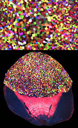
More than 70 Skinbow colors distinguish hundreds of live cells from a tiny bit (0.0003348 square inches) of skin on the tail fin of an adult zebrafish. The bottom image shows the cells on the outer surface of a scale. Credit: Chen-Hui Chen, Duke University.

Zebrafish, blue-and-white-striped fish that are about 1.5 inches long, can regrow injured or lost fins. This feature makes the small fish a useful model organism for scientists who study tissue regeneration.

To better understand how zebrafish skin recovers after a scrape or amputation, researchers led by Kenneth Poss of Duke University tracked thousands of skin cells in real time. They found that lifespans of individual skin cells on the surface were 8 to 9 days on average and that the entire skin surface turned over in 20 days.

The scientists used an imaging technique they developed called “Skinbow,” which essentially shows the fish’s outer layer of skin cells in a spectrum of colors when viewed under a microscope. Skinbow is based on a technique created to study nerve cells in mice, another model organism.

The research team’s color-coded experiments revealed several unexpected cellular responses during tissue repair and replacement. The scientists plan to incorporate additional imaging techniques to generate an even more detailed picture of the tissue regeneration process.

The NIH director showcased the Skinbow technique and these images on his blog, writing: “You can see more than 70 detectable Skinbow colors that make individual cells as visually distinct from one another as jellybeans in a jar.”

This work was funded in part by NIH under grant R01GM074057.

The Proteasome: The Cell’s Trash Processor in Action

2 comments

Our cells are constantly removing and recycling molecular waste. On the occasion of Earth Day, we put together this narrated animation to show you one way cells process their trash. The video features the proteasome, a cellular machine that breaks down damaged or unwanted proteins into bits that the cell can re-use to make new proteins. For this reason, the proteasome is as much a recycling plant as it is a garbage disposal.

For more details about the proteasome and other cellular disposal systems, check out our article How Cells Take Out the Trash.

Another Piece to a Century-Old Evolutionary Puzzle

0 comments
After mating about 55,000 pairs of fruit flies and sifting through 333,000 daughter flies, a research team found six sons that each had mutations in the same gene that helped make two fruit fly species unique from each other. Credit: Jim Woolace, Fred Hutch News Service.

Nitin Phadnis and Harmit Malik set out to conduct an experiment that could solve a century-old evolutionary puzzle: How did two related fruit fly species arise from one? Years after they began their quest, they finally have an answer.

The existence of a gene that helps make each of these fruit fly species unique and separate from each other had been guessed at since 1940, following experiments decades earlier in which geneticists first noticed that the two types of flies, when mated, had only daughters—no sons.

Scientists had previously discovered two other genes involved in driving the fruit fly species apart, but they knew those two genes weren’t the full story.

Continue reading “Another Piece to a Century-Old Evolutionary Puzzle”

Finding Adventure: Blake Wiedenheft’s Path to Gene Editing

1 comment
Blake Wiedenheft
Blake Wiedenheft
Grew up in: Fort Peck, Montana
Fields: Microbiology, biochemistry, structural biology
Job site: Montana State University
Secret talent: Being a generalist; enjoying many different subjects and activities
When not in the lab, he’s: Running, biking, skiing or playing scrabble with his grandmother

Scientific discoveries are often stories of adventure. This is the realization that set Blake Wiedenheft on a path toward one of the hottest areas in biology.

His story begins in Montana, where he grew up and now lives. Always exploring different interests, Wiedenheft decided in his final semester at Montana State University (MSU) in Bozeman to volunteer for Mark Young, a scientist who studies plant viruses. Even though he majored in biology, Wiedenheft had spent little time in a lab and hadn’t even considered research as a career option. Continue reading “Finding Adventure: Blake Wiedenheft’s Path to Gene Editing”

New Views on What the Cell’s Parts Can Do

2 comments

Studying some of the most well-tread territory in science can turn up surprising new findings. Take, for example, the cell. You may have read in textbooks how the cell’s parts look and function during important biological processes like cellular movement and division. You may have even built models of the cell out of gelatin or clay. But scientists continue to learn new facts that require those textbooks to be updated, and those models to be reshaped. Here are a few examples.

Nuclear Envelope: More Than a Protective Barrier

Damaged heterochromatin represented by nucleotides GCAT
Damaged heterochromatin, a tightly packed form of DNA, travels to the inner wall of the nuclear envelope for repair. Credit: Irene Chiolo and Taehyun Ryu, University of Southern California.

Like a security guard checking IDs at the door, the nuclear envelope forms a protective barrier around the cell’s nucleus, only letting specific proteins and chemical signals pass through. Scientists recently found that this envelope may also act as a repair center for broken strands of heterochromatin, a tightly packed form of DNA.

Irene Chiolo of the University of Southern California and Gary Karpen of the University of California, Berkeley, and the Lawrence Berkeley National Laboratory were part of a team that learned that healthy fruit fly cells mend breaks in heterochromatin by moving the damaged DNA strands to the inner wall of the nuclear envelope. There, proteins embedded in the envelope make the necessary repairs in a safe place where the broken DNA can’t accidentally get fused to the wrong chromosome. Continue reading “New Views on What the Cell’s Parts Can Do”

Cool Image: A Circadian Circuit

1 comment
Clock neurons (middle right, right corner and edge), leucokinin (LK) neurons (top left, top right and bottom middle), leucokinin receptor (LK-R) neurons (top left, top right and bottom middle)

This image, taken with a confocal microscope, shows how time-of-day information flows through the fruit fly brain. Clock neurons (stained in blue) communicate with leucokinin (LK) neurons (stained in red at the top left, top right and bottom middle), which, in turn, signal to leucokinin receptor (LK-R) neurons (stained in green). This circuit helps regulate daily activity in the fly. Credit: Matthieu Cavey and Justin Blau, New York University.

Feeling sleepy and dazed after the switch to daylight savings time this weekend? Your internal clocks are probably a little off and need some time to adjust.

Researchers have been studying biological clocks for decades to figure out how they control circadian rhythms, the natural 24-hour pattern of physical, mental and behavioral changes that affect sleep, appetite and metabolism. Knowing more about what makes our clocks tick could help researchers develop better therapies for sleep problems, metabolic conditions and other disorders associated with mistimed internal clocks. Continue reading “Cool Image: A Circadian Circuit”

Four Ways Inheritance Is More Complex Than Mendel Knew

3 comments
Original edition of Gregor Mendel’s 1866 publication, Experiments in Plant Hybridization
An original edition of Gregor Mendel’s 1866 publication, “Experiments in Plant Hybridization,” housed in NIH’s National Library of Medicine. Credit: Alisa Machalek.

This year marks the 150th anniversary of Gregor Mendel’s publication that—after sitting ignored for a few decades—helped launch the field of modern genetics. Mendel didn’t know about DNA. But after painstakingly cross-fertilizing tens of thousands of pea plants over the course of 8 years, this Austrian monk came very close to describing genes.

By picking a species with a handful of visible characteristics that occur in two easily identifiable forms, Mendel was able to pinpoint what he called “factors.” These factors determine traits like a pea’s shape or color, for instance, and are passed down from parents to offspring. He also observed that factors can be dominant or recessive.

Today, we know that inheritance is far more complex than what Mendel saw in his pea plants. Here are some of the things scientists have learned about how traits are passed from one generation to the next. Continue reading “Four Ways Inheritance Is More Complex Than Mendel Knew”

Evolution and Health: A Conversation with Evolutionary Geneticist Dr. Dan Janes on the Occasion of Charles Darwin’s Birthday

5 comments

Today, February 12, is Darwin Day—an occasion to recognize the scientific contributions of 19th-century naturalist Charles Darwin. In this video, our own evolutionary geneticist, Dan Janes, answers questions about Darwin and the role of evolution in health and biomedicine.

For more details about evolution and you, check out our articles Evolution and Health and Everyday Evolution.