Month: January 2015

A Bright New Method for Rapidly Screening Cancer Drugs

0 comments
Illustration of red, green and blue fluorescent proteins.
Chemists have devised a new approach to screening cancer drugs that uses gold nanoparticles with red, green and blue outputs provided by fluorescent proteins. Credit: University of Massachusetts Amherst.

Scientists may screen billions of chemical compounds before uncovering the few that effectively treat a disease. But identifying compounds that work is just the first step toward developing a new therapy. Scientists then have to determine exactly how those compounds function.

Different cancer therapies attack cancer cells in distinct ways. For example, some drugs kill cancer cells by causing their outer membranes to rapidly rupture in a process known as necrosis. Others cause more subtle changes to cell membranes, which result in a type of programmed cell death known as apoptosis.

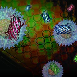
If researchers could distinguish the membrane alterations of chemically treated cancer cells, they could quickly determine how that chemical compound brings about the cells’ death. A new sensor developed by a research team led by Vincent Rotello of the University of Massachusetts Amherst can make these distinctions in minutes.

Continue reading “A Bright New Method for Rapidly Screening Cancer Drugs”

New Streamlined Technique for Processing Biological Samples

0 comments
Illustration of Slug flow microextraction.
Researchers have discovered a faster, easier and more affordable technique for processing biological samples. Credit: Weldon School of Biomedical Engineering, Purdue University.

It’s not unusual for the standard dose of a drug to work well for one person but be less effective for another. One reason for such differences is that individuals can break down drugs at different rates, leading to different concentrations of drugs and of their breakdown products (metabolites) in the bloodstream. A promising new process called slug-flow microextraction could make it faster, easier and more affordable to regularly monitor drug metabolites so that medication dosages could be tailored to each patient’s needs, an approach known as personalized medicine. This technique could also allow researchers to better monitor people’s responses to new drug treatments during clinical trials. Continue reading “New Streamlined Technique for Processing Biological Samples”

Delivering Gene-Editing Proteins to Living Cells

0 comments
Illustration of a DNA strand being cut by a pair of scissors.
Researchers are testing new ways to get gene editing proteins into living cells to potentially modify human genes associated with disease. Credit: Stock image.

Over the last two decades, exciting tools have emerged that allow researchers to cut and paste specific sequences of DNA within living cells, a process called gene editing. These tools, including one adapted from a bacterial defense system called CRISPR, have energized the research community with the possibility of using them to modify human genes associated with disease.

A major barrier to testing medical applications of gene editing has been getting the proteins that do the cutting into the cells of living animals. The main methods used in the laboratory take a roundabout route: Researchers push the DNA templates for making the proteins into cells, and then the cells’ own protein factories produce the editing proteins.

Researchers led by David Liu Exit icon from Harvard University are trying to cut out the middleman, so to speak, by ferrying the editing proteins, not the DNA instructions, directly into cells. In a proof-of-concept study, their system successfully delivered three different types of editing proteins into cells in the inner ears of live mice. Continue reading “Delivering Gene-Editing Proteins to Living Cells”