Month: June 2017

The Drama of Cell Death

1 comment
spermatids

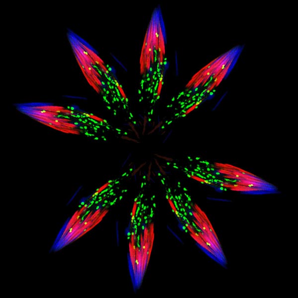
Spermatids—one stage in the formation of sperm—in the fruit fly (Drosophila). Credit: Sigi Benjamin-Hong, Rockefeller University (modified).

Although it looks like a bursting firework from a Fourth of July celebration, this image actually was created from pictures of spermatids—one stage in the formation of sperm—in the fruit fly (Drosophila). Drosophila is an organism that scientists often use as a model for studying how cells accomplish their amazing tasks. Drosophila studies can help reveal where an essential cellular process goes wrong in diseases such as autoimmune conditions or cancer. Cell death, or apoptosis, is one of these processes.

Almost every animal cell has the ability to destroy itself via apoptosis. Apoptosis is important because it allows the body both to develop normally and get rid of dangerous and unwanted cells when it needs to later in life, such as when cells become cancerous. Many different signals both within and outside the cell influence whether apoptosis happens when it should, and abnormal regulation of this process is associated with some diseases. Hermann Steller Exit icon and colleagues at Rockefeller University in New York City study Drosophila and mammalian cells to tease apart the steps of apoptosis and the many molecular signals that regulate it. Continue reading “The Drama of Cell Death”

Flipping the Switch on Controlling Disease-Carrying Insects

4 comments
Illustration of some of the jobs that the ER performs in the cell.

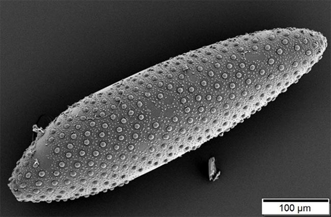
This image shows a mosquito egg. Wolbachia bacteria, which infect many species of insects including mosquitos, move from one generation to the next inside insect eggs. Credit: Wikimedia Commons, Mogana Das Murtey and Patchamuthu Ramasamy, Universiti Sains, Malaysia.

Suppressing insects that spread disease is an essential public health effort, and scientists are testing a possible new tool to use in this challenging arena. They’re harnessing a microbe capable of controlling insects’ reproductive processes.

The microbes, called Wolbachia, live inside the cells of about two-thirds of insect species worldwide, and they can manipulate the host’s reproductive cells in ways that boost their own survival. Scientists think they can use Wolbachia’s methods to reduce populations of insects that spread disease among humans.

A Switch to Control Fertility

Wolbachia have evolved complex ways to control insect reproduction so as to infect increasing numbers of an insect species—such as those prolific disease-spreaders, mosquitos. One method Wolbachia uses is called cytoplasmic incompatibility, or CI. The end result of CI, basically, is that the sperm of infected male insects cause sterility in uninfected females.

Wolbachia that have infected male insects can insert proteins that produce a kind of infertility switch into the host’s sperm. When the sperm later fuses with an egg from an uninfected female, the switch is triggered and renders the egg sterile. If the female is already infected, her eggs will contain Wolbachia, which can turn off the switch and allow the egg to develop. This trick ensures that more Wolbachia-infected insects will survive and continue to reproduce, while uninfected ones will be less successful.

Already, some states Exit icon and countries Exit icon are releasing Wolbachia-infected male mosquitoes into wild mosquito populations that carry disease-causing viruses to test this strategy for insect control. Males carrying a Wolbachia strain that strongly induces infertility in uninfected females should reduce the numbers of mosquito eggs that mature, leading to fewer mosquitos. Continue reading “Flipping the Switch on Controlling Disease-Carrying Insects”

Researchers Score Goal with New Atomic-Scale Model of Salmonella-Infecting Virus

1 comment

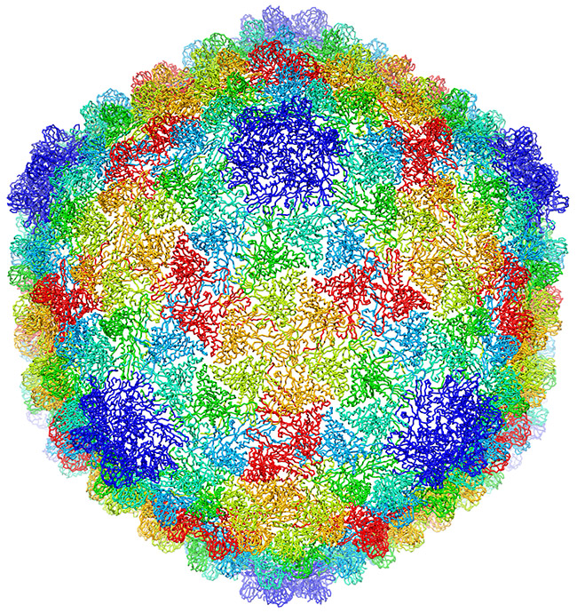
An atomic-scale model of a virus that infects the Salmonella bacterium. Credit: C. Hryc and the Chiu Lab, Baylor College of Medicine.

This sphere could be a prototype design for the 2018 World Cup official match soccer ball, but you won’t see it dribbled around any soccer fields. The image is actually an atomic-scale model of a virus that infects the Salmonella bacterium. Like a soccer ball, both are approximately spherical shapes created by a combination of hexagonal (six-sided) and pentagonal (five-sided) units. Wah Chiu, a biochemist at Baylor College of Medicine, and his colleagues used new computational methods to construct the model from more than 20,000 cryo-electron microscopy (cryo-EM) images. Cryo-EM is a sophisticated technique that uses electron beams for visualizing frozen samples of proteins and other biological specimens.

The researchers’ model, published in a recent issue of PNAS, shows the virus’ protein shell, or capsid, that encloses the virus’ genetic material. Each color shows capsid proteins having the same interactions with their neighbors. The fine resolution allowed researchers to identify the protein interactions essential to building a stable shell. They developed a new approach to checking the accuracy and reliability of the virus model and reporting what parts are the most certain. The approach could be used to evaluate other complex biological structures, potentially leading to better quality models and new avenues for drug design and development.

This research is funded in part by NIH under grants R01GM079429, P01GM063210, P41GM103832, PN2EY016525, T15LM007093.