Month: July 2018

Interview With a Scientist—Julius Lucks: Shape Seeker

0 comments

While DNA acts as the hard drive of the cell, storing the instructions to make all of the proteins the cell needs to carry out its various duties, another type of genetic material, RNA, takes on a wide variety of tasks, including gene regulation, protein synthesis, and sensing of metals and metabolites. Each of these jobs is handled by a slightly different molecule of RNA. But what determines which job a certain RNA molecule is tasked with? Primarily its shape. Julius LucksLink to external web site, a biological and chemical engineer at Northwestern University, and his team study the many ways in which RNA can bend itself into new shapes and how those shapes dictate the jobs the RNA molecule can take on.

Continue reading “Interview With a Scientist—Julius Lucks: Shape Seeker”

Interview With a Scientist—Elhanan Borenstein: Metagenomics Systems Biology

0 comments


Cataloging the human microbiome—the complete collection of bacteria, fungi, archaea, protists, and viruses that live in and on our bodies—is an enormous task. Most estimates put the number of organisms who call us home on par with the number of our own cells. Imagine trying to figure out how the billions of critters influence each other and, ultimately, impact our health. Elhanan Borenstein,Link to external web site a computer scientist-cum-genomicist at the University of Washington, and his team are not only tackling this difficult challenge, they are also trying to obtain a systems-level understanding of the collective effect of all of the genes, proteins, and metabolites produced by the numerous species within the microbiome.

Continue reading “Interview With a Scientist—Elhanan Borenstein: Metagenomics Systems Biology”

Molecular Fireworks: How Microtubules Form Inside Cells

1 comment
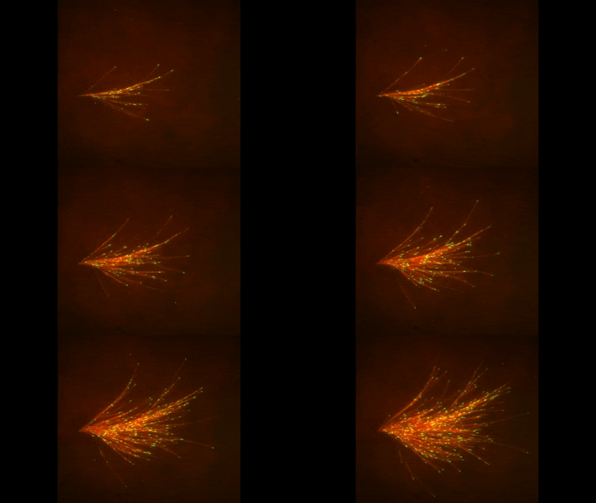
A video depicting red strands of various lengths exploding outward from a focal point at the left. The strands are tipped in neon green.
       Microtubules sprout from one another. Credit: Petry lab, Princeton University.

The red spray pictured here may look like fireworks erupting across the night sky on July 4th, but it’s actually a rare glimpse of tiny protein strands called microtubules sprouting and growing from one another in a lab. Microtubules are the largest of the molecules that form a cell’s skeleton. When a cell divides, microtubules help ensure that each daughter cell has a complete set of genetic information from the parent. They also help organize the cell’s interior and even act as miniature highways for certain proteins to travel along.

As their name suggests, microtubules are hollow tubes made of building blocks called tubulins. Scientists know that a protein called XMAP215 adds tubulin proteins to the ends of microtubules to make them grow, but until recently, the way that a new microtubule starts forming remained a mystery.

Sabine Petry Link to external web site and her colleagues at Princeton University developed a new imaging method for watching microtubules as they develop and found an important clue to the mystery. They adapted a technique called total internal reflection fluorescence (TIRF) microscopy, which lit up only a tiny sliver of a sample from frog egg (Xenopus) tissue. This allowed the scientists to focus clearly on a few of the thousands of microtubules in a normal cell. They could then see what happened when they added certain proteins to the sample.

Continue reading “Molecular Fireworks: How Microtubules Form Inside Cells”