Tag: Wound Healing

Interview With a Worm: We’re Not So Different

2 comments
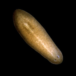
Planarian
Credit: Alejandro Sánchez Alvarado, Stowers Institute for Medical Research.
Schmidtea mediterranea
Home: Freshwater habitats along the Mediterranean
Party trick: Regenerating its head
Most charismatic feature: Eyespots
Work site: Science labs worldwide

The planarian has a power few creatures can match. Remove its head, its tail or nearly any of its body parts, and this aquatic flatworm will simply grow it back. Humans can’t do that, of course. And yet many of the genes that help the planarian regenerate are also found in us. To learn more about this tiny marvel, we “interviewed” a representative. Continue reading “Interview With a Worm: We’re Not So Different”

Molecules Known to Damage Cells May Also Have Healing Power

0 comments
Free radicals in an ying-yang symbol
Biology in balance: Molecules called free radicals—like the peroxide molecules illustrated here—have a reputation for being dangerous. Now, they’ve revealed healing powers. In worms, at least. Credit: Stock image

When our health is concerned, some molecules are widely labeled “good,” while others are considered “bad.” Often, the truth is more complicated.

Consider free radical molecules. These highly reactive, oxygen-containing molecules are well known for damaging DNA, proteins and other molecules in our bodies. They are suspected of contributing to premature aging and cancer. But now, new research shows they might also have healing powers.

Using the oft-studied laboratory roundworm known as C. elegans, a research group led by Andrew Chisholm at the University of California, San Diego, made a surprising discovery. Free radicals, specifically those made in cell structures called mitochondria, appear necessary for skin wounds to heal. In fact, higher (but not dangerously high) levels of the molecules can actually speed wound closure.

If further research shows the same holds true in humans, the work could point to new ways to treat hard-to-heal wounds, like diabetic foot ulcers.

This work was funded in part by NIH under grants R01GM054657 and P40OD010440.

Meet Elizabeth Grice

0 comments
Elizabeth Grice
Elizabeth Grice
First job: Detasseling corn
Favorite food: Chocolate
Pets: An adopted shelter cat, Dolce
Favorite city: Athens, Greece
Hidden talent: Baking creative desserts
Credit: Bill Branson, NIH

Imagine a landscape with peaks and valleys, folds and niches, cool, dry zones and hot, wet ones. Every inch is swarming with diverse communities, but there are no cities, no buildings, no fields and no forests.

You’ve probably thought little about the inhabitants, but you see their environment every day. It’s your largest organ—your skin.

Elizabeth Grice, an assistant professor at the University of Pennsylvania, studies the skin microbiome to learn how and why bacteria colonize particular places on the body. Already, she’s found that the bacterial communities on healthy skin are different from those on diseased skin.

She hopes her work will point to ways of treating certain skin diseases, especially chronic wounds. “I like to think that I am making discoveries that will impact the way medicine is practiced,” she says.

Grice’s Findings

To investigate what role bacteria play in diabetic wounds, Grice and her colleagues took skin swabs from both diabetic and healthy mice, and then compared the two. They found that diabetic mice had about 40 times more bacteria on their skin, but it was concentrated into few species. A more diverse array of bacteria colonized the skin of healthy mice.

The researchers then gave each mouse a small wound and spent 28 days swabbing the sites to collect bacteria and observing how the skin healed. They found that wounds on diabetic mice started to increase in size at the same time as wounds on healthy mice began to heal. In about 2 weeks, most healthy mice looked as good as new. But most of the wounds on diabetic mice had barely healed even after a month.

Interestingly, bacterial communities in the wounds became more diverse in both groups of mice as they healed—although the wounds on diabetic mice still had less diversity than the ones on healthy mice.

“Bacterial diversity is probably a good thing, especially in wounds,” says Grice. “Often, potentially infectious bacteria are found on normal skin and are kept in check by the diversity of bacteria surrounding them.”

Grice and her colleagues also found distinctly different patterns of gene activity between the two groups of mice. As a result, the diabetic mice put out a longer-lasting immune response, including inflamed skin. Scientists believe prolonged inflammation might slow the healing process.

Grice’s team suspects that one of the main types of bacteria found on diabetic wounds, Staphylococcus, makes one of the inflammation-causing genes more active.

Now that they know more about the bacteria that thrive on diabetic wounds, Grice and her colleagues are a step closer to looking at whether they could reorganize these colonies to help the wounds heal.

Content adapted from the NIGMS Findings magazine article Body Bacteria.
Good Vibrations

3 comments
A knot-like structure in a section of RNA from a flavivirus
Findings in mice may lead to a drug-free, noninvasive way to treat chronic wounds in people with type 2 diabetes. Credit: Stock image.

For people living with type 2 diabetes, wounds often heal slowly, sometimes even becoming chronic. Now, scientists have shown that low-intensity vibrations can speed up the healing process in a strain of diabetic mice commonly used to study delayed wound healing. The research team, led by Timothy Kohof the University of Illinois at Chicago, found that exposing the mice to barely perceptible vibrations five times a week for just 30 minutes promoted wound healing by increasing the formation of new blood vessels and of granulation tissue, a type of tissue critical in the early stages of healing. If researchers can show that the vibration technique also works in humans, this approach could one day offer a drug-free, non-invasive therapy for chronic wounds in people with diabetes.

This work also was funded by NIH’s National Institute of Dental and Craniofacial Research.

Learn more:
University of Illinois at Chicago News Release exit icon