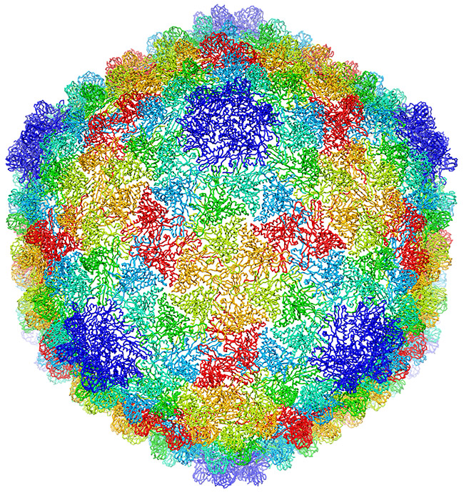
This sphere could be a prototype design for the 2018 World Cup official match soccer ball, but you won’t see it dribbled around any soccer fields. The image is actually an atomic-scale model of a virus that infects the Salmonella bacterium. Like a soccer ball, both are approximately spherical shapes created by a combination of hexagonal (six-sided) and pentagonal (five-sided) units. Wah Chiu, a biochemist at Baylor College of Medicine, and his colleagues used new computational methods to construct the model from more than 20,000 cryo-electron microscopy (cryo-EM) images. Cryo-EM is a sophisticated technique that uses electron beams for visualizing frozen samples of proteins and other biological specimens.
The researchers’ model, published in a recent issue of PNAS, shows the virus’ protein shell, or capsid, that encloses the virus’ genetic material. Each color shows capsid proteins having the same interactions with their neighbors. The fine resolution allowed researchers to identify the protein interactions essential to building a stable shell. They developed a new approach to checking the accuracy and reliability of the virus model and reporting what parts are the most certain. The approach could be used to evaluate other complex biological structures, potentially leading to better quality models and new avenues for drug design and development.
This research is funded in part by NIH under grants R01GM079429, P01GM063210, P41GM103832, PN2EY016525, T15LM007093.


Very interesting.