Author: Alisa Zapp Machalek

Alisa Machalek Headshot.

Posts by Alisa Zapp Machalek

Visualizing Vessels

0 comments
Blood vessels in a mouse retina
Blood vessels in a mouse retina visualized using cutting-edge imaging technology. Credit: Tom Deerinck and Mark Ellisman, NCMIR.

For poets and lovers, the eyes are the windows of the soul. For scientists and doctors, blood vessels at the back of the eye are windows into many diseases.

Blood vessel abnormalities can indicate a variety of serious conditions such as atherosclerosis (hardening of the arteries), heart attacks and strokes. But most vessels are buried beneath skin and other tissues, making them difficult to examine without surgery.

There’s one exception—in the eye. Unlike anywhere else in the body, larger vessels on the retina at the back of the eye are directly visible through the pupil, requiring essentially only light and magnifying lenses to view.

These vessels are used to diagnose glaucoma and diabetic eye disease. Because they display characteristic changes in people with high blood pressure, some researchers hope retinal vessels might one day help predict an impending stroke, congestive heart failure or other diseases stemming from dangerously high blood pressure.

The medical importance of retinal vessels piqued the interest of scientists funded by the National Institutes of Health at the National Center for Microscopy and Imaging Research (NCMIR) at the University of California, San Diego, who captured this micrograph image of mouse retinal vessels.

Continue reading this new Inside Life Science article

Meet Jasmine Johnson and Gabe Vela

0 comments
Jasmine Johnson and Gabriel 'Gabe' Vela
Jasmine Johnson and Gabriel “Gabe” Vela
Field: Genetics of sleep and obesity
Worked as researchers at: The Jackson Laboratory, Bar Harbor, Maine
Graduated from high school at: Rockdale Magnet School for Science and Technology in Conyers, Ga.
Now freshman at: Stanford University in Palo Alto, Calif. (Johnson) and Southern Polytechnic State University in Marietta, Ga. (Vela)
Fascinating fact: Johnson presented her research at the 2013 White House Science Fair
Credit: Joe Piergrossi

Jasmine Johnson and Gabe Vela might still be teenagers, but they are also seasoned scientists. It all started 3 years ago, when, as high school juniors, they took the research course Independent Studies in Computational Biology at The Jackson Laboratory in Bar Harbor, Maine. They were hooked. They continued to do research until they graduated, working part-time for 2 academic years and full-time for 2 summers.

They worked with statistical geneticist Gary Churchill, using computational biology to explore the relationship between sleep and obesity. They focused on finding genes that regulate sleep and understanding how sleep affects the body. One goal of the research is to tease out a genetic explanation for why sleep deprivation increases the risk of obesity.

Working in a lab “completely changed what I thought I was going to do with my life,” said Vela. “Now I’m going to focus more on research than anything else.”

For Johnson, the experience provided the opportunity to present her research at the 2013 White House Science Fair, where she hobnobbed with some political hot shots.

“It was an amazing experience,” she said. Having “important White House officials be interested in my project … inspired me.”

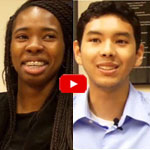
Johnson and Vela visited NIH a few months ago and talked with us about their research experiences, their lives and their future goals. Jasmine Johnson & Gabriel Vela on their experience as high school researchers at The Jackson Laboratory in Bar Harbor, Maine.

Learn More
Article Exit icon about Johnson and Vela and other young researchers, from The Jackson Laboratory’s magazine The Search.
Article about the work of Gary Churchill, from NIH’s Findings magazine.

Neanderthal DNA—Still Among Us

0 comments
Map of Europe and Asia showing the presumed range of where Neanderthals lived.
For tens of thousands of years, Neanderthals lived in Europe and Asia (presumed range shown in blue) and interbred with humans, passing on some DNA to present-day people. Credit: Ryulong, Wikimedia Commons.
View larger image

Many of us have some Neanderthal genes. Before Neanderthals went extinct about 30,000 years ago, they interbred with humans living in Europe and Asia. Today’s descendants of those pairings inherited about 2 percent of their genomes from the big-brained hominids.

A research group led by David Reich at Harvard Medical School recently completed an analysis to determine the extent and identity of Neanderthal DNA in modern-day human populations. The group found that many traits in present-day people—including skin characteristics and susceptibility to various diseases—can be traced to Neanderthal DNA.

It also appears that, genetically speaking, Neanderthals and humans weren’t completely compatible. Based on the uneven distribution of Neanderthal DNA in today’s genomes, the scientists concluded that many of the male offspring of Neanderthal-human unions were infertile. In the animal world, this phenomenon is known as hybrid infertility, where the offspring of a male from one subspecies and a female from another have low or no fertility.

Studying human genes passed down through Neanderthals—as well as regions of the human genome notably devoid of Neanderthal DNA—provides an increasingly complete picture of the genetic landscape that contributed to health, disease and diversity among humans today.

Learn more:
Harvard Medical School News Release
Reich Lab Exit icon

One Mutation Leads to Another—At Least in Yeast

0 comments
DNA mutation. Credit: Stock image.
Newly discovered genetic effect in yeast could shed light on carcinogenesis. Credit: Stock image.

Cancer cells typically include many gene mutations, extra or missing genes, or even the wrong number of chromosomes. Scientists know that certain genetic changes lead to ones elsewhere. But they’ve had a chicken-and-egg problem trying to figure out which changes trigger which others—or whether mutations accumulate randomly in tumors.

New research led by J. Marie Hardwick Exit icon of Johns Hopkins University sheds light on the issue. She found that incapacitating a single gene in yeast cells—regardless of which gene it was—spurred mutations in one or two other genes. The process was anything but random: If, say, gene X was knocked out, yeast cells almost always developed a secondary mutation in gene Y. It’s as if knocking out one gene disrupts the genomic balance enough that the cell must alter a different gene to compensate.

Significantly, the secondary mutations—but not the original ones—caused altered yeast cell characteristics, including traits linked to cancer. Also, many of the secondary mutations occurred in genes associated with cancer in humans, further suggesting that these secondary changes might play a role in carcinogenesis.

This new information will help researchers better understand the chain of genetic events that lead to cancer. It might also prompt scientists to reevaluate years of research that attributed changes in cell behavior or appearance to a given gene knockout.

This work also was funded by NIH’s National Institute of Neurological Disorders and Stroke.

Learn more:
Johns Hopkins University News Release Exit icon

Genetic Discovery Could Enable More Precise Prescriptions

0 comments
Prescription pad with DNA illustration on it. Credit: Jane Ades, NIH’s National Human Genome Research Institute.
New insight into the genes that affect drug responses may help doctors prescribe the medications and doses best suited for each individual. Credit: Jane Ades, NIH’s National Human Genome Research Institute.

Scientists know that variations in certain genes can affect the way a person responds to medications. New research by Wolfgang Sadee Exit icon at Ohio State University shows that drug responses also depend on previously overlooked parts of DNA—sections that regulate genes, but are not considered genes themselves. This study focused on an important enzyme abbreviated CYP2D6 that processes about one-fourth of all prescription drugs. Differences in the enzyme’s performance, which range from zilch to ultra-rapid, can dramatically alter the effectiveness and safety of certain medications. Researchers discovered two new genetic variants that impact CYP2D6 performance. One of these, located in a non-gene, regulatory region of DNA, doubles or even quadruples enzyme activity. Coupling these findings with genetic tests could help doctors better identify each patient’s CYP2D6 activity level, enabling more precise prescriptions. The findings also open up a whole new area of investigation into genetic factors that impact drug response.

This work also was funded by NIH’s Eunice Kennedy Shriver National Institute of Child Health and Human Development.

Learn more:

Ohio State University News Release (no longer available)

Healing Wounds, Growing Hair

1 comment
Wound healing in process. Credit: Yaron Fuchs and Samara Brown in the lab of Hermann Steller, Rockefeller University.

Credit: Yaron Fuchs and Samara Brown in the lab of Hermann Steller, Rockefeller University.

Whether injured by a scrape, minor burn or knife wound, skin goes through the same steps to heal itself. Regrowing hair over new skin is one of the final steps. All the hair you can see on your body is non-living, made up of “dead” cells and protein. It sprouts from living cells in the skin called hair follicle stem cells, shown here in red and orange. For more pictures of hair follicle stem cells—and many other stunning scientific images and videos—go to the NIGMS Image and Video Gallery.

Learn more:

Rockefeller University News Release Exit icon
Steller Lab Exit icon

Cell Biology Advances and Computational Techniques Earn Nobels for NIGMS Grantees

0 comments
The winners of the 2013 Nobel Prize in physiology or medicine discovered that cells import and export materials using fluid-filled sacs called vesicles. Credit: Judith Stoffer.

Every October, a few scientists receive a call from Sweden that changes their lives. From that day forward, they will be known as Nobel laureates. This year, five of the new Nobelists have received funding from NIGMS.

In physiology or medicine, NIGMS grantees James Rothman (Yale University) and Randy Schekman (University of California, Berkeley) were honored “for their discoveries of machinery regulating vesicle traffic, a major transport system in our cells.” They share the prize with Thomas C. Südhof of Stanford University.

Rothman and Schekman started out working separately and in different systems—Schekman in yeast and Rothman in reconstituted mammalian cells—and their conclusions validated each others’. It’s yet another example of the power of investigator-initiated research and the value of model systems.

Highly accurate molecular models like this one are based on computational techniques developed by the winners of the 2013 Nobel Prize in chemistry. Credit: Rommie E. Amaro, University of California, San Diego.

The three Nobelists in chemistry are NIGMS grantees Martin Karplus (Harvard University), Michael Levitt (Stanford University) and Arieh Warshel (University of Southern California), who developed “multiscale models for complex chemical systems.” They used computational techniques to obtain, for the first time, detailed structural information about proteins and other large molecules. Because of that work, scientists around the world are now able to access, with a few keystrokes, highly accurate models of nearly 100,000 molecular structures. Studying these structures has advanced our understanding of countless diseases, pharmaceuticals and basic biological processes.

Learn more:

NIGMS Nobel Prize News Announcement
NIGMS Nobelists Fact Sheet

Cool Video: How Bee Venom Toxin Kills Cells

0 comments
Credit: Huey Huang, Rice University.

Credit: Huey Huang, Rice University.

A new video, starring the toxin in bee venom, might help scientists design new drugs to combat bacterial infections. The video, by Rice University biophysicist Huey Huang Exit icon, condenses 6.5 minutes into less than a minute to show how the toxin, called melittin, destroys an animal or bacterial cell.

What looks like a red balloon is an artificial cell filled with red dye. Melittin molecules are colored green and float on the cell’s surface like twigs on a pond. As melittin accumulates on the cell’s membrane, the membrane expands to accommodate it. In the video, the membrane stretches into a column on the left.

When melittin levels reach a critical threshold, countless pinhole leaks burst open in the membrane. The cell’s vital fluids—red dye in the video—leak out through these pores. Within minutes, the cell collapses.

Many organisms use such a pore-forming technique to kill attacking bacterial cells. This research reveals molecular-level details of the strategy, bringing pharmaceutical scientists a step closer to harnessing it in the design of new antibiotics.

Chemist Phil Baran Joins “Genius” Ranks as MacArthur Fellow

1 comment
Cake decorated with a two-dimensional structure of the molecule, stephacidin B
When Baran’s research team succeeds in synthesizing an important natural product, the group sometimes celebrates with a cake decorated with a two-dimensional structure of the molecule. This molecule, stephacidin B, was isolated from a fungus and has anticancer properties. See images of other Baran lab cakes.

As a newly appointed MacArthur Fellow, Phil Baran is now officially a genius. The MacArthur award recognizes “exceptionally creative” individuals who have made significant contributions to their field and are expected to continue doing so. Baran, a synthetic organic chemist at Scripps Research Institute in La Jolla, Calif., was recognized today for “inventing efficient, scalable, and environmentally sound methods” for building, from scratch, molecules produced in nature. Many of these natural products have medicinal properties. Baran has already concocted a host of natural products, including those with the ability to kill bacteria or cancer cells. In addition to emphasizing the important pharmaceutical applications of his work, Baran embraces its creative aspects: “The area of organic chemistry is such a beautiful one because one can be both an artist and an explorer at the same time,” he said in the MacArthur video interview Exit icon.

Learn more:

NIGMS “Meet a Chemist” Profile of Baran
NIH Director’s Blog Post on Baran’s Recent Work