Author: Anne Oplinger

Anne Oplinger

Posts by Anne Oplinger

Six Things to Know About DNA and DNA Repair

3 comments

Deoxyribonucleic acid, better known as DNA, was first identified on a discarded surgical bandage almost 150 years ago. Increasingly sophisticated tools and techniques have allowed scientists to learn more about this chemical compound that includes all the instructions necessary for building a living organism. From among the dozens of fascinating things known about DNA, here are six items touching on the make up of DNA’s double helix, the vast amounts of DNA packed into every human’s cells, common DNA errors and a few ways DNA can repair itself.

1. DNA is in every living thing.

Nucleotide
DNA consists of two long, twisted chains made of nucleotides. Each nucleotide contains one base, one phosphate molecule and the sugar molecule deoxyribose. The bases in DNA nucleotides are adenine, cytosine, guanine and thymine. Credit: NIGMS.

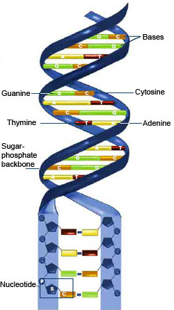
The chemical instructions for building a person—and every other creature on Earth—are contained in DNA. DNA is shaped like a corkscrew-twisted ladder, called a double helix. The two ladder rails are referred to as backbones, made of alternating groups of sugar and phosphate. The ladder’s rungs are made from four different building blocks called bases, arranged in pairs: adenine (A) paired with thymine (T), and cytosine (C) paired with guanine (G). Humans have about 3 billion base pairs in each cell. The order of the base pairs determines the exact instructions encoded in that part of the DNA molecule. Also, the sequence of DNA base pairs in one person is about 99.9 percent identical to that of everyone else.

2. Humans have a lot of DNA.

Humans begin as a single fertilized cell containing (with some rare exceptions) the full complement of DNA—the genome—arranged into 46 discrete chromosomes (23 pairs, with mom and dad each contributing half of each pair) in the cell’s nucleus. There are 6 feet of DNA coiled up tightly in that first cell. All the information in the DNA is replicated each time the cell divides. The amount of DNA packed into all of an adult’s cells is on the order of 100 trillion feet (about 19 billion miles)—so that if the DNA chain was stretched out, it would be long enough to reach back and forth between the Earth and the Sun more than 200 times. Continue reading “Six Things to Know About DNA and DNA Repair”

Cool Image: Adding Color to the Gray World of Electron Microscopy

1 comment

Color electron micrograph of an endosome, a cell organelle. Credit: Ranjan Ramachandra, UCSD

As his Christmas gift to himself each year, the late biochemist Roger Tsien treated himself to two weeks of uninterrupted research in his lab. This image is a product of those annual sojourns. While it may look like a pine wreath dotted with crimson berries, it is in fact one of the world’s first color electron micrographs—and the method used to create it may dramatically advance cell imaging.

Electron microscopy (EM) is a time-honored technique for visualizing cell structures that uses beams of accelerated electrons to magnify objects up to 10 million times their actual size. Standard EM images are in grayscale and any color is added in with computer graphics programs after the image is made. With their new technique, Tsien, who received a Nobel Prize for his development of green fluorescent protein into a tool for visualizing details in living cells using light microscopes, and his colleagues have found a way to incorporate color labeling directly into EM. Continue reading “Cool Image: Adding Color to the Gray World of Electron Microscopy”

You’ve Got Questions, We’ve Got Answers: Cell Day 2016

0 comments

Students from Connecticut to Washington State and points in between peppered our experts with questions during the recent live Cell Day web chat. They fielded questions about cell structures, microscopes and other tools, life as a scientist, and whether there are still discoveries to be made in cell biology. One of the Cell Day moderators, Jessica Faupel-Badger, even gave a shout-out to the Biomedical Beat blog as a great way to keep up with new and exciting discoveries being made every day. Thanks!

The full transcript with all the questions and answers is now available. We’ve recapped some of the highlights below.

[Check out our Facebook Live post-chat video Exit icon for a bonus answer to the question “If you put lizard DNA into human cells, could humans regrow their limbs?”]


Being a Scientist

Patrick Brown
Prior to joining NIGMS in 2016 as a program director, Patrick Brown, was a high school chemistry teacher in Maryland.

What do you think is the best thing about being a biologist? Why do you love your job so much? (Assuming you do!)

Patrick Brown answered: I love that question! And, I love being a scientist. There are so many things that I like about my career choice. The answer is simple—I like learning! I like learning about different living organisms and how they may be the same or different. I also really enjoy the multi-cultural aspect of science. I get to interact with so many different people from different parts of the world who are all studying different aspects of science that are just as interesting as my own, and we are all interested in knowing more about life.

Gram stained cerebrospinal fluid with gram-positive anthrax bacilli. Credit: Wikimedia Commons, Yuval Madar.

How did you know that biology was the career for you? In other words, what motivated you to become a biologist?

Amy Kullas answered: I remember being in my high school biology class, gazing through a microscope, and seeing the mixture of beautiful purple and pink cocci after performing my first Gram stain Exit icon. It was at that moment that I got hooked on science. I majored in microbiology in college and then went on to graduate school.

What does a typical day at work look like?

Amicia Elliott answered: The truth is that every day at work is an adventure. A typical day includes some of the following things: reading scientific papers, thinking about and designing experiments (my favorite part!), carrying out those experiments, data analysis and discussing results. Scientists work long hours to accomplish all of these things, but it is mostly a labor of love!

In a 2014 Molecular Cell Exit icon paper, NIGMS Director Jon Lorsch and colleagues determined the structure of initiation complexes.

What was the most interesting experiment you have conducted?

Jon Lorsch answered: In my lab, we study how proteins are synthesized by the eukaryotic ribosome. We have learned a great deal about how the ribosome and the proteins that help it (called translation factors) find the start codon in the messenger RNA. Recently, in collaboration with a group in the UK, we used cryo-electron microscopy to determine the three-dimensional structure of various ‘initiation complexes’ – the small subunit of the ribosome bound to mRNA, tRNA and initiation factors. Being able to see how this process works in three dimensions is amazing!

Continue reading “You’ve Got Questions, We’ve Got Answers: Cell Day 2016”