I can still remember that giddy feeling I had seven years ago, when I first read about the “zombie ant.” The story was gruesome and fascinating, and it was everywhere. Even friends and family who aren’t so interested in science knew the basics: in a tropical forest somewhere there’s a fungus that infects an ant and somehow takes control of the ant’s brain, forcing it to leave its colony, crawl up a big leaf, bite down and wait for the sweet relief of death. A grotesque stalk then sprouts from the poor creature’s head, from which fungal spores rain down to infect a new batch of ants.
A fungal fruiting body erupts through the head of a carpenter ant infected by a parasitic fungus in Thailand. Credit: David Hughes, Penn State University.
The problem is, it doesn’t happen quite like that. David Hughes, the Penn State University entomologist who reported his extensive field observations of the fungus/ant interactions in BMC Ecology, which caused much excitement back in 2011, has continued to study the fungus, Ophiocordyceps unliateralis, and its carpenter ant host, Camponotus leonardi.
In late 2017, Hughes and his colleagues published an article in PNAS in which they used sophisticated microscopy and image-processing techniques to describe in great detail how the fungus invades various parts of the ant’s body including muscles in its legs and head.
Although Hughes’s earlier BMC Ecology paper showed fungus in the head of an ant, the new study reveals that the fungus never actually enters the brain.
To me, the new finding somehow made the fungus’ control over the ant even more baffling. What exactly was going on?
To find out, I spoke with Hughes and his graduate student Maridel Fredericksen.
Every one of our thoughts, emotions, sensations, and movements arise from changes in the flow of electricity in the brain. Disruptions to the normal flow of electricity within and between cells is a hallmark of many diseases, especially neurological and cardiac diseases.
The source of electricity within nerve cells (i.e., neurons) is the separation of charge, referred to as voltage, across neuronal membranes. In the past, scientists weren’t able to identify all the molecules that control neuronal voltage. They simply lacked the tools. Now, University of Colorado biologist Joel Kralj has developed a way to overcome this hurdle. His new technique—combining automated imaging tools and genetic manipulation of cells—is designed to measure the electrical contribution of every protein coded by every gene in the human genome. Kralj believes this technology will usher in a new field of “electromics” that will be of enormous benefit to both scientists studying biological processes and clinicians attempting to treat disease.
In 2017, Kralj won a New Innovator Award from the National Institutes of Health for his work on studying voltage in neurons. He is using the grant money to develop a new type of microscope that will be capable of measuring neuronal voltage from hundreds of cells simultaneously. He and his research team will then attempt to identify the genes that encode any of the 20,000 proteins in the human body that are involved in electrical signaling. This laborious process will involve collecting hundreds of nerve cells, genetically removing a single protein from each cell, and using the new microscope to see what happens. If the voltage within a cell is changed in any way when a specific protein is removed, the researchers can conclude that the protein is essential to electrical signaling.
In this video, Kralj discusses how he plans to use his electromics platform to study electricity-generating cells throughout the body, as well as in bacterial cells (see our companion blog post “Feeling Out Bacteria’s Sense of Touch” featuring Kralj’s research for more details).
Dr. Kralj’s work is funded in part by the NIH under grant 1DP2GM123458-01.
Although not as well-known as other medical conditions, sepsis kills more people in the United States than AIDS, breast cancer, or prostate cancer combined. Sepsis is body-wide inflammation, usually triggered by an overwhelming immune response to infection. Though doctors and medical staff are well-aware of the condition—it is involved in 1 in 10 hospital deaths—the condition is notoriously hard to diagnose. In this video, sepsis expert Sarah Dunsmore, a program director with the National Institute of General Medical Sciences (NIGMS), describes what sepsis is and how to recognize it, what kinds of patients are most at risk, and what NIGMS is doing to reduce the impact of this deadly condition.
Sepsis is a serious medical condition caused by an overwhelming immune response to infection. The body’s infection-fighting chemicals trigger widespread inflammation, which can lead to blood clots and leaky blood vessels. As a result, blood flow is impaired, depriving organs of nutrients and oxygen. In severe cases, one or more organs fail. In the worst cases, blood pressure drops, the heart weakens, and the patient spirals toward septic shock. Once this happens, multiple organs—lungs, kidneys, liver—may quickly fail, and the patient can die.
Because sepsis is traditionally hard to diagnose, doctors do not always recognize the condition in its early stages. In the past, it has been unclear how quickly sepsis needs to be diagnosed and treated to provide patients with the best chance of surviving.
Credit: University of Pittsburgh.
Now we may have an answer: A large-scale clinical study, published recently in the New England Journal of Medicine, found that for every hour treatment is delayed, the odds of a patient’s survival are reduced by 4 percent. Christopher Seymour, assistant professor of critical care and emergency medicine at the University of Pittsburgh, and his team analyzed the medical records of nearly 50,000 sepsis patients at 149 clinical centers to determine whether administering the standard sepsis treatment—antibiotics and intravenously administered fluids—sooner would save more lives.
I spoke with Seymour about his experience treating sepsis patients and his research on the condition, including the new study.
CP: How big a public health problem is sepsis?
CS: Our recent work with the Centers for Disease Control and Prevention suggests there might be as many as 2 million sepsis cases in the United States each year. I can share personally that sepsis, or septic shock, is far and away the most common life-threatening condition that I treat in the ICU (intensive care unit). It’s quite devastating, particularly among our elders, and it requires prompt care. Although the mortality rate may be decreasing, it’s still quite high. About 1 in 10 patients with sepsis don’t survive their hospital stay. Even young, healthy people can succumb from sepsis. And if you’re fortunate to survive, you can have significant problems with cognitive and physical function for many months to years down the line.
Unfortunately, the incidence of sepsis may even be increasing. More patients are surviving serious illnesses that used to be fatal. They’re alive, but their health is compromised, so they are at higher risk for sepsis. Also—and this is a positive—we are seeing greater recognition and increased reporting of sepsis. Both factors probably contribute to the higher numbers of reported sepsis cases.
CP: What are some of the biggest challenges in fighting sepsis?
CS: The first challenge is public awareness. It’s important that the public knows the word sepsis, that they’re familiar with sepsis being a life-threatening condition that results from an infection, and that they know it can strike anyone—young, old, healthy, or sick. But it’s also important to know that not every infection is septic, nor will every cut or abrasion lead to life-threatening organ dysfunction.
Another part of the problem is that sepsis is not as easy for patients to recognize as, say, myocardial infarction (heart attack). When patients clutch their chest in pain, they intuitively recognize what’s happening. Patients frequently don’t recognize that they’re septic. People should know that when they have an infection or take antibiotics as an outpatient, and they’re starting to feel worse or having other new symptoms, they may be at risk of sepsis. They should go to the emergency department or seek medical help.
The second challenge in fighting sepsis is that it’s just hard to diagnose, even for well-trained clinicians. Both issues can lead to delays in care, the most important of which is the delay in treatment with antibiotics.
CP: Tell me about your recent clinical trial. What question did you set out to answer?
CS: There’s been a lot of interest in the early recognition and treatment of sepsis over the past decade. Recently, the National Institutes of Health/National Institute of General Medical Sciences funded a large, multicenter trial called ProCESS, which tested various strategies for treating sepsis. This trial told us that a standardized sepsis protocol among people who had already received antibiotics didn’t necessarily change survival rates. But what it left unanswered was the very important question of when the patient first arrives at the emergency department, how fast do we need to provide antibiotics and fluids for the best possible outcome?
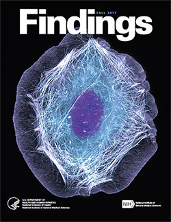
It’s back! Check out the new issue of Findings magazine.
Findings presents cutting-edge research from scientists in diverse biomedical fields. The articles are aimed at high school students with the goal of making science—and the people who do it—interesting and exciting, and to inspire young readers to pursue careers in biomedical research. In addition to putting a face on science, Findings offers activities such as quizzes and crossword puzzles and, in its online version, video interviews with scientists.
The Fall 2017 issue profiles Yale University biologist Enrique De La Cruz, who studies how actin—a protein chain that supports cell structure—breaks so easily. Also profiled is University of California, Berkeley, biologist Rebecca Heald and her study of developmental factors that control an animal’s size.
This issue also features:
A virtual reality program designed to help burn patients manage pain
The promise of gene therapy for glaucoma
The many ways scientists categorize the biological world using “omics”
What researchers know—and don’t know—about how general anesthetics work
How animation helps researchers visualize interactions between biological molecules
How cells use sugary outer coatings to distinguish friend from foe
What makes our tissues stiff, squishy, solid, or see-through (hint: its initials are ECM)
How super-powerful microscopes are revealing views of biology never possible before
View Findings online, or order a print copy (classroom sets of up to 30 copies are available for educators).
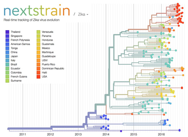
Credit: Trevor Bedford and Richard Neher, nextstrain.org.
Over the past decade, scientists and clinicians have eagerly deposited their burgeoning biomedical data into publicly accessible databases. However, a lack of computational tools for sharing and synthesizing the data has prevented this wealth of information from being fully utilized.
In an attempt to unleash the power of open-access data, the National Institutes of Health, in collaboration with the Howard Hughes Medical Institute and Britain’s Wellcome Trust, launched the Open Science Prize. Last week, after a multi-stage public voting process, the inaugural award was announced. The winner of the grand prize—and $230,000—is a prototype computational tool called nextstrain that tracks the spread of emerging viruses such as Ebola and Zika. This tool could be especially valuable in revealing the transmission patterns and geographic spread of new outbreaks before vaccines are available, such as during the 2013-2016 Ebola epidemic and the current Zika epidemic.
An international team of scientists—led by NIGMS grantee Trevor Bedford of the Fred Hutchinson Cancer Research Center, Seattle, and Richard Neher of Biozentrum at the University of Basel, Switzerland—developed nextstrain as an open-access system capable of sharing and analyzing viral genomes. The system mines viral genome sequence data that researchers have made publicly available online. nextstrain then rapidly determines the evolutionary relationships among all the viruses in its database and displays the results of its analyses on an interactive public website.
The image here shows nextstrain’s analysis of the genomes from Zika virus obtained in 25 countries over the past few years. Plotting the relatedness of these viral strains on a timeline provides investigators a sense of how the virus has spread and evolved, and which strains are genetically similar. Researchers can upload genome sequences of newly discovered viral strains—in this case Zika—and find out in short order how their new strain relates to previously discovered strains, which could potentially impact treatment decisions.
Nearly 100 interdisciplinary teams comprising 450 innovators from 45 nations competed for the Open Science Prize. More than 3,500 people from six continents voted online for the winner. Other finalists for the prize focused on brain maps, gene discovery, air-quality monitoring, neuroimaging and drug discovery.
nextstrain was funded in part by NIH under grant U54GM111274.
Inside our bodies is a surprising amount of metal. Not enough to set off the scanners at the airport or make us rich, but enough to fill each of our cells with billions of metal ions, including calcium, iron, copper and zinc. These ions facilitate critical biological functions.
However, too much of any metal can be toxic, while too little can cause disease. Our cells carefully monitor and control their metal content using a whole series of proteins that bind, sense and transport metal ions.
Keeping tabs on why and how metals flow into and out of our cells is a passion of Thomas O’Halloran, professor of chemistry and molecular biosciences at Northwestern University in Illinois. For the past three decades, O’Halloran has investigated how fluctuations in the amount of metal ions inside cells influence gene expression, cell growth and other vital functions. Using a variety of approaches, he has uncovered new types of proteins that bind metal ions and investigated the role that imbalances in these ions play in a number of disease-related physiological processes.
One recent focus of his studies has been how zinc regulates oocyte (egg cell) maturation and fertilization. Ultimately, his work could help us better understand infertility, cancer and certain bacterial infections.
In the 13 years since the sequencing of the human genome, the list of “omes” has proliferated. Drop us a comment with your favorite ome—we may feature it in a follow-up post next month.
Have you ever collected coins, cards, toy trains, stuffed animals? Did you feel the need to complete the set? If so, then you may be a completist. A completist will go to great lengths to acquire a complete set of something.
Scientists can also be completists who are inspired to identify and catalog every object in a particular field to further our understanding of it. For example, a comprehensive parts list of the human body—and of other organisms that are important in biomedical research—could aid in the development of novel treatments for diseases in the same way that a parts list for a car enables auto mechanics to build or repair a vehicle.
More than 15 years ago, scientists figured out how to catalog every gene in the human body. In the years since, rapid advances in technology and computational tools have allowed researchers to begin to categorize numerous aspects of the biological world. There’s actually a special way to name these collections: Add “ome” to the end of the class of objects being compiled. So, the complete set of genes in the body is called the “genome,” and the complete set of proteins is called the “proteome.”
Below are three -omes that NIH-funded scientists work with to understand human health.
Genome
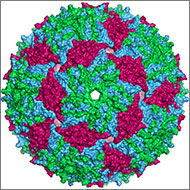
Illustration of the entire outer shell of the bacteriophage MS2. Credit: Wikimedia Commons, Naranson.
The genome is the original -ome. In 1976, Belgium scientists identified all 3,569 DNA bases—the As, Cs, Gs and Ts that make up DNA’s code—in the genes of bacteriophage MS2, immortalizing this bacteria-infecting virus as possessing the first fully sequenced genome.
Over the next two decades, a small handful of additional genomes from other microorganisms followed. The first animal genome was completed in 1998. Just 5 years later, scientists identified all 3.2 billion DNA bases in the human genome, representing the work of more than 1,000 researchers from six countries over a period of 13 years. Continue reading “There’s an “Ome” for That”
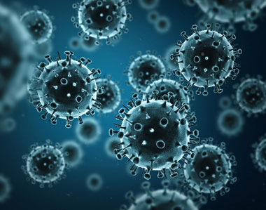
Nearly 10 percent of the human genome is derived from the genes of viruses. Credit: Stock image.
When viruses infect us, they can embed small chunks of their genetic material in our DNA. Although infrequent, the incorporation of this material into the human genome has been occurring for millions of years. As a result of this ongoing process, viral genetic material comprises nearly 10 percent of the modern human genome. Over time, the vast majority of viral invaders populating our genome have mutated to the point that they no longer lead to active infections. But they are not entirely dormant.
Sometimes, these stowaway sequences of viral genes, called “endogenous retroviruses” (ERVs), can contribute to the onset of diseases such as cancer. They can also make their hosts susceptible to infections from other viruses. However, scientists have identified numerous cases of viral hitchhikers bestowing crucial benefits to their human hosts—from protection against disease to shaping important aspects of human evolution, such as the ability to digest starch.
For a virus to successfully make copies of itself inside a host cell, it needs molecular tools similar to the ones its host normally uses to translate genes into proteins. As a result, viruses have tools meticulously shaped by evolution to commandeer the protein-producing machinery of human cells.
An exhibit called “Minerals in Medicine” opened at the NIH Clinical Center last month (see slideshow). The display features a fascinating overview of how dozens of minerals are used to create drugs and medical instruments useful in treating disease and maintaining health. The minerals ranged from commonplace ones like quartz, which is used to make medical instruments, to more exotic ones like hubnerite, a source of the metal tungsten, which is used in radiation shielding.
Inspired by this collection, which is co-sponsored by NIH and the Smithsonian Institution, we highlight here examples of “Metals in Medicine.”
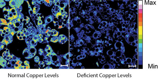
Copper and Fat Metabolism
Fluorescent imaging of copper in white fat cells from mice. The left panel shows fat cells with normal levels of copper, and the right panel shows fat cells deficient in copper. Credit: Lakshmi Krishnamoorthy and Joseph Cotruvo Jr., University of California, Berkeley.
What does a metal like copper have to do with our ability to breakdown fat? Researchers explored this question by observing mice with Wilson’s disease—a rare, inherited condition that causes copper to accumulate in the liver, brain and other vital organs. The mice with the condition usually have larger deposits of fat compared to healthy mice. To confirm that fat metabolism is somehow compromised in these mice, the researchers treated them with a drug that induces the breakdown of fat. And indeed they found that less fat was metabolized in mice with the disease.
In an effort to investigate what role copper may be playing in fat metabolism, the researchers examined adipose tissue, or fat, cells under a microscope to track the metal’s interactions with various proteins in the cell. They discovered that copper inhibits an enzyme called PDE3. This enzyme usually prevents another enzyme called cAMP from helping to break down fat. The researchers concluded that copper actually promotes fat metabolism. This work shows that transition metal nutrients can play signaling roles, which has been previously thought to be restricted to alkali and alkaline earth metals like sodium, potassium and calcium.


 Findings presents cutting-edge research from scientists in diverse biomedical fields. The articles are aimed at high school students with the goal of making science—and the people who do it—interesting and exciting, and to inspire young readers to pursue careers in biomedical research. In addition to putting a face on science, Findings offers activities such as quizzes and crossword puzzles and, in its online version, video interviews with scientists.