Tag: HIV/AIDS Structural Biology

Interview with a Scientist: Michael Summers, Using Nuclear Magnetic Resonance to Study HIV

0 comments

For more than 30 years, NIGMS has supported the structural characterization of human immunodeficiency virus (HIV) enzymes and viral proteins. This support has been instrumental in the development of crucial drugs for antiretroviral therapy such as protease inhibitors. NIGMS continues to support further characterization of viral proteins as well as cellular and viral complexes. These complexes represent the fundamental interactions between the virus and its host target cell and, as such, represent potential new targets for therapeutic development.

In this third in a series of three video interviews with NIGMS-funded researchers probing the structure of HIV, Michael Summers,Link to external web site professor of biochemistry at the University of Maryland, Baltimore County, discusses his use of nuclear magnetic resonance (NMR) technology to study HIV. Of recent interest to Summers has been using NMR to investigate how HIV’s RNA enables the virus to reproduce. His goals for this line of research are to develop treatments against HIV as well as learning how to best engineer viruses to deliver helpful therapies to individuals with a variety of diseases.

Continue reading “Interview with a Scientist: Michael Summers, Using Nuclear Magnetic Resonance to Study HIV”

Interview with a Scientist: Wes Sundquist, How the Host Immune System Fights HIV

1 comment

For more than 30 years, NIGMS has supported the structural characterization of human immunodeficiency virus (HIV) enzymes and viral proteins. This support has been instrumental in the development of crucial drugs for antiretroviral therapy such as protease inhibitors. NIGMS continues to support further characterization of viral proteins as well as cellular and viral complexes. These complexes represent the fundamental interactions between the virus and its host target cell and, as such, represent potential new targets for therapeutic development.

In this second in a series of three video interviews with NIGMS-funded researchers probing the structure of HIV, Wes Sundquist, professor of biochemistry at the University of Utah, discusses his lab’s studies of how HIV uses factors in host cells to replicate itself. In particular, Sundquist focuses on the ESCORT pathway that enables HIV to leave infected cells and spread infection elsewhere.

Continue reading “Interview with a Scientist: Wes Sundquist, How the Host Immune System Fights HIV”

Interview With a Scientist: Irwin Chaiken, Rendering HIV Inert

0 comments

For more than 30 years, NIGMS has supported the structural characterization of human immunodeficiency virus (HIV) enzymes and viral proteins. This support has been instrumental in the development of crucial drugs for antiretroviral therapy such as protease inhibitors. NIGMS continues to support further characterization of viral proteins as well as cellular and viral complexes. These complexes represent the fundamental interactions between the virus and its host target cell and, as such, represent potential new targets for therapeutic development.

Continue reading “Interview With a Scientist: Irwin Chaiken, Rendering HIV Inert”

Viral Views: New Insights on Infection Strategies

0 comments

The following images show a few ways in which cutting-edge research tools are giving us clearer views of viruses—and possible ways to disarm them. The examples, which highlight work involving HIV and the coronavirus, were funded in part by our Biomedical Technology Research Resources program.

Uncloaking HIV’s Camouflage

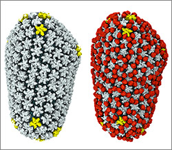
HIV capsid with (right, red) and without (left) a camouflaging human protein.
HIV capsid with (right, red) and without (left) a camouflaging human protein. Credit: Juan R. Perilla, Klaus Schulten and the Theoretical and Computational Biophysics Group, University of Illinois at Urbana-Champaign.

To sneak past our immune defenses and infect human cells, HIV uses a time-honored strategy—disguise. The virus’ genome is enclosed in a protein shell called a capsid (on left) that’s easily recognized and destroyed by the human immune system. To evade this fate, the chrysalis-shaped capsid cloaks itself with a human protein known as cyclophilin A (in red, on right). Camouflaged as human, the virus gains safe passage into and through a human cell to deposit its genetic material in the nucleus and start taking control of cellular machinery.

Biomedical and technical experts teamed up to generate these HIV models at near-atomic resolution. First, structural biologists at the Pittsburgh Center for HIV Protein Interactions Exit icon used a technique called cryo-electron microscopy (cryo-EM) to get information on the shape of an HIV capsid as well as the capsid-forming proteins’ connections to each other and to cyclophilin A. Then experts at the Resource for Macromolecular Modeling and Bioinformatics fed the cryo-EM data into their visualization and simulation programs to computationally model the physical interactions among every single atom of the capsid and the cyclophilin A protein. The work revealed a previously unknown site where cyclophilin A binds to the capsid, offering new insights on the biology of HIV infection. Continue reading “Viral Views: New Insights on Infection Strategies”

Unprecedented Views of HIV

0 comments

Visualizations can give scientists unprecedented views of complex biological processes. Here’s a look at two new ones that shed light on how HIV enters host cells.

Animation of HIV’s Entry Into Host Cells

Screen shot of the video
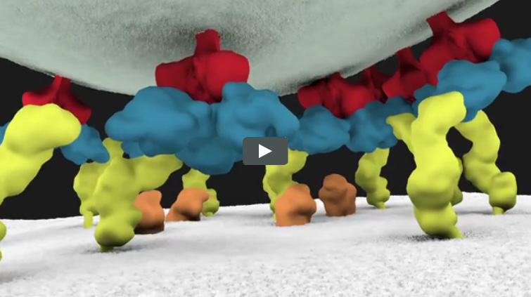
This video animation of HIV’s entry into a human immune cell is the first one released in Janet Iwasa’s current project to visualize the virus’ life cycle. As they’re completed, the animations will be posted at http://scienceofhiv.org Exit icon.

We previously introduced you to Janet Iwasa, a molecular animator who’s visualized complex biological processes such as cells ingesting materials and proteins being transported across a cell membrane. She has now released several animations from her current project of visualizing HIV’s life cycle Exit icon. The one featured here shows the virus’ entry into a human immune cell.

“Janet’s animations add great value by helping us consider how complex interactions between viruses and their host cells actually occur in time and space,” says Wes Sundquist, who directs the Center for the Structural Biology of Cellular Host Elements in Egress, Trafficking, and Assembly of HIV Exit icon at the University of Utah. “By showing us how different steps in viral replication must be linked together, the animations suggest hypotheses that hadn’t yet occurred to us.”

Continue reading “Unprecedented Views of HIV”

Meet Janet Iwasa

0 comments
Janet Iwasa
Credit: Janet Iwasa
Janet Iwasa
Fields: Cell biology and molecular animation
Works at: University of Utah
Raised in: Indiana and Maryland
Studied at: University of California, San Francisco, and Harvard Medical School
When not in the lab she’s: Keeping up with her two preschool-aged sons
Something she’s proud of that she’ll never try again: Baking a multi-tiered wedding cake, complete with sugar flowers, for a friend’s wedding.

Janet Iwasa wouldn’t have described herself as an artistic child. She didn’t carry around a sketch pad, pencils or paintbrushes. But she remembers accompanying her father, a scientist at the National Institutes of Health, to his lab on the weekends. She’d spend hours doodling in a drawing program on his old Macintosh computer while he worked on experiments.

“I always remember wanting to be a scientist, and that’s probably highly inspired by my dad,” says Iwasa. Her early affinity for art and technology set her on an unusual career path to become a molecular animator. A typical work day now finds her adapting computer programs originally designed to bring characters like Buzz Lightyear to life to help researchers probe complicated, dynamic interactions within cells.

Iwasa’s interest in animation was sparked when she was a graduate student in cell biology, studying a protein called actin, which helps cells to move and change shape. At the time, the only visual representations she had of actin networks were flat, two-dimensional drawings on paper. When she saw an animation of the dynamic movement of a molecule called kinesin, she thought, “Why are we relying on oversimplified, static illustrations [of molecules], when we can be doing something like this video?”

Within a year, she was taking an animation class at a local college. She quickly realized that she would need more intensive instruction to be able to animate complex biological processes. A few summers later, she flew to Hollywood for a 3-month training program in industry-standard animation technology.

The oldest student in that course—and the only woman—Iwasa immediately began thinking about how to adapt a standard animator’s toolkit to illustrate the inner life of cells. A technique used to create the effect of human hair blowing in the wind could also show the movement of an RNA molecule. A chunk of computer code used to make the facets of a soccer ball fall apart and come back together in a different order could be adapted to model virus assembly and disassembly.

Her Findings

Following her training, Iwasa spent 2 years as a National Science Foundation Discovery Corps fellow, producing the Exploring Life’s Origins exhibit with the Boston Museum of Science and the Szostak Lab at Massachusetts General Hospital/Harvard Medical School. As part of the multi-media exhibit, she created animations to illustrate how the simplest living organisms may have evolved on early Earth.

Since then, Iwasa has helped researchers model such complex actions as how cells ingest materials, how proteins are transported across a cell membrane, and how the motor protein dynein helps cells divide.

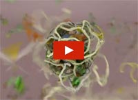
Screenshot from the video that shows how a protein called clathrin forms a cage-like container that cells use to engulf and ingest materials
Iwasa developed this video to show how a protein called clathrin forms a cage-like container that cells use to engulf and ingest materials.

Iwasa calls her animations “visual hypotheses”: The end results may be beautiful, but the process of animation itself is what encapsulates, clarifies and communicates the science.

“It’s really building the animated model that brings insights,” she says. “When you’re creating an animation, you’re really grappling with a lot of issues that don’t necessarily come up by any other means. In some cases, it might raise more questions, and make people go back and do some more experiments when they realize there might be something missing” in their theory of how a molecular process works.

Now she’s working with an NIH-funded research team at the University of Utah to develop a detailed animation of how HIV enters and exits human immune cells.

Abbreviated CHEETAH, the full name of the group is the Center for the Structural Biology of Cellular Host Elements in Egress, Trafficking, and Assembly of HIV.

“In the HIV life cycle, there are a number of events that aren’t really well understood, and people have different ideas of how things happen,” says Iwasa. She plans to animate the stages of viral infection in ways that reflect different proposals for how the process works, to give researchers a new way to visualize, communicate—and potentially harmonize—their hypotheses.

The full set of Iwasa’s HIV-related animations will be available online as they are completed, at https://scienceofhiv.org, with the first set launching in the fall of 2014.

Learn more:
Janet Iwasa’s TED Talk: How animations can help scientists test a hypothesis
Janet Iwasa’s 3D model of an HIV particle was a winner in the 2014 BioArt contest sponsored by Federation of American Societies for Experimental Biology
NIH Director’s blog post about Iwasa and her HIV video animation

Detailing Key Structures of HIV

1 comment
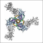
Structure of Env. Credit: The Scripps Research Institute.
This model shows a birds-eye view of the structure of Env, a protein on the HIV surface that enables it to infect cells, bound to neutralizing antibodies. Credit: The Scripps Research Institute.

In a statement on World AIDS Day 2013, NIH leaders wrote, “In the 25 years that have passed since the first annual commemoration of World AIDS Day, extraordinary scientific progress has been made in the fight against HIV/AIDS. That progress has turned an HIV diagnosis from an almost-certain death sentence to what is now for many, a manageable medical condition and nearly normal lifespan. We have come far, yet not far enough.”

One area of progress is in understanding the structural biology of HIV, the virus that causes AIDS. Capturing details about the virus’ shape has helped scientists better understand how HIV operates and pinpoint its Achilles’ heels. Recently, scientists got a closer look at two key pieces of the virus.

In one study, researchers developed the most detailed picture yet of Env, a three-segment protein on the HIV surface that allows the virus to infect cells. The work illuminated the complex process by which the protein assembles, undergoes radical shape changes during infection and interacts with neutralizing antibodies, which can block many strains of HIV from infecting human cells. The findings also may guide the development of HIV vaccines.

In another study, researchers created the best image yet of the cocoon-like container, or capsid, that carries HIV’s genome. Capsids have been difficult to study because individual imaging techniques had not produced high enough detail. By combining several cutting-edge imaging methods, the scientists pieced together the individual polygonal units of the capsid like a jigsaw puzzle to determine its structure in detail. Now that they know how HIV’s inner vessel looks, the research team is searching for its cracks—potential targets for drug development.

Learn more:
Imaging HIV’S Inner Shell Article from Inside Life Science
Key HIV Protein Structure Revealed Article from NIH Research Matters
Interactive HIV Structural Model