Tag: Scientific Process

From Basic Research to Bioelectronic Medicine

3 comments
Kevin Tracey
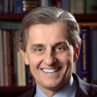
Kevin J. Tracey of the Feinstein Institute for Medical Research, the research branch of the North Shore-LIJ Health System, helped launch a new discipline called bioelectronic medicine. Credit: North Shore-LIJ Studios.

By showing that our immune and nervous systems are connected, Kevin J. Tracey of the North Shore-LIJ Health System’s Feinstein Institute for Medical Research helped launch a new discipline called bioelectronic medicine. In this field, scientists explore how to use electricity to stimulate the body to produce its own disease-fighting molecules.

I spoke with Tracey about his research, the scientific process and where bioelectronic medicine is headed next.

How did you uncover the connection between our immune and nervous systems?

My lab was testing whether a chemical we developed called CNI-1493 could stop immune cells from producing inflammation-inducing molecules called TNFs in the brain of rats during a stroke. It does. But we were surprised to find that this chemical also affects neurons, or brain cells. The neurons sense the chemical and respond by sending an electrical signal along the vagus nerve, which runs from the brain to the internal organs. The vagus nerve then releases molecules that tell immune cells throughout the body to make less TNF. I’ve named this neural circuit the inflammatory reflex. Today, scientists in bioelectronic medicine are exploring ways to use tiny electrical devices to stimulate this reflex to treat diseases ranging from rheumatoid arthritis to cancer. Continue reading “From Basic Research to Bioelectronic Medicine”

5 Reasons Biologists Love Math

4 comments

Biologists use math in a variety of ways, from designing experiments to mapping complex biological systems. Credit: Stock image.

On Saturday (at 9:26:53 to be exact), math lovers and others around the world will celebrate Pi—that really long number that represents the ratio of the circumference of a circle to its diameter. I asked our scientific experts why math is important to biomedical research. Here are a few reasons.

  1. Math allows biologists to describe how molecules move in and out of cells, how bacteria shuttle through blood vessels, how drugs get broken down in the body and many other physiological processes.
  2. Studying the geometry, topology and other physical characteristics of DNA, proteins and cellular structures has shed light on their functions and on approaches for enhancing or disrupting those functions.
  3. Math helps scientists design their experiments, including clinical trials, so they result in meaningful data, a.k.a statistical significance.
  4. Scientists use math to piece together all the different parts of a cell, an organ or an entire organism to better understand how the parts interact and how perturbations in these complex systems may contribute to disease.
  5. Sometimes it’s impossible or too difficult to answer a research question through traditional lab experiments, so biologists rely on math to develop models that represent the system they’re studying, whether it’s a metastasizing cancer cell or an emerging infectious disease. These approaches allow scientists to indicate the likelihood of certain outcomes as well as refine the research questions.

Want more? Here’s a video with 10 reasons biologists should know some math.

The “Virtuous Cycle” of Technology and Science

0 comments
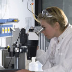
A scientist looking through a  microscope. Credit: Stock image.
Whether it’s a microscope, computer program or lab technique, technology is at the heart of biomedical research. Credit: Stock image.

Whether it’s a microscope, computer program or lab technique, technology is at the heart of biomedical research. Its central role is particularly clear from this month’s posts.

Some show how different tools led to basic discoveries with important health applications. For instance, a supercomputer unlocked the secrets of a drug-making enzyme, a software tool identified disease-causing variations among family members and high-powered microscopy revealed a mechanism allowing microtubules—and a cancer drug that targets them—to work.

Another theme featured in several posts is novel uses for established technologies. The scientists behind the cool image put a new spin on a long-standing imaging technology to gain surprising insights into how some brain cells dispose of old parts. Similarly, the finding related to sepsis demonstrates yet another application of a standard lab technique called polymerase chain reaction: assessing the immune state of people with this serious medical condition.

“We need tools to answer questions,” says NIGMS’ Doug Sheeley, who oversees biomedical technology research resource grants. “When we find the answers, we ask new questions that then require new or improved tools. It’s a virtuous cycle that keeps science moving forward.”

Meet Rhiju Das

1 comment
Rhiju Das
Credit: Rhiju Das
Rhiju Das
Fields: Biophysics and biochemistry
Works at: Stanford University
Born and raised in: The greater Midwest (Texas, Indiana and Oklahoma)
Studied at: Harvard University, Stanford University
When he’s not in the lab he’s: Enjoying the California outdoors with his wife and 3-year-old daughter
If he could recommend one book about science to a lay reader, it would be: “The Eighth Day of Creation,” about the revolution in molecular biology in the 1940s and 50s.

At the turn of the 21st century, Rhiju Das saw a beautiful picture that changed his life. Then a student of particle physics with a focus on cosmology, he attended a lecture unveiling an image of the ribosome—the cellular machinery that assembles proteins in every living creature. Ribosomes are enormous, complicated machines made up of many proteins and nucleic acids similar to DNA. Deciphering the structure of a ribosome—the 3-D image Das saw—was such an impressive feat that the scientists who accomplished it won the 2009 Nobel Prize in chemistry.

Das, who had been looking for a way to apply his physics background to a research question he could study in a lab, had found his calling.

“It was an epiphany—it was just flabbergasting to me that a hundred thousand atoms could find their way into such a well-defined structure at atomic resolution. It was like miraculously a bunch of nuts and bolts had self-assembled into a Ferrari,” recounted Das. “That inspired me to drop everything and learn everything I could about nucleic acid structure.”

Das focuses on the nucleic acid known as RNA, which, in addition to forming part of the ribosome, plays many roles in the body. As is the case for most proteins, RNA folds into a 3-D shape that enables it to work properly.

Das is now the head of a lab at Stanford University that unravels how the structure and folding of RNA drives its function. He has taken a unique approach to uncovering the rules behind nucleic acid folding: harnessing the wisdom of the crowd.

Together with his collaborator, Adrien Treuille of Carnegie Mellon University, Das created an online, multiplayer video game called EteRNA Exit icon. More than a mere game, it does far more than entertain. With its tagline “Played by Humans, Scored by Nature,” it’s upending how scientists approach RNA structure discovery and design.

Das’ Findings

Treuille and Das launched EteRNA after working on another computer game called Foldit, which lets participants play with complex protein folding questions. Like Foldit, EteRNA asks players to assemble, twist and revise structures—this time of RNA—onscreen.

But EteRNA takes things a step further. Unlike Foldit, where the rewards are only game points, the winners of each round of EteRNA actually get to have their RNA designs synthesized in a wet lab at Stanford. Das and his colleagues then post the results—which designs resulted in a successful, functional RNAs and which didn’t—back online for the players to learn from.

In a paper published in the Proceedings of the National Academy of Sciences Exit icon, Das and his colleagues showed how effective this approach could be. The collective effort of the EteRNA participants—which now number over 100,000—was better and faster than several established computer programs at solving RNA design problems, and even came up with successful new structural rules never before proposed by scientists or computers.

“What was surprising to me was their speed,” said Das. “I had just assumed that it would take a year or so before players were really able to analyze experimental data, make conclusions and come up with robust rules. But it was one of the really shocking moments of my life when, about 2 months in, we plotted the performance of players against computers and they were out-designing the computers.”

“As far as I can tell, none of the top players are academic scientists,” he added. “But if you talk to them, the first thing they’ll tell you is not how many points they have in the game but how many times they’ve had a design synthesized. They’re just excited about seeing whether or not their hypotheses were correct or falsified. So I think the top players truly are scientists—just not academic ones. They get a huge kick out of the scientific method, and they’re good at it.”

To capture lessons learned through the crowd-sourcing approach, Das and his colleagues incorporated successful rules and features into a new algorithm for RNA structure discovery, called EteRNABot, which has performed better than older computer algorithms.

“We thought that maybe the players would react badly [to EteRNABot], that they would think they were going to be automated out of existence,” said Das. “But, as it turned out, it was exciting for them to have their old ideas put into an algorithm so they could move on to the next problems.”

You can try EteRNA for yourself at http://eternagame.org Exit icon. Das and Treuille are always looking for new players and soliciting feedback.
Basic Research Fuels Medical Advances

0 comments
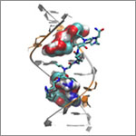
Genetic defect that causes myotonic dystrophy type 2 and used that information to design drug candidates to counteract the disease. Credit: Ilyas Yildirim, Northwestern University.
Scientists revealed a detailed image of the genetic change that causes myotonic dystrophy type 2 and used that information to design drug candidates to counteract the disease. Credit: Ilyas Yildirim, Northwestern University. View larger image

This image may look complicated, but it tells a fairly straightforward tale about basic research: Learning more about basic life processes can pave the way for medical and other advances.

In this example, researchers led by Matthew Disney of the Scripps Research Institute’s Florida campus focused on better understanding the structural underpinnings of myotonic dystrophy type 2, a relatively rare, inherited form of adult-onset muscular dystrophy. While this work is still in the preliminary stages, it may hold potential for someday treating the disorder.

Some 300,000 NIH-funded scientists are working on projects aimed at improving disease diagnosis, treatment and prevention, often through increasing understanding of basic life processes.

Read the complete Inside Life Science article.

Meet Brad Duerstock

0 comments
Brad Duerstock
Brad Duerstock
Fields: Neuroscience, assistive technology design
Works at: Purdue University, West Lafayette, IN
Hobbies: Gadgetry, architectural design
Bizarre collectible: Ecuadorian shrunken head (not a real one—it’s a replica made from goatskin)
Credit: Andrew Hancock, Purdue University

At the age of 18, Brad Duerstock had a devastating accident. A star member of his high school swim team, Duerstock hit his head during practice in a way that broke his neck and paralyzed all of his limbs. Today, he studies spinal cord injuries much like his own, investigating how the damage occurs and how it could possibly be repaired.

Duerstock has worked to make science accessible to people with disabilities, whether they use wheelchairs, as he does, or have visual or other impairments. For example, he has redesigned laboratory space to make it easier for people with disabilities to navigate and perform tasks.

“I like knowing that what I do can ultimately impact others,” Duerstock says.

Duerstock’s Findings

Much of Duerstock’s research deals with what occurs immediately following a nerve injury. In a spinal cord injury, nerve tissue becomes severed or dies. The immune response and bleeding in the injured area can cause extra damage to nerves in the spinal cord. Duerstock and his team have found that a molecule called acrolein is produced in spinal cord injuries and that it kills the nerves it encounters as it spreads around the injury site. They have been investigating a compound called polyethylene glycol (PEG), a polymer that could seal ruptured nerve cell membranes, possibly protecting nerve tissue from further damage immediately following a spinal cord injury.

Duerstock also founded and leads the Institute for Accessible Science (IAS), a community of scientists, students, parents and teachers whose goal is to promote better accommodations for people with disabilities who are studying or working in the sciences. The IAS looks into how to redesign lab spaces and equipment to increase accessibility for people with disabilities, particularly those with limited mobility or vision.

Although Duerstock originally wanted to be a doctor, he believes his true calling is in research. “The sense of discovery and the impact on others are big motivations for me,” says Duerstock. “Being a researcher, you might have a broader impact on society than you would as a practicing physician.”

Content adapted from the NIGMS Findings magazine article Opening Up the Lab.