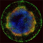
Each fluorescent point of light making up the multicolored rings in this image is an individual human embryonic cell in the early stages of development. Scientists seeking to understand the molecular cues responsible for early embryonic patterning found that human embryonic cells confined to areas of precisely controlled size and shape begin to specialize, migrate and organize into distinct layers just as they would under natural conditions.
Read the Inside Life Science article to learn more about this research, which has opened a new window for studying early development and could advance efforts aimed at using human stem cells to replace diseased cells and regenerate lost or injured body parts.


The article states the diameter as “1 mm”
Could there be an error there?
Thanks for your interest in the post! The 1 millimeter (1,000 micrometers) figure is correct. In their experiments, the researchers grew human embryonic stem (ES) cells in circular micropatterns measuring 250, 500 or 1,000 micrometers in diameter. They found that only human ES cells confined to the largest of these circles organized into the three main germ layers: endoderm, mesoderm and ectoderm. If you’re interested in more details, the journal article on this study was published in the Aug. 11, 2014, issue of Nature Methods.