
Transformations aren’t just for people or pets around Halloween. Scientific images also can look different than you might expect, depending on how they’re photographed. Check out these tricky-looking images and learn more about the science behind them.
 Credit: Nilay Taneja, Vanderbilt University, and Dylan T. Burnette, Ph.D., Vanderbilt University School of Medicine.
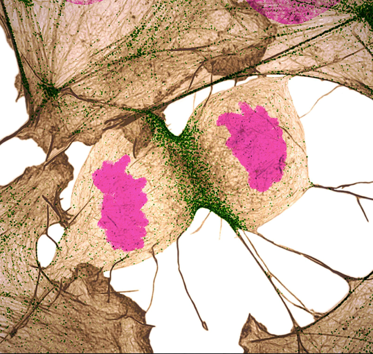
Do you have a hunch about what this image is? Perhaps something to do with dry leaves? It’s a human fibroblast cell undergoing cell division, or cytokinesis, into two daughter cells. Cytokinesis is essential for the growth and development of new cells. And fibroblasts play a big role in wound healing by helping with contraction and closure.
 Credit: Scott Chimileski, Ph.D., and Roberto Kolter, Ph.D., Harvard Medical School.
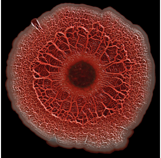
Any guesses for this circular image? Look closely. It’s a biofilm, a highly organized community of microorganisms that develops naturally on certain surfaces. Bacteria, fungi, and protists (one-celled eukaryotes) all can form biofilms. Many biofilms can have positive effects on people, but they also can be harmful. The dime-sized example shown here, for instance, was formed by a bacterium that can cause antibiotic-resistant infections in people. Scary stuff. This probably isn’t something you’d want to see in real life.
 Credit: Dylan T. Burnette, Ph.D., Vanderbilt University School of Medicine.
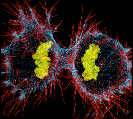
Though the image shown here may look futuristic to some, its subject has a long past. This colorful shot shows a human HeLa cell dividing into two daughter cells. HeLa cells are immortal cell lines used in scientific research. They come from cervical cancer cells that were obtained in 1951 from Henrietta Lacks, a patient at the Johns Hopkins Hospital. These cells were the first that could be shared and easily multiplied in a lab setting. Although initially collected without permission, these cells have contributed to medical research and scientific breakthroughs, from the study of leukemia and cancer to the polio vaccine. In recent years, the National Institutes of Health (NIH) has worked with the Lacks family to establish a process for controlled access to data derived from HeLa cells. These cells continue to help researchers today.
 Credit: Crystal D. Rogers, Ph.D., University of California, Davis, School of Veterinary Medicine, and Mariano A. Loza -Coll, Ph.D., California State University, Northridge.
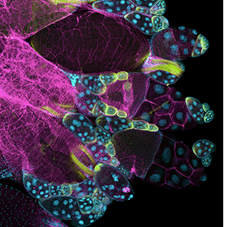
If you thought this was part of a delicate brooch or some other piece of jewelry, we wouldn’t blame you. It’s really an image of a stained fruit fly ovary. And although you can’t see this specimen with the naked eye, here it’s surprisingly pretty. To capture this view, researchers stained the ovary to examine it closer. They then used this image to show molecular staining and high-resolution imaging techniques to students.
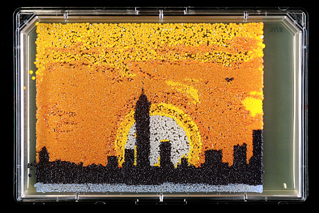 Credit: Michael Shen, Ph.D., Jasmine Temple, Leslie Mitchell, Ph.D., and Jef Boeke, Ph.D., New York University School of Medicine; and Nick Phillips, James Chuang, Ph.D., and Jiarui Wang, Johns Hopkins University.
Can you guess what this artistic image is made from? It’s yeast! Yes, that ingredient people use to bake. Researchers created this skyline of New York City by “printing” nanodroplets containing baker’s yeast onto a large plate. Yeast is a single-celled fungus, and each dot is a separate yeast colony. As the colonies grew, a picture took shape, which became “yeast art.” To make the colors (especially fitting for Halloween), the yeast strains were genetically engineered to produce pigments naturally made by bacteria, fungi, and sea creatures such as coral and sea anemones. Using genes from other organisms to make biological compounds sets the stage to someday use yeast to make other useful things, from food and fuels to drugs.
Visit the NIGMS Image and Video Gallery to peek at many more cool images.