A biofilm is a highly organized community of microorganisms that develops naturally on certain surfaces. Typically, biofilms are made up of microbes and an extracellular matrix that they produce. This matrix can include polysaccharides (chains of sugars), proteins, lipids, DNA, and other molecules. The matrix gives the biofilm structure and helps it stick to a surface.
Formation of a biofilm often involves a process called quorum sensing. In this process, microbes detect when they reach a certain population density and change their behavior in ways that help them function as a community.
Research on how diet impacts the gut microbiota has rapidly expanded in the last several years. Studies show that diets rich in red meat are linked to diseases such as colon cancer and heart disease. In both mice and humans, researchers have recently discovered differences in the gut microbiota of those who eat diets rich in red meat compared with those who don’t. This is likely because of a sugar molecule in the red meat, called N-glycolylneuraminic acid (Neu5Gc), that our bodies can’t break down. Researchers believe the human immune system sees Neu5Gc as foreign. This triggers the immune system, causing inflammation in the body, and possibly leads to disease over time.
Transformations aren’t just for people or pets around Halloween. Scientific images also can look different than you might expect, depending on how they’re photographed. Check out these tricky-looking images and learn more about the science behind them.
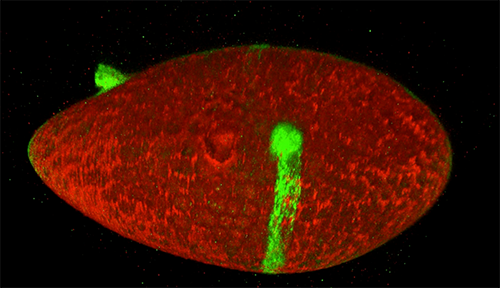
Credit: Nilay Taneja, Vanderbilt University, and Dylan T. Burnette, Ph.D., Vanderbilt University School of Medicine.
Do you have a hunch about what this image is? Perhaps something to do with dry leaves? It’s a human fibroblast cell undergoing cell division, or cytokinesis, into two daughter cells. Cytokinesis is essential for the growth and development of new cells. And fibroblasts play a big role in wound healing by helping with contraction and closure.
What do you have in common with rodents, birds, and reptiles? A lot more than you might think. These creatures have organs and body systems very similar to our own: a skeleton, digestive tract, brain, nervous system, heart, network of blood vessels, and more. Even so-called “simple” organisms such as insects and worms use essentially the same genetic and molecular pathways we do. Studying these organisms provides a deeper understanding of human biology in health and disease, and makes possible new ways to prevent, diagnose, and treat a wide range of conditions.
Historically, scientists have relied on a few key organisms, including bacteria, fruit flies, rats, and mice, to study the basic life processes that run bodily functions. In recent years, scientists have begun to add other organisms to their toolkits. Many of these newer research organisms are particularly well suited for a specific type of investigation. For example, the small, freshwater zebrafish grows quickly and has transparent embryos and see-through eggs, making it ideal for examining how organs develop. Organisms such as flatworms, salamanders, and sea urchins can regrow whole limbs, suggesting they hold clues about how to improve wound healing and tissue regeneration in humans.
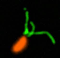
An engineered cell (green) in a fruit fly follicle (red), or egg case, leaves a trail of fluorescent material as it moves across a fruit fly egg chamber, allowing scientists to trace its path and measure how long it took to complete its journey. Credit: David Bilder, University of California, Berkeley.
Cells are the basis of the living world. Our cells make up the tissues and organs of our bodies. Bacteria are also cells, living sometimes alone and sometimes in groups called biofilms. We think of cells mostly as staying in one spot, quietly doing their work. But in many situations, cells move, often very quickly. For example, when you get a cut, infection-fighting cells rally to the site, ready to gobble up bacterial intruders. Then, platelet cells along with proteins from blood gather and form a clot to stop any bleeding. And finally, skin cells surrounding the wound lay down scaffolding before gliding across the cut to close the wound.
This remarkable organization and timing is evident right from the start. Cells migrate within the embryo as it develops so that body tissues and organs end up in the right places. Harmful cells use movement as well, as when cells move and spread (metastasize) from an original cancer tumor to other parts of the body. Learning how and why cells move could give scientists new ways to guide those cells or turn off or slow down the movement when needed.
Glowing Breadcrumbs
Scientists studying how humans and animals form, from a single cell at conception to a complex body at birth, are particularly interested in how and when cells move. They use research organisms like the fruit fly, Drosophila, to watch movements by small populations of cells. Still, watching cells migrate inside a living fly is challenging because the tissue is too dense to see individual cell movement. But moving those cells to a dish in the lab might cause them to behave differently than they do inside the fly. To solve this problem, NIGMS-funded researcher David Bilder and colleagues at the University of California, Berkeley, came up with a way to alter fly cells so they could track how the cells behave without removing them from the fly. They engineered the cells to lay down a glowing track of proteins behind them as they moved, leaving a traceable path through the fly’s tissue. The technique, called M-TRAIL (matrix-labeling technique for real-time and inferred location), allows the researchers to see where a cell travels and how long it takes to get there.
Bilder and his team first used M-TRAIL in flies to confirm the results of past studies of Drosophila ovaries in the lab using other imaging techniques. In addition, they found that M-TRAIL could be used to study a variety of cell types. The new technique also could allow a cell’s movement to be tracked over a longer period than other imaging techniques, which become toxic to cells in just a few hours. This is important, because cells often migrate for days to reach their final destinations.
Every one of our thoughts, emotions, sensations, and movements arise from changes in the flow of electricity in the brain. Disruptions to the normal flow of electricity within and between cells is a hallmark of many diseases, especially neurological and cardiac diseases.
The source of electricity within nerve cells (i.e., neurons) is the separation of charge, referred to as voltage, across neuronal membranes. In the past, scientists weren’t able to identify all the molecules that control neuronal voltage. They simply lacked the tools. Now, University of Colorado biologist Joel Kralj has developed a way to overcome this hurdle. His new technique—combining automated imaging tools and genetic manipulation of cells—is designed to measure the electrical contribution of every protein coded by every gene in the human genome. Kralj believes this technology will usher in a new field of “electromics” that will be of enormous benefit to both scientists studying biological processes and clinicians attempting to treat disease.
In 2017, Kralj won a New Innovator Award from the National Institutes of Health for his work on studying voltage in neurons. He is using the grant money to develop a new type of microscope that will be capable of measuring neuronal voltage from hundreds of cells simultaneously. He and his research team will then attempt to identify the genes that encode any of the 20,000 proteins in the human body that are involved in electrical signaling. This laborious process will involve collecting hundreds of nerve cells, genetically removing a single protein from each cell, and using the new microscope to see what happens. If the voltage within a cell is changed in any way when a specific protein is removed, the researchers can conclude that the protein is essential to electrical signaling.
In this video, Kralj discusses how he plans to use his electromics platform to study electricity-generating cells throughout the body, as well as in bacterial cells (see our companion blog post “Feeling Out Bacteria’s Sense of Touch” featuring Kralj’s research for more details).
Dr. Kralj’s work is funded in part by the NIH under grant 1DP2GM123458-01.
Our sense of touch provides us with bits of information about our surroundings that inform the decisions we make. When we touch something, our nervous system transmits signals through nerve endings that feed information to our brain. This enables us to sense the stimulus and take the appropriate action, like drawing back quickly when we touch a hot stovetop.
Bacteria are single cells and lack a nervous system. But like us, they rely on their “sense” of touch to make decisions—or at least change their behavior. For example, bacteria’s sense of touch is believed to trigger the cells to form colonies, called biofilms, on surfaces they make contact with. Bacteria may form biofilms as a way to defend themselves, share limited nutrients, or simply to prevent being washed away in a flowing liquid.
Humans can be harmed by biofilms because these colonies serve as a reservoir of disease-causing cells that are responsible for high rates of human infection. Biofilms can protect at least some cells from being affected by antibiotics. The surviving reservoir of bacteria then have more time to evolve resistance to antibiotics.
At the same time, some biofilms can be valuable; for example, they help to break down waste in water treatment plants and to drive electrical current as part of microbial fuel cells.
Until recently, scientists thought that bacteria formed biofilms and caused infections in response to chemical signals they received from their environments. But research in 2014 showed that the bacterium Pseudomonas aeruginosa could infect a variety of living tissues—from plants to many kinds of animals—simply by making contact with them. In the past year, multiple groups of investigators have learned more about how bacteria sense that they have touched a surface and how that sense translates to changes in their behavior. This understanding could lead to new ways of preventing infections or harmful biofilm formation.
Making Contact
Pili (green) on cells from the bacterium Caulobacter crescentus (orange). Scientists used a fluorescent dye to stain pili so they could watch the structures extend and retract. Credit: Courtney Ellison, Indiana University.
When they first make contact with a surface, bacteria change from free-ranging, swimming cells to stationary ones that secrete a sticky substance, tethering them in one place. To form a biofilm, they begin replicating, creating an organized mass stable enough to resist shaking and to repel potential invaders (see https://biobeat.nigms.nih.gov/2017/01/cool-image-inside-a-biofilm-build-up/).
How do swimming bacteria sense that they have found a potential surface to colonize? Working with the bacterium Caulobacter crescentus, Indiana University Ph.D. student Courtney Ellison and her colleagues, under the direction of professor of biology and NIGMS grantee Yves Brun, recently showed that hair-like structures on the cell’s surface, called pili, play a role here. The researchers found that as a bacterial cell swims in a fluid, its pili are constantly stretching out and retracting. When the cell makes contact with a surface, the pili stop moving, start producing a sticky substance and use it to hold onto the surface.
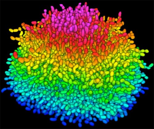
A growing Vibrio cholerae biofilm. Each slightly curved comma shape represents an individual bacterium from assembled confocal microscopy images. Different colors show each bacterium’s position in the biofilm in relation to the surface on which the film is growing. Credit: Jing Yan, Ph.D., and Bonnie Bassler, Ph.D., Department of Molecular Biology, Princeton University, Princeton, NJ.
Bacteria use many methods to overcome threats in their environment. One of these ways is forming colonies called biofilms on surfaces of objects. Often appearing like the bubble-shaped fortress represented in this image, biofilms enable bacteria to withstand attacks, compete for space and survive fluctuations in nutrient supply. Bacteria aggregated within biofilms inside our bodies, for example, resist antibiotic therapy more effectively than free swimming cells, making infections difficult to treat. On the other hand, biofilms are also useful for making microbial fuel cells and for waste-water treatment. Learning how biofilms work, therefore, could provide essential tools in our ongoing battle against disease-causing agents and in our efforts to harness beneficial bacterial behaviors. Researchers are now using new imaging techniques to watch how biofilms grow, cell by cell, and to identify more effective ways of disrupting or fostering them.
Until now, poor imaging resolution meant that scientists could not see what individual bacteria in the films are up to as the biofilms grow. The issue is that bacteria are tiny, making imaging each cell, as well as the ability to distinguish each cell in the biofilm community, problematic. Continue reading “Cool Image: Inside a Biofilm Build-up”
Researchers created an apparatus to study quorum sensing, a communication system that allows some bacteria to cause dangerous infections. Their findings suggest that blocking bacterial communication might lead to a new way to combat such infections. Credit: Minyoung Kevin Kim and Bonnie Bassler, Princeton University.
If you’ve ever felt a slimy coating on your teeth, scrubbed grime from around a sink drain or noticed something growing between the tiles of a shower, you’ve encountered a biofilm. Made up of communities of bacteria and other microorganisms, biofilms thrive where they can remain moist and relatively undisturbed. As they enlarge, biofilms can block narrow passages like medical stents, airways, pipes or intestines. Continue reading “Cool Video: Watching Bacteria Turn Virulent”

 Microbiota in the intestines. Credit: iStock.
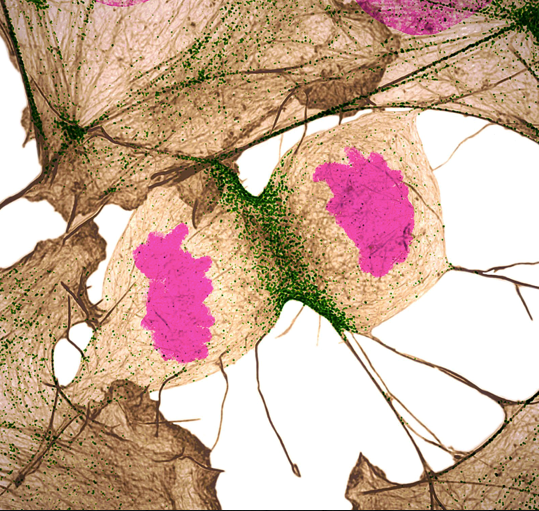 Credit: Nilay Taneja, Vanderbilt University, and Dylan T. Burnette, Ph.D., Vanderbilt University School of Medicine.