Author: Sharon Reynolds

avatar

Posts by Sharon Reynolds

An Experimental Contact Lens to Prevent Glaucoma-Induced Blindness

3 comments
Contact lens. Credit: Peter Mallen, Massachusetts Eye and Ear Laboratory/Kohane Laboratory, Boston Children's Hospital.
An experimental contact lens design releases a glaucoma medicine at a steady rate for up to a month. Credit: Peter Mallen, Massachusetts Eye and Ear Laboratory/Kohane Laboratory, Boston Children’s Hospital.

Like a miniature donut stuffed inside a tiny pita pocket, a common glaucoma medicine held within a biomaterial ring is sandwiched inside this contact lens. In laboratory experiments, the lens, which can also correct vision, releases the eyesight-saving medication at a steady rate for up to a month. Its construction offers numerous potential clinical advantages over the standard glaucoma treatment and may have additional applications, such as delivering anti-inflammatory drugs or antibiotics to the eye. Led by Daniel Kohane and Joseph Ciolino at Harvard Medical School, the researchers who developed the lens are now gearing up to test its effectiveness in additional laboratory studies. They hope a Phase I clinical trial to evaluate the safety and ability of the lens to reduce pressure in the human eye could begin in about a year.

This work also was funded by NIH’s National Eye Institute.

Learn more
:
NEI Glaucoma Awareness Month Resources

Cool Images: Holiday Season Cells

0 comments
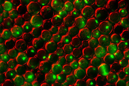
Yeast cells deficient in zinc and the Tsa1 protein have protein tangles. Credit: Colin MacDiarmid and David Eide, University of Wisconsin-Madison.

Yeast cells deficient in zinc and the Tsa1 protein have protein tangles. Credit: Colin MacDiarmid and David Eide, University of Wisconsin-Madison.

Just in time for the holidays, we’ve wrapped up a few red and green cellular images from basic research studies. In this snapshot, we see a group of yeast cells that are deficient in zinc, a metal that plays a key role in creating and maintaining protein shape. The cells also lack a protein called Tsa1, which normally keeps proteins from sticking together. Green areas highlight protein tangles caused by the double deficiency. Red outlines the cells. Protein clumping plays a role in many human diseases, including Parkinson’s and Alzheimer’s, so knowledge of why it happens—and what prevents it in healthy cells—could aid the development of treatments.

See more festive images!

Stop the (Biological) Clock

0 comments
Molecular structure of the three proteins in blue-green algae’s circadian clock.  Credit: Johnson Lab, Vanderbilt University.
Molecular structure of the three proteins in blue-green algae’s circadian clock. Credit: Johnson Lab, Vanderbilt University.

Many microorganisms can sense whether it’s day or night and adjust their activity accordingly. In tiny blue-green algae, the “quartz-crystal” of the time-keeping circadian clock consists of only three proteins, making it the simplest clock found in nature. Researchers led by Carl Johnson of Vanderbilt University recently found that, by manipulating these clock proteins, they could lock the algae into continuously expressing its daytime genes, even during the nighttime.

Why would one want algae to act like it’s always daytime? The kind used in Johnson’s study is widely harnessed to produce commercial products, from drugs to biofuels. But even when grown in constant light, algae with a normal circadian clock typically decrease production of biomolecules when nighttime genes are expressed. When the researchers grew the algae with the daytime genes locked “on” in constant light, the microorganism’s output increased by as much as 700 percent. This proof of concept experiment may be applicable to improving the commercial production of compounds such as insulin and some anti-cancer drugs.

Learn more:

Vanderbilt University Press Release Exit icon
Johnson Laboratory Exit icon
Circadian Rhythms Fact Sheet

Meet Shanta Dhar

1 comment
Shanta Dhar
Shanta Dhar
Fields: Chemistry and cancer immunotherapy
Works at: University of Georgia, Athens
Born and raised in: Northern India
Studied at: Indian Institute of Science, Bangalore; Johns Hopkins University, Baltimore, Md.; and Massachusetts Institute of Technology, Cambridge, Mass.
To unwind: She hits the gym
Credit: Frankie Wylie, Stylized Portraiture

The human body is, at its most basic level, a giant collection of chemicals. Finding ways to direct the actions of those chemicals can lead to new treatments for human diseases.

Shanta Dhar, an assistant professor of chemistry at the University of Georgia, Athens (UGA), saw this potential when she was exposed to the field of cancer immunotherapy as a postdoctoral researcher at the Massachusetts Institute of Technology. (Broadly, cancer immunotherapy aims to direct the body’s natural immune response to kill cancer cells.) Dhar was fascinated by the idea and has pursued research in this area ever since. “I always wanted to use my chemistry for something that could be useful [in the clinic] down the line,” she said.

A major challenge in the field has been training the body’s immune system—specifically the T cells—to recognize and attack cancer cells. The process of training T cells to go after cancer is rather like training a rescue dog to find a lost person: First, you present the scent, then you command pursuit.

The type of immune cell chiefly responsible for training T cells to search for and destroy cancer is a called a dendritic cell. First, dendritic cells present T cells with the “scent” of cancer (proteins from a cancer cell). Then they activate the T cells using signaling molecules.

Dhar’s Findings

Dhar’s work focuses on creating the perfect trigger for cancer immunotherapy—one that would provide both the scent of cancer for T cells to recognize and a burst of immune signaling to activate the cells.

Using cells grown in the lab, Dhar’s team recently showed that they could kill most breast cancer cells using a new nanotechnology technique, then train T cells to eradicate the remaining cancer cells.

For the initial attack, the researchers used light-activated nanoparticles that target mitochondria in cancer cells. Mitochondria are the organelles that provide cellular energy. Their destruction sets off a signaling cascade that triggers dendritic cells to produce one of the proteins needed to activate T cells.

Because the strategy worked in laboratory cells, Dhar and her colleague Donald Harn of the UGA infectious diseases department are now testing it in a mouse model of breast cancer to see if it is similarly effective in a living organism.

For some reason, the approach works against breast cancer cells but not against cervical cancer cells. So the team is examining the nanoparticle technique to see if they can make it broadly applicable against other cancer types.

Someday, Dhar hopes to translate this work into a personalized cancer vaccine. To create such a vaccine, scientists would remove cancer cells from a patient’s body during surgery. Next, in a laboratory dish, they would train immune cells from the patient to kill the cancer cells, then inject the trained immune cells back into the patient’s body. If the strategy worked, the trained cells would alert and activate T cells to eliminate the cancer.

Mapping Approach Yields Insulin Secretion Pathway Insights

0 comments
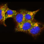
TMEM24 protein (green) and insulin (red) in pancreatic beta cells (yellow).  Credit: Balch Lab, the Scripps Research Institute.
The interactions of TMEM24 protein (green) and insulin (red) in pancreatic beta cells are shown in yellow. Credit: Balch Lab, the Scripps Research Institute.
View larger image

The identities of the proteins that drive insulin production and release from pancreatic beta cells have largely been a mystery. In new work from the lab of William Balch of the Scripps Research Institute, researchers isolated and then identified all the insulin-bound proteins from mouse beta cells. The results provided a roadmap of the protein interactions that lead to insulin production, storage and secretion. The researchers used the roadmap to identify a protein called TMEM24, which was abundant in beta cells and binds readily to insulin. Balch and his team uncovered that TMEM24, whose involvement in insulin secretion was previously unknown, effectively regulates slower insulin release and could have a key role in maintaining control of glucose levels in the blood. The scientists hope that this roadmap of insulin-interacting proteins will lead to the development of new, targeted approaches to treating type 2 diabetes and a similar insulin-related condition called metabolic syndrome.

Learn more:

The Scripps Research Institute News Release Exit icon
Balch Lab Exit icon

Flu Finds a Way In

0 comments
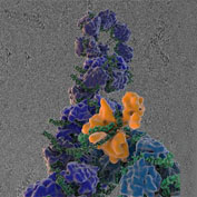
Influenza virus proteins in the act of self-replication. Credit: Wilson, Carragher and Potter labs, Scripps Research Institute.

Flu viruses evolve rapidly, often staying one step ahead of efforts to vaccinate against infections or treat them with antiviral drugs. Work led by Jesse Bloom of the Fred Hutchinson Cancer Research Center has uncovered a surprising new flu mutation that allows influenza to infect cells in a novel way. Normally, a protein called hemagglutinin lets flu viruses attach to cells, and a protein called neuraminidase lets newly formed viruses escape from infected cells. Bloom’s lab has characterized a mutant flu virus where neuraminidase can enable the virus to attach to host cells even when hemagglutinin’s binding is blocked. Although the researchers generated the neuraminidase mutant studied in these experiments in their lab, the same mutation occurs naturally in strains from several recent flu outbreaks. There’s a possibility that flu viruses with such mutations may be able to escape antibodies that block the binding of hemagglutinin.

This work also was funded by NIH’s National Institute of Allergy and Infectious Diseases.

Learn more:

Bloom Lab Exit icon

Fitting a Piece of the Protein-Function Puzzle

0 comments
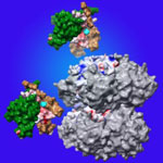
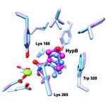
Comparison of the predicted binding of the substrate to the active site of HpbD (blue) with the binding sites determined experimentally by crystallography (magenta). Credit: Matt Jacobson, University of California, San Francisco; Steve Almo, Albert Einstein College of Medicine.
The image shows a comparison of the predicted binding of the substrate to the active site of HpbD (blue) with the binding sites determined experimentally by crystallography (magenta). Credit: Matt Jacobson, University of California, San Francisco; Steve Almo, Albert Einstein College of Medicine.

Sequencing the genomes of almost 7,000 organisms has identified more than 40 million proteins. But how do we figure out what all these proteins do? New results from an initiative led by John Gerlt of the University of Illinois suggest a possible method for identifying the functions of unknown enzymes, proteins that speed chemical reactions within cells. Using high-powered computing, the research team modeled how the structure of a mystery bacterial enzyme, HpbD, might fit like a puzzle piece into thousands of proteins in known metabolic pathways. Since an enzyme acts on other molecules, finding its target or substrate can shed light on its function. The new method narrowed HpbD’s candidate substrate down from more than 87,000 to only four. Follow-up lab work led to the actual substrate, tHypB, and determined the enzyme’s biological role. This combination of computational and experimental methods shows promise for uncovering the functions of many more proteins.

Learn more:

University of Illinois at Urbana-Champaign News Release
Gerlt Lab
Enzyme Function Initiative

Cool Image: Tiny Bacterial Motor

2 comments
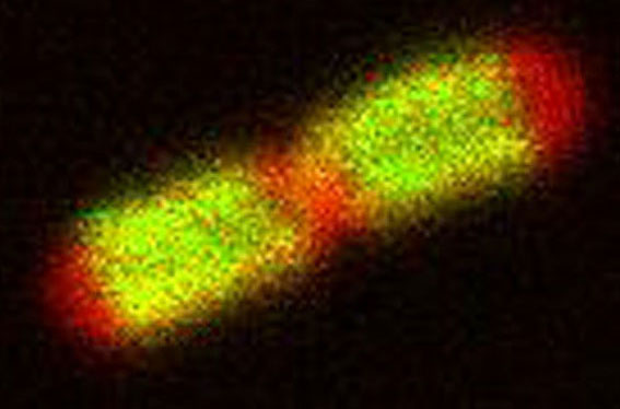
Phillip Klebba, Kansas State University.

Credit: Phillip Klebba, Kansas State University.

It looks like a fluorescent pill, but this image of an E. coli cell actually shows a new potential target in the fight against infectious diseases. The green highlights a protein called TonB, which is produced by many gram-negative bacteria, including those that cause typhoid fever, meningitis and dysentery. TonB lets bacteria take up iron from the host’s body, which they need to survive. New research from Phillip Klebba of Kansas State University and his colleagues shows how TonB powers iron uptake. When TonB spins within the cell envelope (the bacteria’s “skin”) like a tiny motor, it produces energy that lets another protein pull iron into the cell. This knowledge may lead to the development of antibiotics that block the motion of TonB, potentially stopping an infection in its tracks.

Learn more:
Kansas State University News Release Exit icon
Klebba Lab Exit icon
Other Cool Images