Category: Chemistry, Biochemistry and Pharmacology

Epilepsy Drug Improves Health in Animal Model of Obesity

2 comments
Liver cells of obese mice treated with valproic acid (right) and untreated obese mice (left).
Liver cells (magenta) of obese mice treated with valproic acid (right) had much less fat accumulation (white) than those of untreated obese mice (left). Credit: Lindsay B. Avery and Namandjé N. Bumpus, Johns Hopkins University. View larger image

With more than 90 million Americans affected by obesity, developing medications to help combat weight gain and its associated diseases has become a priority. In a study using obese mice, a team led by Namandjé Bumpus of Johns Hopkins University recently showed that a commonly prescribed epilepsy drug, valproic acid, reduced fat accumulation in the liver and lowered elevated blood sugar levels like those associated with type 2 diabetes. Body weight also stabilized in mice given the drug, whereas untreated mice continued to gain weight. Additional experiments in mouse and human liver cells suggested that the byproducts of valproic acid produced as the body breaks down the drug, rather than valproic acid itself, were responsible for the observed effects. These byproducts achieved the same effects in cells at one-fortieth the concentration of valproic acid, making them promising candidates for further drug development.

Learn more:
Johns Hopkins University News Release Exit icon
Bumpus Laboratory

An Experimental Contact Lens to Prevent Glaucoma-Induced Blindness

3 comments
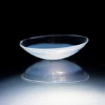
Contact lens. Credit: Peter Mallen, Massachusetts Eye and Ear Laboratory/Kohane Laboratory, Boston Children's Hospital.
An experimental contact lens design releases a glaucoma medicine at a steady rate for up to a month. Credit: Peter Mallen, Massachusetts Eye and Ear Laboratory/Kohane Laboratory, Boston Children’s Hospital.

Like a miniature donut stuffed inside a tiny pita pocket, a common glaucoma medicine held within a biomaterial ring is sandwiched inside this contact lens. In laboratory experiments, the lens, which can also correct vision, releases the eyesight-saving medication at a steady rate for up to a month. Its construction offers numerous potential clinical advantages over the standard glaucoma treatment and may have additional applications, such as delivering anti-inflammatory drugs or antibiotics to the eye. Led by Daniel Kohane and Joseph Ciolino at Harvard Medical School, the researchers who developed the lens are now gearing up to test its effectiveness in additional laboratory studies. They hope a Phase I clinical trial to evaluate the safety and ability of the lens to reduce pressure in the human eye could begin in about a year.

This work also was funded by NIH’s National Eye Institute.

Learn more
:
NEI Glaucoma Awareness Month Resources

Targeting Toxic RNA Molecules in Muscular Dystrophy

0 comments
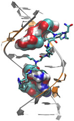
Genetic defect that causes myotonic dystrophy type 2 and used that information to design drug candidates to counteract the disease. Credit: Ilyas Yildirim, Northwestern University.
Scientists revealed a detailed image of the genetic defect that causes myotonic dystrophy type 2 and used that information to design drug candidates to counteract the disease. Credit: Ilyas Yildirim, Northwestern University. View larger image

Myotonic dystrophy type 2 (DM2) is a relatively rare, inherited form of adult-onset muscular dystrophy that has no cure. It’s caused by a genetic defect in which a short series of nucleotides—the chemical units that spell out our genetic code—is repeated more times than normal. When the defective gene is transcribed, the resulting RNA repeat forms a hairpin-like structure that binds to and disables a protein called MBNL1.

Now, research led by Matthew Disney of The Scripps Research Institute (TSRI), Florida Campus, has revealed the detailed, three-dimensional structure of the RNA defect in DM2 and used this information to design small molecules that bind to the aberrant RNA. These designer molecules, even in small amounts, significantly improved disease-associated defects in a cellular model of DM2, and thus hold potential for reversing the disorder.

Drugs that target toxic RNA molecules associated with diseases such as DM2 are few and far between, as developing such compounds is technically challenging. The “bottom-up” approach that the scientists used to design potent new drug candidates, by first studying in detail how the RNA structure interacts with small molecules, is unconventional, noted Jessica Childs-Disney of TSRI, who was lead author of the paper with Ilyas Yildirim of Northwestern University. But it may serve as an effective strategy for pioneering the use of small molecules to manipulate disease-causing RNAs—a central focus of the Disney lab.

This work also was funded by NIH’s National Cancer Institute.

Learn more:
The Scripps Research Institute News Release Exit icon
Disney Lab

Meet Shanta Dhar

1 comment
Shanta Dhar
Shanta Dhar
Fields: Chemistry and cancer immunotherapy
Works at: University of Georgia, Athens
Born and raised in: Northern India
Studied at: Indian Institute of Science, Bangalore; Johns Hopkins University, Baltimore, Md.; and Massachusetts Institute of Technology, Cambridge, Mass.
To unwind: She hits the gym
Credit: Frankie Wylie, Stylized Portraiture

The human body is, at its most basic level, a giant collection of chemicals. Finding ways to direct the actions of those chemicals can lead to new treatments for human diseases.

Shanta Dhar, an assistant professor of chemistry at the University of Georgia, Athens (UGA), saw this potential when she was exposed to the field of cancer immunotherapy as a postdoctoral researcher at the Massachusetts Institute of Technology. (Broadly, cancer immunotherapy aims to direct the body’s natural immune response to kill cancer cells.) Dhar was fascinated by the idea and has pursued research in this area ever since. “I always wanted to use my chemistry for something that could be useful [in the clinic] down the line,” she said.

A major challenge in the field has been training the body’s immune system—specifically the T cells—to recognize and attack cancer cells. The process of training T cells to go after cancer is rather like training a rescue dog to find a lost person: First, you present the scent, then you command pursuit.

The type of immune cell chiefly responsible for training T cells to search for and destroy cancer is a called a dendritic cell. First, dendritic cells present T cells with the “scent” of cancer (proteins from a cancer cell). Then they activate the T cells using signaling molecules.

Dhar’s Findings

Dhar’s work focuses on creating the perfect trigger for cancer immunotherapy—one that would provide both the scent of cancer for T cells to recognize and a burst of immune signaling to activate the cells.

Using cells grown in the lab, Dhar’s team recently showed that they could kill most breast cancer cells using a new nanotechnology technique, then train T cells to eradicate the remaining cancer cells.

For the initial attack, the researchers used light-activated nanoparticles that target mitochondria in cancer cells. Mitochondria are the organelles that provide cellular energy. Their destruction sets off a signaling cascade that triggers dendritic cells to produce one of the proteins needed to activate T cells.

Because the strategy worked in laboratory cells, Dhar and her colleague Donald Harn of the UGA infectious diseases department are now testing it in a mouse model of breast cancer to see if it is similarly effective in a living organism.

For some reason, the approach works against breast cancer cells but not against cervical cancer cells. So the team is examining the nanoparticle technique to see if they can make it broadly applicable against other cancer types.

Someday, Dhar hopes to translate this work into a personalized cancer vaccine. To create such a vaccine, scientists would remove cancer cells from a patient’s body during surgery. Next, in a laboratory dish, they would train immune cells from the patient to kill the cancer cells, then inject the trained immune cells back into the patient’s body. If the strategy worked, the trained cells would alert and activate T cells to eliminate the cancer.

Meet Emily Scott

0 comments
Emily Scott
Emily Scott
Field: Biochemistry
Works at: University of Kansas in Lawrence
Favorite hobby: Scuba diving
Likes watching: “Law & Order”
Likes reading: True-life survival stories
Credit: Chuck France, University of Kansas

With an air tank strapped to her back, college student Emily Scott dove to the bottom of the Gulf of Mexico to examine life in an oxygen-starved area called the Dead Zone. The bottom waters had once teemed with red snapper, croaker and shrimp, but to Scott, the region appeared virtually devoid of life. Then, from out of the mud, appeared the long, undulating arms of a brittle star.

As Scott learned, that particular species of brittle star survived in the Dead Zone because it has something many other marine creatures don’t: an oxygen-carrying protein called hemoglobin. This same protein makes our blood red. Key to hemoglobin’s special oxygen-related properties is a small molecular disk called a heme (pronounced HEEM).

Once she saw what it meant to brittle stars, Scott was hooked on heme and proteins that contain it.

Scott’s Findings

Now an associate professor, Scott studies a family of heme proteins called cytochromes P450 (CYP450s). These proteins are enzymes that facilitate many important reactions: They break down cholesterol, help process vitamins and play an important role in flushing foreign chemicals out of our systems.

To better understand CYP450s, Scott uses a combination of two techniques–X-ray crystallography and nuclear magnetic resonance spectroscopy—for capturing the enzymes’ structural and reactive properties.

She hopes to apply her work to design drugs that block certain CYP450 reactions linked with cancer. One target reaction, carried out by CYP2A13, converts a substance in tobacco into two cancer-causing molecules. Another target reaction is carried out by CYP17A1, an enzyme that helps the body produce steroid sex hormones but that, later in life, can fuel the uncontrolled growth of prostate or breast cancer cells.

“I’m fascinated by these proteins and figuring out how they work,” Scott says. “It’s like trying to put together a puzzle—a very addictive puzzle.” Her drive to uncover the unknown and her willingness to apply new techniques have inspired the students in her lab to do the same.

Content adapted from “Hooked on Heme,” an article in the September 2013 issue of Findings magazine.

Chemist Phil Baran Joins “Genius” Ranks as MacArthur Fellow

1 comment
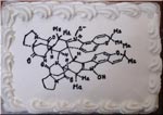
Cake decorated with a two-dimensional structure of the molecule, stephacidin B
When Baran’s research team succeeds in synthesizing an important natural product, the group sometimes celebrates with a cake decorated with a two-dimensional structure of the molecule. This molecule, stephacidin B, was isolated from a fungus and has anticancer properties. See images of other Baran lab cakes.

As a newly appointed MacArthur Fellow, Phil Baran is now officially a genius. The MacArthur award recognizes “exceptionally creative” individuals who have made significant contributions to their field and are expected to continue doing so. Baran, a synthetic organic chemist at Scripps Research Institute in La Jolla, Calif., was recognized today for “inventing efficient, scalable, and environmentally sound methods” for building, from scratch, molecules produced in nature. Many of these natural products have medicinal properties. Baran has already concocted a host of natural products, including those with the ability to kill bacteria or cancer cells. In addition to emphasizing the important pharmaceutical applications of his work, Baran embraces its creative aspects: “The area of organic chemistry is such a beautiful one because one can be both an artist and an explorer at the same time,” he said in the MacArthur video interview Exit icon.

Learn more:

NIGMS “Meet a Chemist” Profile of Baran
NIH Director’s Blog Post on Baran’s Recent Work

New Door Opens in the Effort to Stave off Mosquito-Borne Diseases

1 comment
Mosquito net Pyrethroids are used in mosquito nets distributed around the globe. Credit: Kurt Stepnitz, Michigan State University.

In the past decade, mosquitoes in many countries have become increasingly resistant to pyrethroid insecticides used to fend off mosquito-borne diseases such as malaria and dengue fever. Now, Ke Dong of Michigan State University and her colleagues have discovered a second pyrethroid-docking site in the molecular doorways, or channels, that control the flow of sodium into cells. Pyrethroids paralyze and kill mosquitoes and other insects by propping open the door and causing the pests to overdose on sodium, a critical regulator of nerve function. By providing new insights on pyrethroid action at the molecular level—and how mutations in the dual docking sites cause resistance—the findings open avenues to better monitoring and management of insecticide resistance.

Learn more:
Michigan State University News Release Exit icon