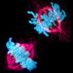
This image shows a cell in two stages of division: prometaphase (top) and metaphase (bottom). To form identical daughter cells, chromosome pairs (blue) separate via the attachment of microtubules made up of tubulin proteins (pink) to specialized structures on centromeres (green). Credit: Lilian Kabeche, Dartmouth.
Chromosome segregation during cell division is like speed dating, according to Geisel School of Medicine at Dartmouth researcher Duane Compton. He and postdoctoral fellow Lilian Kabeche learned that protein cyclin A plays moderator, helping to properly separate chromosomes via the attachment of microtubule fibers to kinetochore structures. Here’s how Compton described the process:
“The chromosomes are testing the microtubules for compatibility—that is, looking for the right match—to make sure there are correct attachments and no errors. The old view of this process held that chromosomes and microtubules pair up individually to find the correct attachment, like conventional dating. However, our results show that the system is more like speed dating. All the chromosomes have to try many connections with microtubules in a short amount of time. Then they all make their final choices at the same time. Cyclin A acts like a timekeeper or referee to make sure no one makes bad connections prematurely.”
Such bad connections can cause chromosome segregation errors that lead to cells with an abnormal number of chromosomes, a hallmark of cancer cells. So in addition to aiding our understanding of a fundamental biological process, the new insights may point to potential ways to correct such errors.
Learn more:
Dartmouth News Release 
Compton Lab