Tag: Cool Images

An Insider’s Look at Life: Magnified, an Airport Exhibit of Stunning Microscopy Images

0 comments
Bubonic plague bacteria on part of the digestive system in a rat flea
What looks like pollen on petals is actually bubonic plague bacteria on the digestive spines of a flea, viewed through a powerful microscope. Credit: B. Joseph Hinnebusch, Elizabeth Fischer and Austin Athman, NIH’s National Institute of Allergy and Infectious Diseases.

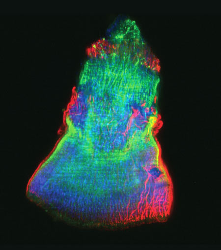
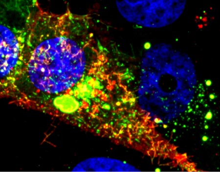
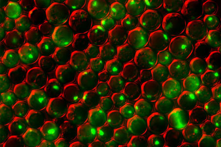
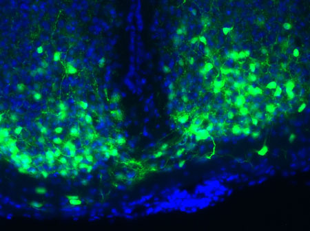
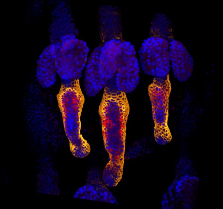
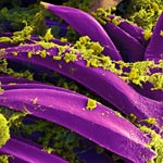
Science. Art. Airports. I’ve never used those three words together before. But I’ve been doing it a lot lately while working on Life: Magnified, an exhibit of 46 striking scientific images created by scientists around the country using state-of-the-art microscopes.

The show is at Washington Dulles International Airport, where more than a million travelers will see it over its 6-month run. As our director said in a recent post on another NIGMS blog, “What a great way to share the complexity and beauty of biomedical science with such a large public audience!”

We’ve also set up an online gallery, where the colorful images can be viewed and freely downloaded for research, educational and news media purposes.

The project itself was quite an adventure. When we asked scientists to send us images, our fears of not getting enough attractive, high-resolution options were drowned by a deluge of more than 600 submissions. Then we worried how to sort through them all and make final selections. Doing so required several rounds of online viewing, various ranking systems and a panel of experts. Then the images were printed as large, digital negatives on transparency film.

Artist's rendition of a network diagram. Credit: Allison Kudla, Institute for Systems Biology.
The midnight installation of Life: Magnified involved five people (that’s me in pink), a ladder and lots of rags and glass cleaner. Credit: Woody Machalek.

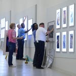
Dulles is a pretty busy place, so we set up the exhibit when it was quietest—the middle of the night (10 p.m. to 1:30 a.m., to be exact). We swapped out images from the previous photography exhibit and installed the Life: Magnified ones in LED lightboxes mounted in the airport’s Gateway Gallery. It was an eerie and exhilarating feeling to be in a noiseless, nearly empty airport without heavy bags and a long walk to a departure gate.

Less than 12 hours later, I was surprised to receive an e-mail from someone who had passed through the exhibit and sent a few photos. He called the images “stunning.” Similar sentiments were expressed by Science, NBC News online Exit icon, The Atlantic Exit icon, The Washington Post Exit icon, National Geographic Exit icon and other publications.

Now I’m being asked about next steps, including whether the exhibit will travel. We’re investigating a variety of options. For now, I hope you’re able to see the exhibit in person. If not, take a look at the images online and see which ones you enjoy most.

Cool Image: Researching Regeneration in a Model Organism

4 comments
The isolated feeding tube of a flatworm.

The feeding tube, or pharynx, of a planarian worm with cilia shown in red and muscle fibers shown in green. Credit: Carrie Adler/Stowers Institute for Medical Research.

This rainbow-hued image shows the isolated feeding tube, or pharynx, of a tiny freshwater flatworm called a planarian, with the hairlike cilia in red and muscle fibers in green. Scientists use these wondrous worms, which have an almost infinite capacity to regrow all organs, as a simple model system for studying regeneration. A research team led by Alejandro Sánchez Alvarado of the Stowers Institute for Medical Research exploited a method known as selective chemical amputation to remove the pharynx easily and reliably. This allowed the team to conduct a large-scale genetic analysis of how stem cells in a planaria fragment realize what’s missing and then restore it. The researchers initially identified about 350 genes that were activated as a result of amputation. They then suppressed those genes one by one and observed the worms until they pinpointed one gene in particular—a master regulator called FoxA—whose absence completely blocked pharynx regeneration. Scientists believe that researching regeneration in flatworms first is a good way to gain knowledge that could one day be applied to promoting regeneration in mammals.

Learn more:
Stowers Institute News Release
Sánchez Lab

Cool Image: Lighting up Brain Cells

3 comments
Neurons activated with red or blue light.

Neurons activated with red or blue light using algae-derived opsins. Credit: Yasunobu Murata/McGovern Institute for Brain Research at MIT.

The nerve cells, or neurons, lit up in blue and red in this image of mouse brain tissue are expressing algae-derived, light-sensitive proteins called opsins. To control neurons with light, scientists engineer the cells to produce particular opsins, most of which respond to light in the blue-green range. Then they shine light on the cell to activate it. Now, a team of researchers led by Ed Boyden of the Massachusetts Institute of Technology and Gane Ka-Shu Wong of the University of Alberta has discovered an opsin that responds to red light preferentially, enabling them to manipulate two groups of neurons simultaneously with different colors of light and get a more comprehensive look at how those two sets of brain cells interact. Other opsins have shown potential for vision restoration in animal studies, and, because red light causes less damage to tissue than blue-green light, this new opsin might eventually be used for such treatments in humans.

Learn more:
MIT News Release Exit icon

Cool Image: Denying Microbial Moochers

0 comments
V. cholerae and V. cholerae


Productive V. cholerae (yellow) and exploitive V. cholerae (red). Credit: Carey Nadell, Princeton University.

What looks like an abstract oil painting is actually an image of several cholera-causing V. cholerae bacterial communities. These communities, called biofilms, include productive and exploitive microbial members. The industrious bacteria (yellow) tend to thrive in denser biofilms (top) while moochers (red) thrive in weaker biofilms (bottom). In an effort to understand this phenomenon, Princeton University researchers led by Bonnie Bassler discovered two ways the freeloaders are denied food. They found that some V. cholerae cover themselves with a thick coating to prevent nutritious carbon- and nitrogen-containing molecules from drifting over to the scroungers. In addition, the natural flow of fluids over biofilms can wash away any leftovers. Encouraging such bacterial fairness could boost the efficient breakdown of organic materials into useful products, such as biofuels. On the other hand, counteracting it could lead to better treatment of illnesses, like cholera, by starving the most productive bacteria and thereby weakening the infection.

Learn more:
Princeton University News Release Exit icon


Visualizing Vessels

0 comments
Blood vessels in a mouse retina
Blood vessels in a mouse retina visualized using cutting-edge imaging technology. Credit: Tom Deerinck and Mark Ellisman, NCMIR.

For poets and lovers, the eyes are the windows of the soul. For scientists and doctors, blood vessels at the back of the eye are windows into many diseases.

Blood vessel abnormalities can indicate a variety of serious conditions such as atherosclerosis (hardening of the arteries), heart attacks and strokes. But most vessels are buried beneath skin and other tissues, making them difficult to examine without surgery.

There’s one exception—in the eye. Unlike anywhere else in the body, larger vessels on the retina at the back of the eye are directly visible through the pupil, requiring essentially only light and magnifying lenses to view.

These vessels are used to diagnose glaucoma and diabetic eye disease. Because they display characteristic changes in people with high blood pressure, some researchers hope retinal vessels might one day help predict an impending stroke, congestive heart failure or other diseases stemming from dangerously high blood pressure.

The medical importance of retinal vessels piqued the interest of scientists funded by the National Institutes of Health at the National Center for Microscopy and Imaging Research (NCMIR) at the University of California, San Diego, who captured this micrograph image of mouse retinal vessels.

Continue reading this new Inside Life Science article

Animal Cells ‘Reach Out and Touch’ to Communicate

0 comments
Cytonemes in the fruit fly tracheal system.
Threadlike cytonemes (at right) convey signals between cells in the developing fruit fly tracheal system. Credit: Sougata Roy, University of California, San Francisco. View larger image

Scientists have long known that multicellular organisms use biological molecules produced by one cell and sensed by another to transmit messages that, for instance, guide proper development of organs and tissues. But it’s been a puzzle as to how molecules dumped out into the fluid-filled spaces between cells can precisely home in on their targets.

Using living tissue from fruit flies, a team led by Thomas Kornberg of the University of California, San Francisco, has shown that typical cells in animals can talk to each other via long, thin cell extensions called cytonemes (Latin for “cell threads”) that may span the length of 50 or 100 cells. The point of contact between a cytoneme and its target cell acts as a communications bridge between the two cells.

Until now, only nerve cells (neurons) were known to communicate this way. “This is an exciting finding,” says NIGMS’ Tanya Hoodbhoy. “Neurons are not the only ‘reach out and touch someone’ cells.”

This work also was funded by NIH’s National Heart, Lung, and Blood Institute.

Learn more:

UCSF News Release Exit icon

Cool Image: Visualizing Viral Activity

0 comments
Viral RNA (red) in an RSV-infected cell. Credit: Eric Alonas and Philip Santangelo, Georgia Institute of Technology and Emory University. 

Viral RNA (red) in an RSV-infected cell. Credit: Eric Alonas and Philip Santangelo, Georgia Institute of Technology and Emory University.

What looks like a colorful pattern produced as light enters a kaleidoscope is an image of a cell infected with respiratory syncytial virus (RSV) illuminated by a new imaging technology. Although relatively harmless in most children, RSV can lead to bronchitis and pneumonia in others. Philip Santangelo of the Georgia Institute of Technology and Emory University, along with colleagues nationwide, used multiply-labeled tetravalent RNA imaging probes (MTRIPS) to observe the entry, assembly and replication of RSV inside a living cell. Once introduced into RSV-plagued cells, the MTRIPS latched onto the viral RNA (in the image, red) without altering the level of infectivity. This led to fluorescent RSV viral particles that let the researchers track the viral RNA in host cells and better understand what the virus was doing. The knowledge gained from this new technique might aid in the development of RSV antiviral drugs and possibly a vaccine. Scientists could also one day use the imaging approach to study other RNA viruses, such as the flu and Ebola.

Learn more:
Georgia Tech News Release Exit icon

Cool Images: Holiday Season Cells

0 comments
Yeast cells deficient in zinc and the Tsa1 protein have protein tangles. Credit: Colin MacDiarmid and David Eide, University of Wisconsin-Madison.

Yeast cells deficient in zinc and the Tsa1 protein have protein tangles. Credit: Colin MacDiarmid and David Eide, University of Wisconsin-Madison.

Just in time for the holidays, we’ve wrapped up a few red and green cellular images from basic research studies. In this snapshot, we see a group of yeast cells that are deficient in zinc, a metal that plays a key role in creating and maintaining protein shape. The cells also lack a protein called Tsa1, which normally keeps proteins from sticking together. Green areas highlight protein tangles caused by the double deficiency. Red outlines the cells. Protein clumping plays a role in many human diseases, including Parkinson’s and Alzheimer’s, so knowledge of why it happens—and what prevents it in healthy cells—could aid the development of treatments.

See more festive images!

Cool Image: Tick Tock, Master Clock

0 comments
Master clock in mouse brain with the nuclei of the clock cells shown in blue and the VIP molecule shown in green. Credit: Cristina Mazuski in the lab of Erik Herzog, Washington University in St. Louis.

Master clock in mouse brain with the nuclei of the clock cells shown in blue and the VIP molecule shown in green. Credit: Cristina Mazuski in the lab of Erik Herzog, Washington University in St. Louis.

Our biological clocks play a large part in influencing our sleep patterns, hormone levels, body temperature and appetite. A small molecule called VIP, shown in green, enables time-keeping neurons in the brain’s central clock to coordinate daily rhythms. New research shows that, at least in mice, higher doses of the molecule can cause neurons to get out of synch. By desynchronizing mouse neurons with an extra burst of VIP, Erik Herzog of Washington University in St. Louis found that the cells could better adapt to abrupt changes in light (day)-dark (night) cycles. The finding could one day lead to a method to reduce jet lag recovery times and help shift workers better adjust to schedule changes.

Learn more:

Washington University in St. Louis News Release Exit icon
Circadian Rhythms Fact Sheet
Tick Tock: New Clues About Biological Clocks and Health Article from Inside Life Science
A Light on Life’s Rhythms Article from Findings Magazine

Healing Wounds, Growing Hair

1 comment
Wound healing in process. Credit: Yaron Fuchs and Samara Brown in the lab of Hermann Steller, Rockefeller University.

Credit: Yaron Fuchs and Samara Brown in the lab of Hermann Steller, Rockefeller University.

Whether injured by a scrape, minor burn or knife wound, skin goes through the same steps to heal itself. Regrowing hair over new skin is one of the final steps. All the hair you can see on your body is non-living, made up of “dead” cells and protein. It sprouts from living cells in the skin called hair follicle stem cells, shown here in red and orange. For more pictures of hair follicle stem cells—and many other stunning scientific images and videos—go to the NIGMS Image and Video Gallery.

Learn more:

Rockefeller University News Release Exit icon
Steller Lab Exit icon