Author: Sharon Reynolds

avatar

Posts by Sharon Reynolds

Cool Image: Outsourcing Cellular Housekeeping

0 comments
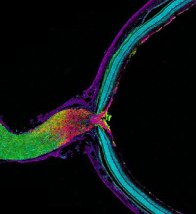
Mouse optic nerve and retina. Credit: Keunyoung Kim, Thomas Deerinck and Mark Ellisman, National Center for Microscopy and Imaging Research, UC San Diego.
This image shows the mouse optic nerve and retina. Credit: Keunyoung Kim, Thomas Deerinck and Mark Ellisman, National Center for Microscopy and Imaging Research Exit icon, UC San Diego.

In this image, the optic nerve (left) leaves the back of the retina (right). Where the retina meets the optic nerve, visual information begins its journey from the eye to the brain. Taking a closer look, axons (purple), which carry electrical and chemical messages, meet astrocytes (yellow), a type of brain cell. Recent research has found a new and surprising role for these astrocytes.

Biologists have long thought that all cells, including neurons, degrade and reuse pieces of their own mitochondria, the little powerhouses that provide energy to cells. Using cutting-edge imaging technology, researchers led by Mark Ellisman of the University of California, San Diego, and Nicholas Marsh-Armstrong of Johns Hopkins University have caught neurons in the mouse optic nerve in the act of passing some of their worn out mitochondria to neighboring astrocytes, which then did the dirty work of recycling.

The researchers also showed that neurons in other regions of the brain appear to outsource mitochondrial breakdown to astrocytes as well. They suggest that it will be important to confirm that this process occurs in other parts of the brain and to determine how possible defects in the outsourcing may contribute to or underlie neuronal dysfunction or neurodegenerative diseases.

This work also was funded by NIH’s National Eye Institute and National Institute on Drug Abuse.

Learn more:
University of California, San Diego News Release and Blog Posting Exit icon
How Cells Take Out the Trash Article from Inside Life Science

Meet Rhiju Das

1 comment
Rhiju Das
Credit: Rhiju Das
Rhiju Das
Fields: Biophysics and biochemistry
Works at: Stanford University
Born and raised in: The greater Midwest (Texas, Indiana and Oklahoma)
Studied at: Harvard University, Stanford University
When he’s not in the lab he’s: Enjoying the California outdoors with his wife and 3-year-old daughter
If he could recommend one book about science to a lay reader, it would be: “The Eighth Day of Creation,” about the revolution in molecular biology in the 1940s and 50s.

At the turn of the 21st century, Rhiju Das saw a beautiful picture that changed his life. Then a student of particle physics with a focus on cosmology, he attended a lecture unveiling an image of the ribosome—the cellular machinery that assembles proteins in every living creature. Ribosomes are enormous, complicated machines made up of many proteins and nucleic acids similar to DNA. Deciphering the structure of a ribosome—the 3-D image Das saw—was such an impressive feat that the scientists who accomplished it won the 2009 Nobel Prize in chemistry.

Das, who had been looking for a way to apply his physics background to a research question he could study in a lab, had found his calling.

“It was an epiphany—it was just flabbergasting to me that a hundred thousand atoms could find their way into such a well-defined structure at atomic resolution. It was like miraculously a bunch of nuts and bolts had self-assembled into a Ferrari,” recounted Das. “That inspired me to drop everything and learn everything I could about nucleic acid structure.”

Das focuses on the nucleic acid known as RNA, which, in addition to forming part of the ribosome, plays many roles in the body. As is the case for most proteins, RNA folds into a 3-D shape that enables it to work properly.

Das is now the head of a lab at Stanford University that unravels how the structure and folding of RNA drives its function. He has taken a unique approach to uncovering the rules behind nucleic acid folding: harnessing the wisdom of the crowd.

Together with his collaborator, Adrien Treuille of Carnegie Mellon University, Das created an online, multiplayer video game called EteRNA Exit icon. More than a mere game, it does far more than entertain. With its tagline “Played by Humans, Scored by Nature,” it’s upending how scientists approach RNA structure discovery and design.

Das’ Findings

Treuille and Das launched EteRNA after working on another computer game called Foldit, which lets participants play with complex protein folding questions. Like Foldit, EteRNA asks players to assemble, twist and revise structures—this time of RNA—onscreen.

But EteRNA takes things a step further. Unlike Foldit, where the rewards are only game points, the winners of each round of EteRNA actually get to have their RNA designs synthesized in a wet lab at Stanford. Das and his colleagues then post the results—which designs resulted in a successful, functional RNAs and which didn’t—back online for the players to learn from.

In a paper published in the Proceedings of the National Academy of Sciences Exit icon, Das and his colleagues showed how effective this approach could be. The collective effort of the EteRNA participants—which now number over 100,000—was better and faster than several established computer programs at solving RNA design problems, and even came up with successful new structural rules never before proposed by scientists or computers.

“What was surprising to me was their speed,” said Das. “I had just assumed that it would take a year or so before players were really able to analyze experimental data, make conclusions and come up with robust rules. But it was one of the really shocking moments of my life when, about 2 months in, we plotted the performance of players against computers and they were out-designing the computers.”

“As far as I can tell, none of the top players are academic scientists,” he added. “But if you talk to them, the first thing they’ll tell you is not how many points they have in the game but how many times they’ve had a design synthesized. They’re just excited about seeing whether or not their hypotheses were correct or falsified. So I think the top players truly are scientists—just not academic ones. They get a huge kick out of the scientific method, and they’re good at it.”

To capture lessons learned through the crowd-sourcing approach, Das and his colleagues incorporated successful rules and features into a new algorithm for RNA structure discovery, called EteRNABot, which has performed better than older computer algorithms.

“We thought that maybe the players would react badly [to EteRNABot], that they would think they were going to be automated out of existence,” said Das. “But, as it turned out, it was exciting for them to have their old ideas put into an algorithm so they could move on to the next problems.”

You can try EteRNA for yourself at http://eternagame.org Exit icon. Das and Treuille are always looking for new players and soliciting feedback.
Meet Elizabeth Grice

0 comments
Elizabeth Grice
Elizabeth Grice
First job: Detasseling corn
Favorite food: Chocolate
Pets: An adopted shelter cat, Dolce
Favorite city: Athens, Greece
Hidden talent: Baking creative desserts
Credit: Bill Branson, NIH

Imagine a landscape with peaks and valleys, folds and niches, cool, dry zones and hot, wet ones. Every inch is swarming with diverse communities, but there are no cities, no buildings, no fields and no forests.

You’ve probably thought little about the inhabitants, but you see their environment every day. It’s your largest organ—your skin.

Elizabeth Grice, an assistant professor at the University of Pennsylvania, studies the skin microbiome to learn how and why bacteria colonize particular places on the body. Already, she’s found that the bacterial communities on healthy skin are different from those on diseased skin.

She hopes her work will point to ways of treating certain skin diseases, especially chronic wounds. “I like to think that I am making discoveries that will impact the way medicine is practiced,” she says.

Grice’s Findings

To investigate what role bacteria play in diabetic wounds, Grice and her colleagues took skin swabs from both diabetic and healthy mice, and then compared the two. They found that diabetic mice had about 40 times more bacteria on their skin, but it was concentrated into few species. A more diverse array of bacteria colonized the skin of healthy mice.

The researchers then gave each mouse a small wound and spent 28 days swabbing the sites to collect bacteria and observing how the skin healed. They found that wounds on diabetic mice started to increase in size at the same time as wounds on healthy mice began to heal. In about 2 weeks, most healthy mice looked as good as new. But most of the wounds on diabetic mice had barely healed even after a month.

Interestingly, bacterial communities in the wounds became more diverse in both groups of mice as they healed—although the wounds on diabetic mice still had less diversity than the ones on healthy mice.

“Bacterial diversity is probably a good thing, especially in wounds,” says Grice. “Often, potentially infectious bacteria are found on normal skin and are kept in check by the diversity of bacteria surrounding them.”

Grice and her colleagues also found distinctly different patterns of gene activity between the two groups of mice. As a result, the diabetic mice put out a longer-lasting immune response, including inflamed skin. Scientists believe prolonged inflammation might slow the healing process.

Grice’s team suspects that one of the main types of bacteria found on diabetic wounds, Staphylococcus, makes one of the inflammation-causing genes more active.

Now that they know more about the bacteria that thrive on diabetic wounds, Grice and her colleagues are a step closer to looking at whether they could reorganize these colonies to help the wounds heal.

Content adapted from the NIGMS Findings magazine article Body Bacteria.

Multitarget Drugs to Challenge Microbial Resistance

0 comments
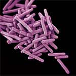
A group of purple, rod-shaped bacterial cells rendered by computer at Centers for Disease Control and Prevention by Melissa Brower.
Computer-generated image of drug-resistant Mycobacterium tuberculosis bacteria. Credit: Melissa Brower, Centers for Disease Control and Prevention.

Drugs that target a single essential protein in a microbial invader can be effective treatments. But the genomes of pathogens—including bacteria, fungi and parasites—mutate rapidly, and resistance can develop if a mutation changes a target protein’s structure. Molecules that interfere with multiple microbial proteins at once have the potential to overcome the growing problem of antimicrobial drug resistance.

Researchers led by Eric Oldfield of the University of Illinois recently explored whether an experimental drug called SQ109, developed to treat tuberculosis (TB), could be tweaked to attack multiple enzymes, as well as to kill different types of microbes. The scientists succeeded in creating several multitarget analogs of SQ109 that were more effective than the original drug at killing their target pathogens in laboratory experiments. These analogs included one compound that was five times more potent against the bacterium that causes TB while also being less toxic to a human cell line tested.

This work was also funded by the National Cancer Institute; the National Heart, Lung, and Blood Institute; the National Institute of Allergy and Infectious Diseases and the NIH Office of the Director.

Learn more:
University of Illinois, Urbana-Champaign News Release exit icon

A Medicine’s Life Inside the Body

0 comments
Heart
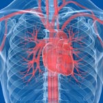
Most often, the bloodstream is the vehicle for carrying medicines throughout the body. Credit: Stock image.

Pharmacology is the scientific field that studies how the body reacts to medicines and how medicines affect the body. Scientists funded by the National Institutes of Health are interested in many aspects of pharmacology, including one called pharmacokinetics, which deals with understanding the entire cycle of a medicine’s life inside the body.

Knowing more about each of the four main stages of pharmacokinetics—absorption, distribution, metabolism and excretion—aids the design of medicines that are more effective and that produce fewer side effects.

Read more about a medicine’s life inside the body in this Inside Life Science article.

The Inner Life of Nerve Cells

2 comments
“Before this research, we didn’t even know that neurons had this special mechanism to control neuropeptide function. This is why we do basic research. This is why it’s important to understand how neurons work, down to the subcellular and molecular levels.”—Kenneth Miller”

Nerve cells (neurons) in the brain use small molecules called neuropeptides to converse with each other. Disruption of this communication can lead to problems with learning, memory and other brain functions. Through genetic studies in a model organism, the tiny worm C. elegans, a team led by Kenneth Miller of the Oklahoma Medical Research Foundation has uncovered a previously unknown mechanism that nerve cells use to package, move and release neuropeptides. The researchers found that a protein called CaM kinase II, which plays many roles in the brain, helps control this mechanism. Neuropeptides in worms lacking CaM kinase II spilled out from their packages before they reached their proper destinations. A more thorough understanding of how neurons work, provided by studies like this, may help researchers better target drugs to treat memory disorders and other neurological problems in humans.

This work also was funded by NIH’s National Institute of Mental Health.

Learn more:
Oklahoma Medical Research Foundation News Release Exit icon

New Life for Toxic Antibiotics?

0 comments
Pills and a bottle
Researchers found that the antibiotic trovafloxacin cuts off a channel for communication between cells and interferes with a cell-death process. Credit: Stock image.

Many compounds that show promise as new antibiotics for treating bacterial infections never make it to the clinic because they turn out to be toxic to humans as well as to bacteria. A research team led by Kodi Ravichandran of the University of Virginia recently gained insights into why one such antibiotic, trovafloxacin, harms human cells. They found that the compound cuts off a channel for communication between cells, which in turn interferes with how dying cells are broken down and recycled by the body. Roughly 200 billion cells in the human body die and are replaced every day as part of a routine cleanup process, and interference in this process by trovafloxacin may have contributed to the serious liver damage seen in some patients in clinical trials of the drug. Understanding how trovafloxacin causes toxicity in people may help researchers re-engineer this and related compounds to make them safe and effective for use in fighting bacterial infections.

Learn more:
Ravichandran Lab

Bleach vs. Bacteria

0 comments
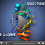
Screenshot of the video showing how chlorine affects a bacterial protein. Exposure to hypochlorous acid causes bacterial proteins to unfold and stick to one another, leading to cell death. Credit: Video segment courtesy of the American Chemistry Council. View video
Spring cleaning often involves chlorine bleach, which has been used as a disinfectant for hundreds of years. But our bodies have been using bleach’s active component, hypochlorous acid, to help clean house for millennia. As part of our natural response to infection, certain types of immune cells produce hypochlorous acid to help kill invading microbes, including bacteria.

Researchers funded by the National Institutes of Health have made strides in understanding exactly how bleach kills bacteria—and how bacteria’s own defenses can protect against the cellular stress caused by bleach. The insights gained may lead to the development of new drugs to breach these microbial defenses, helping our bodies fight disease.

Learning More About Our Partners in Digestion

0 comments
Bacteroides ovatus
Bacteroides ovatus. Credit: Eric Martens, University of Michigan Medical School.

After eating, we don’t do all the work of digestion on our own. Trillions of gut bacteria help us break food down into the simple building blocks our cells need to function. New research from an international team co-led by Eric Martens of the University of Michigan Medical School has uncovered how a strain of beneficial gut bacteria, Bacteroides ovatus, digests complex carbohydrates called xyloglucans that are found in fruits and vegetables. The researchers traced the microorganism’s digestive ability to a single piece of the genome. They also examined a publicly available set of genomic data, which included information from both humans and their resident bacteria, and found that more than 90 percent of 250 adults harbored at least one Bacteroides strain with xyloglucan-digesting capabilities. These results underscore the importance of the bacteria to human health and nutrition.

This work also was funded by the National Institute of Diabetes and Digestive and Kidney Diseases.

Learn more:
University of Michigan News Release
University of Michigan Host Microbiome Initiative
Gut Reactions and Other Findings About Our Resident Microbes from Inside Life Science
Body Bacteria from Findings Magazine

Epilepsy Drug Improves Health in Animal Model of Obesity

2 comments
Liver cells of obese mice treated with valproic acid (right) and untreated obese mice (left).
Liver cells (magenta) of obese mice treated with valproic acid (right) had much less fat accumulation (white) than those of untreated obese mice (left). Credit: Lindsay B. Avery and Namandjé N. Bumpus, Johns Hopkins University. View larger image

With more than 90 million Americans affected by obesity, developing medications to help combat weight gain and its associated diseases has become a priority. In a study using obese mice, a team led by Namandjé Bumpus of Johns Hopkins University recently showed that a commonly prescribed epilepsy drug, valproic acid, reduced fat accumulation in the liver and lowered elevated blood sugar levels like those associated with type 2 diabetes. Body weight also stabilized in mice given the drug, whereas untreated mice continued to gain weight. Additional experiments in mouse and human liver cells suggested that the byproducts of valproic acid produced as the body breaks down the drug, rather than valproic acid itself, were responsible for the observed effects. These byproducts achieved the same effects in cells at one-fortieth the concentration of valproic acid, making them promising candidates for further drug development.

Learn more:
Johns Hopkins University News Release Exit icon
Bumpus Laboratory