Month: March 2014

Cool Image: Lighting up Brain Cells

3 comments
Neurons activated with red or blue light.

Neurons activated with red or blue light using algae-derived opsins. Credit: Yasunobu Murata/McGovern Institute for Brain Research at MIT.

The nerve cells, or neurons, lit up in blue and red in this image of mouse brain tissue are expressing algae-derived, light-sensitive proteins called opsins. To control neurons with light, scientists engineer the cells to produce particular opsins, most of which respond to light in the blue-green range. Then they shine light on the cell to activate it. Now, a team of researchers led by Ed Boyden of the Massachusetts Institute of Technology and Gane Ka-Shu Wong of the University of Alberta has discovered an opsin that responds to red light preferentially, enabling them to manipulate two groups of neurons simultaneously with different colors of light and get a more comprehensive look at how those two sets of brain cells interact. Other opsins have shown potential for vision restoration in animal studies, and, because red light causes less damage to tissue than blue-green light, this new opsin might eventually be used for such treatments in humans.

Learn more:
MIT News Release Exit icon

Study Comparing Sepsis Treatment Methods Shows Equivalent Survival Rates

0 comments
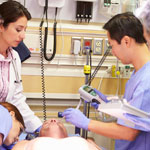
Doctors and a patient in a hospital
A 5-year, randomized clinical trial helped resolve a long-standing debate about how best to manage sepsis patients.

For years, doctors have debated the best ways to identify, predict and treat sepsis. The condition, which is usually triggered by infection, is marked by body-wide inflammation and can lead to a dangerous drop in blood pressure known as septic shock. Sepsis affects more than 800,000 people each year and kills about 20 to 30 percent of them. It’s the most expensive condition treated in U.S. hospitals, costing more than $20 billion a year.

Now, a nationwide, 5-year clinical trial that set out to compare three different treatment approaches has shown that survival of patients with septic shock was the same regardless of whether they received treatment based on structured, standardized medical plans (protocols) or the usual high-level standard of care. If patients were diagnosed shortly after the onset of sepsis and treated promptly with fluids and antibiotics, they did equally well whether they received treatment based on either of two specific protocols—one less invasive than the other—or got the usual, high-level care provided by the academic hospitals where the study was conducted.

According to the study’s leaders, the trial “helps resolve a long-standing clinical debate about how best to manage sepsis patients, particularly during the critical first few hours of treatment,” and shows that “there is not a mandated need for more invasive care in all patients.”

Learn more:
NIGMS News Release
University of Pittsburg News Release Exit icon
New England Journal of Medicine Article
Sepsis Fact Sheet

Capitalizing on Cellular Conversations

0 comments
Fat cells
Fat cells such as these listen for incoming signals like FGF21, which tells them to burn more fat. Credit: David Gregory and Debbie Marshall. All rights reserved by Wellcome Images.

Living things are chatty creatures. Even when they’re not making actual sounds, organisms constantly communicate using chemical signals that course through their systems. In multicellular organisms like people, brain cells might call, “I’m in trouble!” signaling others to help mount a protective response. Single-celled organisms like bacteria may broadcast, “We have to stick together to survive!” so they can coordinate certain activities that they can’t carry out solo. In addition to sending out signals, cells have to receive information. To help them do this, they use molecular “ears” called receptors on their surfaces. When a chemical messenger attaches to a receptor, it tells the cell what’s going on and causes a response.

Scientists are following the dialogue, learning how cell signals affect health and disease. They’re also starting to take part in the cellular conversations, inserting their own comments with the goal of developing therapies that set a diseased system right.

Continue reading this new Inside Life Science article

Nanoparticles Developed to Stick to Damaged Blood Vessels, Deliver Drugs

0 comments
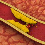
Artery with fat deposits and a formed clot. Credit: Stock image.
Artery with fat deposits and a formed clot. Credit: Stock image. View larger image

Heart disease is the leading cause of death for both men and women in the United States, according to the Centers for Disease Control and Prevention. One treatment challenge is developing non-invasive ways to direct medication to damaged or clogged arteries, which can block blood flow and increase the risk for heart attack and stroke. A team led by Naren Vyavahare at Clemson University has engineered extremely tiny particles—nanoparticles—that offer a promising step forward.

Healthy arteries have elastic fibers that make the arteries flexible. But, in most cardiovascular diseases, the fibers get damaged. The new nanoparticles, which can deliver drugs, attach only to damaged fibers and could enable site-specific drug delivery to minimize off-target side effects. The nanoparticles also allow drugs to be released over longer periods of time, potentially increasing the drugs’ effectiveness. The new biomaterial was tested in rodent models for studying vascular disease, so it is still in the early stages of development.

This work also was funded by NIH’s National Heart, Lung, and Blood Institute.

Learning More About Our Partners in Digestion

0 comments
Bacteroides ovatus
Bacteroides ovatus. Credit: Eric Martens, University of Michigan Medical School.

After eating, we don’t do all the work of digestion on our own. Trillions of gut bacteria help us break food down into the simple building blocks our cells need to function. New research from an international team co-led by Eric Martens of the University of Michigan Medical School has uncovered how a strain of beneficial gut bacteria, Bacteroides ovatus, digests complex carbohydrates called xyloglucans that are found in fruits and vegetables. The researchers traced the microorganism’s digestive ability to a single piece of the genome. They also examined a publicly available set of genomic data, which included information from both humans and their resident bacteria, and found that more than 90 percent of 250 adults harbored at least one Bacteroides strain with xyloglucan-digesting capabilities. These results underscore the importance of the bacteria to human health and nutrition.

This work also was funded by the National Institute of Diabetes and Digestive and Kidney Diseases.

Learn more:
University of Michigan News Release
University of Michigan Host Microbiome Initiative
Gut Reactions and Other Findings About Our Resident Microbes from Inside Life Science
Body Bacteria from Findings Magazine

Anti-Clotting Drugs: The Next Generation

1 comment
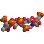
Form of heparin
Scientists created a tailor-made form of the anti-clotting drug heparin that offers several advantages.
View larger image

The low molecular weight (LMW) form of the drug heparin is commonly used to prevent unwanted blood clots that can lead to heart attacks and strokes. It’s usually derived from pig intestines and normally cleared from the human body by the kidneys. In individuals with impaired kidney function, the drug can build up in the circulation and cause excessive bleeding. Impurities and the risk of contamination are also concerns with pig-derived heparin.

Now, Robert Linhardt of Rensselaer Polytechnic Institute and Jian Liu of the University of North Carolina at Chapel Hill have created a synthetic, tailor-made form of LMW heparin that offers several advantages over the animal-derived version, including alleviating the risk of contamination from natural sources. Studies in the test tube and in mice showed that the activity of this customized heparin molecule is easily reversible in cases of overdose or uncontrolled bleeding. And, since it is cleared from the body by the liver rather than the kidneys, this form of heparin would be safer for people with impaired kidney function. Additional research, including testing in humans, will be needed before this new version of LMW heparin can be considered for medical use.

This work also was funded by NIH’s National Heart, Lung, and Blood Institute.

Learn more:

Rensselaer Polytechnic Institute News Release Exit icon
University of North Carolina at Chapel Hill News Release
Linhardt Labs
Liu Lab

Resetting Our Clocks: New Details About How the Body Tells Time

0 comments
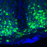
VIP in time-keeping brain cells
Boosting doses of a molecule called VIP (green) in time-keeping brain cells (blue) helped mice adjust quickly to major shifts in light-dark cycles. Credit: Cristina Mazuski in the lab of Erik Herzog, Washington University in St. Louis.

Springing clocks forward by an hour this Sunday, traveling across time zones, staring at a computer screen late at night or working the third shift are just a few examples of activities that can disrupt our daily, or circadian, rhythms. These roughly 24-hour cycles influence our physiology and behavior, and they’re driven by our body’s network of tiny timekeepers. If our daily routines fall out of sync with our body clocks, sleep, metabolic and other disorders can result.

Researchers funded by the National Institutes of Health have spent decades piecing together the molecular mechanisms of our biological clocks. Now, they’re building on that basic knowledge to better understand the intricate relationship among these clocks, circadian rhythms and physiology—and ultimately, find ways to manipulate the moving parts to improve our modern-day lives.

Continue reading this new Inside Life Science article

Meet Ravi Iyengar

0 comments
Ravi Iyengar
Ravi Iyengar
Fields: Systems pharmacology and systems biology
Works at: Mount Sinai School of Medicine, New York, NY
Favorite sports team: Yankees
Favorite subject in high school: Math
Recently read book: The Signal and the Noise by Nate Silver
Credit: Pedro Martinez, Systems Biology Center New York

Ravi Iyengar, a professor at Mount Sinai School of Medicine, stood in an empty lecture hall, primed to tell thousands of students about systems biology, a holistic approach to studying fundamental life processes. To prepare for this moment, he had spent 4 months reading hundreds of scientific papers and distilling the research into understandable nuggets. But that day, his only student was a videographer.

Together, they recorded 15 different lectures about systems biology—many related to Iyengar’s own research—that thousands of people would stream or download as part of a MOOC, or massive open online course.

Trained in biochemistry, Iyengar built his research career around studying molecules and developing a list of all the parts that help nerve, kidney and skin cells to function. As he obtained more information, he realized he needed to know how all the components worked together. To achieve this comprehensive understanding, Iyengar turned to computational techniques and mathematical analyses—cornerstones of systems biology.

For more than a decade, he has been using and developing systems biology approaches to explore a range of biomedical questions, from very basic to translational ones with immediate relevance to human health.

Iyengar’s Findings

In his earlier work, Iyengar used mathematical analyses to show that molecules within cells connect with one another to form switches that produce cellular memory. This may allow, for instance, an immune cell to remember a foreign object and secrete an antibody. In recent work, he and his team developed a mathematical model showing that the shape of a cell influences the flow of information across the membrane, possibly contributing to disease states and offering a way to study and identify them under the microscope. In another study, they analyzed a database of drug side effects to find combinations of medications that produce fewer adverse reactions and then created a cell biology interaction network that explains why a certain drug pair had this beneficial outcome. The approach could point to other combinations of FDA-approved drugs that reduce serious side effects and thereby guide clinical practice.

“Systems biology is a powerful way to explore important biological and medical questions, and it’s relevant to many fields of science,” said Iyengar. But he added that the majority of educational institutions, including liberal arts and community colleges, don’t have systems biology courses. So, Iyengar teamed with colleagues to create a series of MOOCs.

The first course, offered last summer and taught by Iyengar, presented all the facets of systems biology. The syllabus included lessons on genomics and bioinformatics, fields that have contributed to systems biology; gathering and integrating data; and the use of modeling in drug development.

“My goal was for the students to get the general gestalt of systems biology,” explained Iyengar, who directs an NIH-funded center focused on the systems-level study of medicine and therapeutics.

In total, more than 12,000 participants watched at least one video lecture, 3,000 submitted one or more of the weekly quizzes and 1,800 took a mid-term or final exam. The online discussions forum included nearly 400 topics with about 5,000 posts. The students, most enrolled in a graduate program or working full-time, had some training in the biological, biomedical, computer and information sciences.

“The stats tell me that many people are in fields adjacent to systems biology and don’t have access to more traditional systems biology courses,” concluded Iyengar. “Through the MOOC, we can reach them in a substantial way.”

The second course, which covers network analysis, wrapped up in early December, and the third course, which covers dynamical modeling methods, began in January. Iyengar plans to offer the intro course again in late March.

Learn more:
MOOC Systems Biology Courses Exit icon