Year: 2014

Anesthesia and Brain Cells: A Temporary Disruption?

1 comment
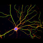
Hippocampal neuron in culture.
Hippocampal neuron in culture. Dendrites are green, dendritic spines are red, and DNA in cell’s nucleus is blue. Credit: Shelley Halpain, University of California, San Diego.

Anesthetic drugs are vital to modern medicine, allowing patients to undergo even the longest and most invasive surgeries without consciousness or pain. Unfortunately, studies have raised the concern that exposing patients, particularly children and the elderly, to some anesthetics may increase risk of long-term cognitive and behavioral issues.

A scientific team led by Hugh Hemmings Exit icon of Weill Cornell Medical College and Shelley Halpain Exit icon of the University of California, San Diego, examined the effects of anesthesia on neurons isolated from juvenile rats. Given at doses and durations frequently used during surgery, the commonly administered general anesthetic isoflurane did in fact reduce the number and size of important structures within neurons called dendritic spines. Dendritic spines help pass information from neuron to neuron, and disruption of these structures can be associated with dysfunction in thinking and behavior.

Promisingly, the shrinkage observed by the researchers appeared to be temporary: After the researchers washed the anesthetic out of the cell cultures, the dendritic spines grew back. But because neurons in culture do not reproduce all aspects of intact neuronal networks, the scientists explain that the findings should be verified in more complex models. Other molecular mechanisms may also potentially contribute to late effects of anesthesia exposure.

This work also was funded by NIH’s National Institute of Mental Health.

Learn more:
University of California, San Diego News Release
Understanding Anesthesia from Inside Life Science

Intercepting Amyloid-Forming Proteins

0 comments
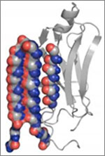
Structure of a protein involved in disease-associated amyloid fibrils.
A molecule targets the intermediary structure of a protein involved in disease-associated amyloid fibrils. Credit: University of Washington.

Alzheimer’s disease, type 2 diabetes and many other illnesses are linked to the buildup of proteins whose structures have changed into shapes that enable the formation of cell-entangling threads called amyloid fibrils. About 10 years ago, researchers led by Valerie Daggett of the University of Washington used computer simulations to suggest that such proteins, on their way to creating fibrils, form an intermediary structure called an alpha sheet that’s even more toxic to cells than fibrils. Now Daggett’s team has experimentally investigated this possibility. The scientists made alpha sheet molecules expected to bind to amyloid-forming proteins in the computationally predicted intermediate state. When they tested the molecules on two amyloid disease-related proteins, they observed a substantial reduction in fibril formation. The work is still very preliminary, but it highlights a potential new avenue for treating a range of amyloid-related diseases.

This work also was funded by NIH’s National Institute of Allergy and Infectious Diseases.

Learn more:
University of Washington News Release Exit icon
Daggett Lab Exit icon
Monster Mash: Protein Folding Gone Wrong Article from Inside Life Science

Improving the Odds of Surviving Sepsis

1 comment
Acupuncture
A form of acupuncture—or a drug that mimics its effect—may one day lead to an anti-inflammatory therapy for people with sepsis. Credit: Stock image.

A leading cause of death in U.S. intensive care units is sepsis, an overwhelming immune response to infection that triggers body-wide inflammation and can cause organ failure.

Sepsis is challenging to diagnose and treat. Many of its early signs, such as fever and difficulty breathing, are similar to those of other conditions. When doctors do not detect sepsis until a more advanced stage, they are often unable to stop its progression or prevent its complications.

“Sepsis is a complex problem,” says Sarah Dunsmore of the National Institutes of Health (NIH). “We need more research at all levels—from the molecular to the patient—to improve sepsis diagnosis and treatment and to enhance the quality of life for sepsis survivors.”

NIH-funded scientists use a variety of tools, including blood tests and acupuncture, in their quest to detect sepsis early, treat it quickly and reduce its later effects.

Read more about sepsis research in this Inside Life Science article.

Meet Janet Iwasa

0 comments
Janet Iwasa
Credit: Janet Iwasa
Janet Iwasa
Fields: Cell biology and molecular animation
Works at: University of Utah
Raised in: Indiana and Maryland
Studied at: University of California, San Francisco, and Harvard Medical School
When not in the lab she’s: Keeping up with her two preschool-aged sons
Something she’s proud of that she’ll never try again: Baking a multi-tiered wedding cake, complete with sugar flowers, for a friend’s wedding.

Janet Iwasa wouldn’t have described herself as an artistic child. She didn’t carry around a sketch pad, pencils or paintbrushes. But she remembers accompanying her father, a scientist at the National Institutes of Health, to his lab on the weekends. She’d spend hours doodling in a drawing program on his old Macintosh computer while he worked on experiments.

“I always remember wanting to be a scientist, and that’s probably highly inspired by my dad,” says Iwasa. Her early affinity for art and technology set her on an unusual career path to become a molecular animator. A typical work day now finds her adapting computer programs originally designed to bring characters like Buzz Lightyear to life to help researchers probe complicated, dynamic interactions within cells.

Iwasa’s interest in animation was sparked when she was a graduate student in cell biology, studying a protein called actin, which helps cells to move and change shape. At the time, the only visual representations she had of actin networks were flat, two-dimensional drawings on paper. When she saw an animation of the dynamic movement of a molecule called kinesin, she thought, “Why are we relying on oversimplified, static illustrations [of molecules], when we can be doing something like this video?”

Within a year, she was taking an animation class at a local college. She quickly realized that she would need more intensive instruction to be able to animate complex biological processes. A few summers later, she flew to Hollywood for a 3-month training program in industry-standard animation technology.

The oldest student in that course—and the only woman—Iwasa immediately began thinking about how to adapt a standard animator’s toolkit to illustrate the inner life of cells. A technique used to create the effect of human hair blowing in the wind could also show the movement of an RNA molecule. A chunk of computer code used to make the facets of a soccer ball fall apart and come back together in a different order could be adapted to model virus assembly and disassembly.

Her Findings

Following her training, Iwasa spent 2 years as a National Science Foundation Discovery Corps fellow, producing the Exploring Life’s Origins exhibit with the Boston Museum of Science and the Szostak Lab at Massachusetts General Hospital/Harvard Medical School. As part of the multi-media exhibit, she created animations to illustrate how the simplest living organisms may have evolved on early Earth.

Since then, Iwasa has helped researchers model such complex actions as how cells ingest materials, how proteins are transported across a cell membrane, and how the motor protein dynein helps cells divide.

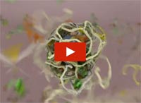
Screenshot from the video that shows how a protein called clathrin forms a cage-like container that cells use to engulf and ingest materials
Iwasa developed this video to show how a protein called clathrin forms a cage-like container that cells use to engulf and ingest materials.

Iwasa calls her animations “visual hypotheses”: The end results may be beautiful, but the process of animation itself is what encapsulates, clarifies and communicates the science.

“It’s really building the animated model that brings insights,” she says. “When you’re creating an animation, you’re really grappling with a lot of issues that don’t necessarily come up by any other means. In some cases, it might raise more questions, and make people go back and do some more experiments when they realize there might be something missing” in their theory of how a molecular process works.

Now she’s working with an NIH-funded research team at the University of Utah to develop a detailed animation of how HIV enters and exits human immune cells.

Abbreviated CHEETAH, the full name of the group is the Center for the Structural Biology of Cellular Host Elements in Egress, Trafficking, and Assembly of HIV.

“In the HIV life cycle, there are a number of events that aren’t really well understood, and people have different ideas of how things happen,” says Iwasa. She plans to animate the stages of viral infection in ways that reflect different proposals for how the process works, to give researchers a new way to visualize, communicate—and potentially harmonize—their hypotheses.

The full set of Iwasa’s HIV-related animations will be available online as they are completed, at https://scienceofhiv.org, with the first set launching in the fall of 2014.

Learn more:
Janet Iwasa’s TED Talk: How animations can help scientists test a hypothesis
Janet Iwasa’s 3D model of an HIV particle was a winner in the 2014 BioArt contest sponsored by Federation of American Societies for Experimental Biology
NIH Director’s blog post about Iwasa and her HIV video animation
A Data Bank Built for Discovery

0 comments
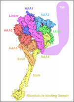
Dynein, a motor protein. Credit: David S. Goodsell, The Scripps Research Institute and the RCSB PDB.
The PDB archive holds structural data for dynein, a motor protein, and more than 100,000 other molecules. Credit: David S. Goodsell, The Scripps Research Institute and the RCSB PDB. Click for larger image

Meet dynein, the August Molecule of the Month presented by the Research Collaboratory for Structural Bioinformatics Protein Data Bank (RCSB PDB). This motor protein travels along the cables of our cellular skeleton, delivering cargo throughout the cell. The structure of dynein’s stalk enables it to bind to regular grooves along its path.

Dynein’s shape is just one of more than 100,000 structures that scientists have deposited in the PDB archive, a freely available digital repository. Because understanding a protein’s shape helps researchers better understand its function, the structural information in the PDB can lead to additional scientific advancements. For example, scientists use the structure of HIV protease, a protein that helps the virus replicate in the body, to develop drugs that fit snugly into the protein’s center, shutting it down. And they use the shape of RNA polymerase to learn how this protein fits together with smaller ones to read our genetic code.

The PDB has doubled in size over the last 6 years. As the collection continues to grow, so does our potential for drug discovery and our understanding of basic life processes.

Learn more:
Molecule of the Month Archive from RCSB PDB

How Heat-Loving Organisms Are Helping Advance Medicine

2 comments
Hot spring. Credit: Stock image.
Icelandic hot springs are the natural habitat of Rhodothermus marinus, one of many species of thermophiles that the Gennis Lab studies to better understand membrane proteins. Credit: Stock image.

As the temperature climbs, most humans look for ways to cool down fast. But for some species of microorganisms, a midsummer heat wave isn’t nearly hot enough. These heat lovers, known as thermophiles, thrive at temperatures of 113°F or more. They’re often found in hot springs, geysers and even home water heaters.

Like humans and other organisms, thermophiles rely on proteins to maintain normal cell function. While our protein molecules break down under intense heat, a thermophile’s proteins actually work more efficiently. They also tend to be more stable at room temperature than our own. An NIH-funded research team is taking advantage of this property of thermophiles to better understand a group of human proteins commonly targeted by today’s medicines.

Read more about the team’s thermophile research in this Inside Life Science article.

The “Virtuous Cycle” of Technology and Science

0 comments
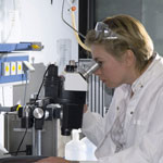
A scientist looking through a  microscope. Credit: Stock image.
Whether it’s a microscope, computer program or lab technique, technology is at the heart of biomedical research. Credit: Stock image.

Whether it’s a microscope, computer program or lab technique, technology is at the heart of biomedical research. Its central role is particularly clear from this month’s posts.

Some show how different tools led to basic discoveries with important health applications. For instance, a supercomputer unlocked the secrets of a drug-making enzyme, a software tool identified disease-causing variations among family members and high-powered microscopy revealed a mechanism allowing microtubules—and a cancer drug that targets them—to work.

Another theme featured in several posts is novel uses for established technologies. The scientists behind the cool image put a new spin on a long-standing imaging technology to gain surprising insights into how some brain cells dispose of old parts. Similarly, the finding related to sepsis demonstrates yet another application of a standard lab technique called polymerase chain reaction: assessing the immune state of people with this serious medical condition.

“We need tools to answer questions,” says NIGMS’ Doug Sheeley, who oversees biomedical technology research resource grants. “When we find the answers, we ask new questions that then require new or improved tools. It’s a virtuous cycle that keeps science moving forward.”

Cool Image: Outsourcing Cellular Housekeeping

0 comments
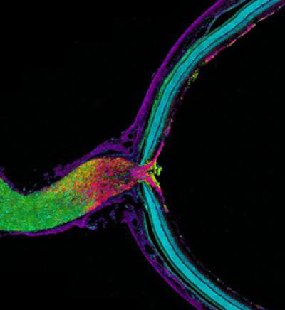
Mouse optic nerve and retina. Credit: Keunyoung Kim, Thomas Deerinck and Mark Ellisman, National Center for Microscopy and Imaging Research, UC San Diego.
This image shows the mouse optic nerve and retina. Credit: Keunyoung Kim, Thomas Deerinck and Mark Ellisman, National Center for Microscopy and Imaging Research Exit icon, UC San Diego.

In this image, the optic nerve (left) leaves the back of the retina (right). Where the retina meets the optic nerve, visual information begins its journey from the eye to the brain. Taking a closer look, axons (purple), which carry electrical and chemical messages, meet astrocytes (yellow), a type of brain cell. Recent research has found a new and surprising role for these astrocytes.

Biologists have long thought that all cells, including neurons, degrade and reuse pieces of their own mitochondria, the little powerhouses that provide energy to cells. Using cutting-edge imaging technology, researchers led by Mark Ellisman of the University of California, San Diego, and Nicholas Marsh-Armstrong of Johns Hopkins University have caught neurons in the mouse optic nerve in the act of passing some of their worn out mitochondria to neighboring astrocytes, which then did the dirty work of recycling.

The researchers also showed that neurons in other regions of the brain appear to outsource mitochondrial breakdown to astrocytes as well. They suggest that it will be important to confirm that this process occurs in other parts of the brain and to determine how possible defects in the outsourcing may contribute to or underlie neuronal dysfunction or neurodegenerative diseases.

This work also was funded by NIH’s National Eye Institute and National Institute on Drug Abuse.

Learn more:
University of California, San Diego News Release and Blog Posting Exit icon
How Cells Take Out the Trash Article from Inside Life Science

A Drug-Making Enzyme in Motion

0 comments
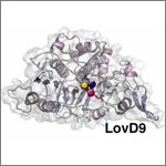
Mutated enzyme, LovD9. Credit: Silvia Osuna and Gonzalo Jiménez-Osés, University of California, Los Angeles.
The movement of this mutated enzyme, LovD9, facilitates rapid production of the cholesterol reducing-drug simvastatin. Credit: Silvia Osuna and Gonzalo Jiménez-Osés, University of California, Los Angeles.

LovD9, a mutated version of an enzyme extracted from mold growing in soil, produces the cholesterol-reducing drug simvastatin 1,000 times faster than its natural predecessor. But scientists didn’t understand why because the enzyme’s mutations are far from the active site, where the drug is actually made. Now they do.

Yi Tang of the University of California, Los Angeles (UCLA), in partnership with the pharmaceutical company Codexis, generated LovD9 by repeatedly inducing random mutations, each time selecting the mutated versions of the enzyme with the most promise for industrial simvastatin production.

Then, the team collaborated with UCLA colleagues Kendall Houk and Todd Yeates to unlock the secret of the enzyme’s speed. Using ANTON, a special-purpose supercomputer at the Pittsburgh Supercomputing Center, they simulated how different parts of the enzyme rotate and twist when synthesizing the drug. The scientists discovered that as LovD9 moves, it forms shapes that facilitate simvastatin production more often than the natural enzyme does.

With their better understanding of how mutations far from an active site may affect an enzyme’s motion, the researchers hope to one day directly engineer enzymes with precise mutations that enhance drug production.

Learn more:
University of California, Los Angeles News Release Exit icon
Houk Exit icon, Tang Exit icon and Yeates Exit icon Labs

Raking the Family Tree for Disease-Causing Variations

1 comment
Silhouettes of people with nucleic acid sequences and a stethoscope.
A new software tool analyzes disease-causing genetic variations within a family. Credit: NIH’s National Human Genome Research Institute.

Changes in your DNA sequence occur randomly and rarely. But when they do happen, they can increase your risk of developing common, complex diseases, such as cancer. One way to identify disease-causing variations is to study the genomes of family members, since the changes typically are passed down to subsequent generations.

To rake through a family tree for genetic variations with the highest probabilities of causing a disease, researchers combined several commonly-used statistical methods into a new software tool called pVAAST. The scientific team, which included Mark Yandell and Lynn Jorde of the University of Utah and Chad Huff of the University of Texas MD Anderson Cancer Center, used the tool to identify the genetic causes of a chronic intestinal inflammation disease and of developmental defects affecting the heart, face and limbs.

The results confirmed previously identified genetic variations for the developmental diseases and pinpointed a previously unknown variation for the intestinal inflammation. Together, the findings confirm the ability of the tool to detect disease-causing genetic changes within a family. Another research team has already used the software tool to discover rare genetic changes associated with family cases of breast cancer. These studies are likely just the beginning for studying genetic patterns of diseases than run in a family.

This work also was funded by NIH’s National Institute of Diabetes and Digestive and Kidney Diseases; National Cancer Institute; National Human Genome Research Institute; National Heart, Lung, and Blood Institute; and National Institute of Mental Health.

Learn more:
University of Utah News Release (no longer available)
Yandell Exit icon, Jorde and Huff Exit icon Labs