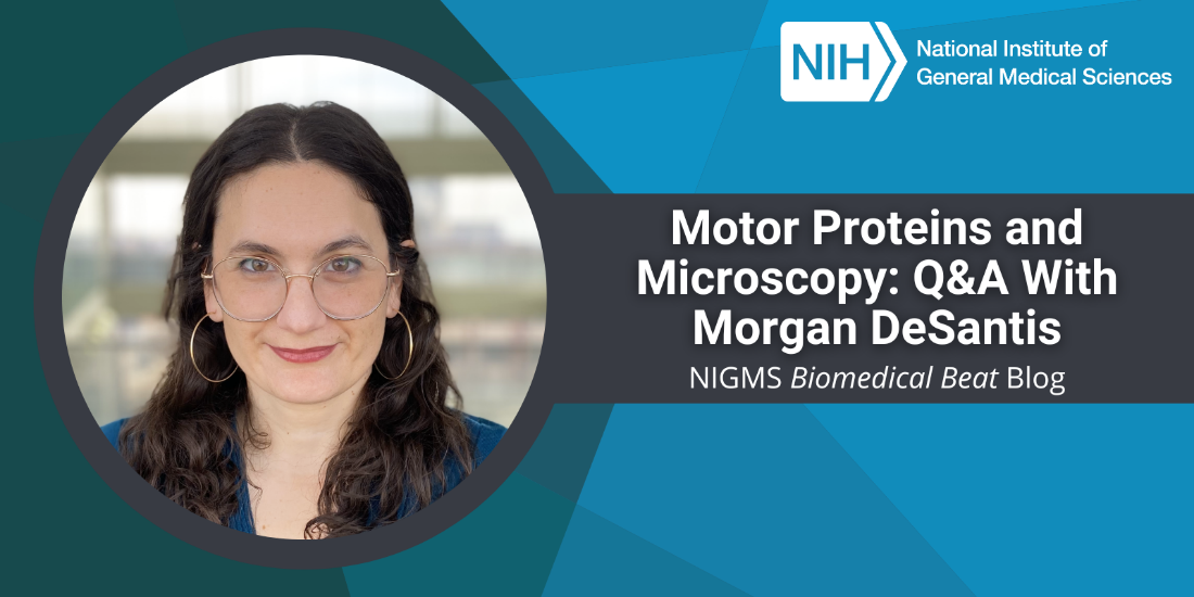
“I remember thinking in my first cellular biology class how impossibly beautiful it is that there are tiny machines in our bodies doing work,” says Morgan DeSantis, Ph.D., an assistant professor of molecular, cellular, and developmental biology at the University of Michigan in Ann Arbor. We talked with Dr. DeSantis about how her career in science almost didn’t happen, how happy she is that it did, and what she’s learning through her research on molecular machines.
Q: How did you become interested in science?
A: I wasn’t remotely interested in science in high school—I was a self-identified artist. I went to the College of Wooster in Ohio with the sole purpose of studying art and doing pottery. But one day during my freshman year, a box with all the pieces I made throughout the year fell, and everything inside broke. It’s hard to describe the emotions I felt that day, but something changed in me, and I realized pottery wasn’t for me. I couldn’t start the projects over, and I didn’t want to drop out and move back home. So, I decided to become a medical doctor.
The next year, I registered for some of the classes I needed as prerequisites for medical school: chemistry, physics, calculus, and cellular biology. It was a dramatic shift; I felt like I was in a different world and didn’t speak the language. I remember needing to read the textbooks closely and looking up the definitions of words constantly. But eventually I started to catch on and became captivated by what I was learning, especially proteins. I’m still amazed by the power and elegance of proteins now, but I felt this childlike awe then. It was like magic.
Because I got a late start, I didn’t have time to join a research lab as an undergrad student. But I knew that it would help with medical school applications, so I applied for a postbaccalaureate position in the structural immunology lab of Sandra Smith-Gill, Ph.D., at the National Cancer Institute (NCI). I didn’t expect to like it—I didn’t like lab classes during undergrad, and I thought this experience would be the same. Within 6 months, I’d changed my mind and knew I was going to be a researcher instead of a doctor. The process of asking questions and devising ways to answer them was addicting.
Q: What steps did you take next in your career progression?
A: After 3 years at NCI, I joined the lab of James Shorter, Ph.D., at the University of Pennsylvania Perelman School of Medicine in Philadelphia for my Ph.D. His lab studies a six-piece protein found in yeast called Hsp104. It has the incredible ability to untangle protein aggregates, which are collections of normally soluble proteins that assemble to form insoluble clumps. These clumps—amyloid aggregates are one example—contribute to some neurodegenerative diseases, like Alzheimer’s disease.
The goal of my Ph.D. project was to understand how the six pieces of Hsp104 assemble into a functional unit and how they untangle protein aggregates. I learned that Hsp104 uses different methods to untangle aggregates depending on the type of protein involved. In some cases, six functional subunits are needed, but in other cases, one functional subunit is enough.
I started my postdoctoral training with Samara Reck-Peterson, Ph.D., at the University of California, San Diego, after I finished my Ph.D. With her, I studied a protein called dynein, which is in the same protein family as Hsp104. Like Hsp104, dynein is made up of six subunits, but instead of untangling aggregates, it acts as a motor protein. Motor proteins are essential for moving cargo, such as vesicles or organelles like mitochondria, within cells.
Dynein “walks” along microtubules (part of the cytoskeleton) and carries its cargo along with it. At the time, we didn’t know the function of all six subunits, so I set out to learn more about the third subunit. By changing it slightly to become nonfunctional, we learned that the third subunit is important in controlling the machine’s overall ability to walk and interact with other proteins, including one called Lis1. Proper interactions between dynein and Lis1 are critical for dynein function, and in fact, some inherited variants of Lis1 cause developmental disorders of the brain, such as lissencephaly. In this condition, newly formed nerve cells in the brain of a developing embryo can’t migrate to the outer regions of the brain, which is necessary for normal development.
Q: What does your lab study?
A: We study dynein and the molecules that interact with it. I want to understand how dynein correctly identifies three things: cargo type, transport timing, and cargo destination. The very broad answer to all these questions is that it uses regulator proteins like Lis1. Dynein forms a large complex with many proteins that help it do its job.
One of our most-used research techniques is fluorescence microscopy. We purify dynein and the molecules it interacts with, combine them with purified microtubules, and visualize dynein walking along the microtubules under the microscope. By adding or removing regulator proteins, we can learn how they change dynein’s behavior.
We also screen proteins to identify ones that interact with dynein or any of its known regulators. Through a technique called proximity-dependent biotinylation, we can identify the proteins that are very close to dynein in a cell. In this way, we found a completely new protein associated with the dynein super complex and showed that it recruits dynein to the leading edge of migrating cells. Its loss causes nerve cell migration defects in zebrafish, a research organism, like some variants of Lis1 do.
Q: What do you consider your biggest accomplishment so far in your career?
A: My biggest accomplishment is that I’ve built a lab full of people who are so passionate, hardworking, thoughtful, and curious that they expand the types of questions we can ask. The mentoring aspect of my job is one of the hardest—we don’t really get training on how to be a good mentor—so I’m proud of the group I’ve put together and the success we’ve had.
Q: How do you broaden the impact of your research?
A: I’m on the scientific advisory board for the DYNC1H1 Association, which is a nonprofit organization started by parents of children affected by disease-causing dynein variants. The organization’s goal is to create opportunities for dynein research that help improve the lives of affected people through the development of new treatments. Unfortunately, it’s hard to study these variants because they cause a wide range of symptoms, so we advise the organization on ways to study them.
Q: In your opinion, what makes a career in science exciting?
A: Everything! I truly love my job. What initially appealed to me was that science allows you to work with your hands just as much as your brain. When I first started, it felt like play, or like solving puzzles. Plus, I found that some of the aspects of pottery that I liked most, such as being creative or practicing to become more skilled at a new technique, translated well to research.
Q: What advice would you give students who are interested in a career in science?
A: Let your curiosity drive you to new scientific fields and questions, because that’s when the coolest findings happen.
Dr. DeSantis’ research is supported by the NIGMS Maximizing Investigators’ Research Award program through grant R35GM146739.