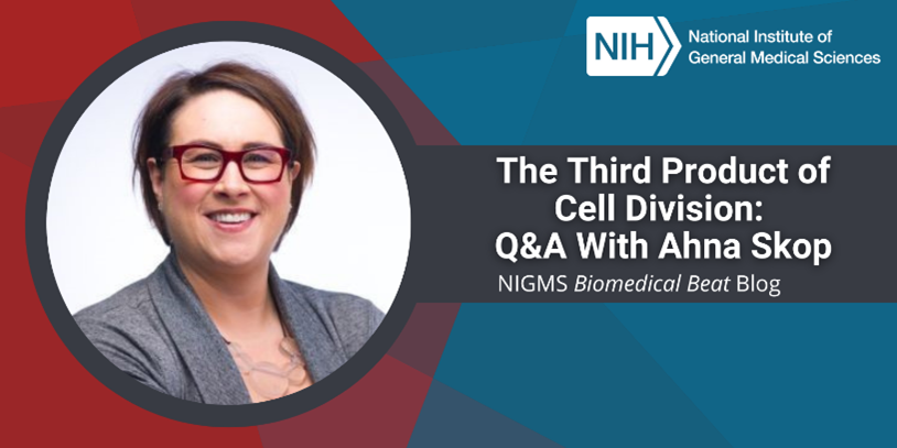
“Throughout my career, I’ve enjoyed studying topics that no one else seems to care about. I always tell people that I like searching through the scientific garbage bin for inspiration,” says Ahna Skop, Ph.D., a professor of genetics at the University of Wisconsin-Madison. We talked with her about the backyard experiment that helped her gain confidence in her scientific abilities, her career-long pursuit to better understand a detail about cell division that others had written off as unimportant, and her desire to build an accessible scientific community.
Q: How did you first become interested in science?
A: Middle school and high school science fairs had a big impact on me. I would develop my ideas, and with the help of my dad, build the experimental setup I needed to answer the scientific question. One of my experiments studied whether ants preferred to eat salt or sugar, so I poured small piles of both all over the backyard and took daily measurements of the height of the piles to figure out which type was shrinking faster. (Spoiler alert for those of you who might try this at home: They liked both but preferred the sugar to the salt.)
Both of my parents nurtured my curiosity, and as a young girl, it gave me the confidence I needed to see that I could become a scientist. Years later, that confidence became even more important when I saw how few women there were in science-related careers at the time.
Q: What was your educational path after high school?
A: I attended Syracuse University in New York as an undergraduate student, with a major in biology and a minor in ceramics. My parents were artists, so I was exposed to the world of art very early on, and I wanted to find a way to combine it with science. One of my dad’s famous quotes is: “Nature is my teacher.” So, I took inspiration from biology in my art and recreated what I saw in the electron microscope, such as a part of a beetle, in my sculptures for my senior ceramics thesis.
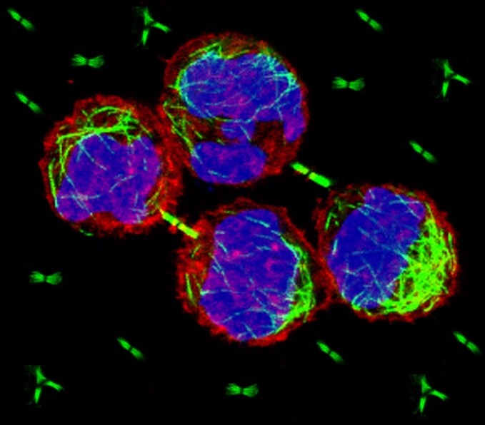
After I finished undergrad, I started at the University of Wisconsin-Madison for my Ph.D. under the mentorship of John White, Ph.D. I studied mitosis—the process by which cells divide into two new identical cells called daughter cells. We used the research organism Caenorhabditis elegans (C. elegans), which is a tiny roundworm. During the last stages of mitosis, an intercellular bridge develops between the newly formed cells. Within that bridge is a compartment called the midbody. The midbody gets pinched off and released as a large membrane-bound vesicle called the midbody remnant (MBR) in the final stage of mitosis, meaning that there are actually three products of mitosis: the two daughter cells and the MBR.
That’s when I first became passionate about the midbody. Previous research suggested that the midbody was unimportant and just a dumping ground for leftover junk the cell had needed during division, but I thought it was more important than that, based on what I saw during my Ph.D. studies. Many cell division proteins localize to the midbody, so I thought using this structure would be a great way to identify more proteins involved in cell division. I started a series of short postdoctoral positions with Rebecca Heald, Ph.D., and Barbara Meyer, Ph.D., at the University of California, Berkeley, and with John Yates, Ph.D., at Scripps Research in La Jolla, California, with the goal of learning more about the midbody, including what’s inside.
I cultured mammalian cells and collected the midbodies that they released. I then used proteomics techniques, such as mass spectrometry, on the midbodies to figure out what proteins were in them. Finally, using genetic engineering, I removed the genes that coded for the midbody-associated proteins from C. elegans to learn how their loss affected cell division. Most played roles in cytokinesis, the last step of mitosis, while a few were involved in functions after mitosis. We learned that the mammalian and C. elegans midbody proteins are more than 90 percent similar despite the evolutionary distance between the species. A proteome being highly preserved like this suggests that it has very important functions, because if an organism didn’t rely on it, we’d expect it to change significantly over time.
Q: What is your lab researching now?
A: My lab is continuing the work I started during my postdoctoral research to further understand midbodies and MBRs. My earlier proteomic studies found around 100 RNA-binding proteins in midbodies, but no one thought that RNA molecules themselves were actually important for mitosis at the time, so I decided to find out. In addition to the RNA-binding proteins, we determined that the midbody contains RNA and is a site of active translation—the process of making proteins based on the genetic information stored in RNA.
We sequenced the midbody RNA molecules and learned that many of them coded for transcription factors (proteins that control the creation of RNA molecules from DNA) important for the phase of the cell cycle after mitosis, including signals that tell cells to continue growing and dividing. We’ve also learned that cells can absorb MBRs and their cargo, making MBRs a unique type of information vesicle that can influence the activity of the cell that absorbs them.
Cancer cells not only produce a lot more MBRs than healthy cells do (because they divide more), but they also absorb more. If we learn that absorption of MBRs is involved in the spread of cancer throughout the body, researchers could design new medicines that block this process.
Q: What’s one of your main goals as a researcher?
A: One thing I’m working on all the time is how to be a better science communicator. I really love getting young kids excited about science. Sometimes I think I was given an eye for both science and art so that I could put the two together to help explain science to the public. The challenge is figuring out how to do that in a fun and accessible way. Currently, I’m working on a project with members of the blind and visually impaired community in the Madison area. We want to create tactile models that depict the cell cycle to make scientific imagery accessible to more students. All people who come to science are of value, and their perspectives are important for discovery, so it’s important for us to build a community that is accessible to all.
Dr. Skop’s research is supported by NIGMS through grant R01GM139695.