Category: Genes

Learning How Mosquito-Borne Viruses Use Knot-like RNA to Cause Disease

0 comments
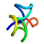
A knot-like structure in a section of RNA from a flavivirus
A knot-like structure in RNA enables flaviviruses to cause diseases like yellow fever, West Nile virus and dengue fever, which threaten roughly half the world’s population. Credit: Jeffrey Kieft.

Roughly half the world’s population is now at risk for mosquito-borne diseases other than malaria, such as yellow fever, West Nile virus and dengue fever. These three diseases are caused by flaviviruses, a type of virus that carries its genetic material as a single strand of RNA.

Flaviviruses have found a way not only to thwart our bodies’ normal defenses, but also to harness a human enzyme—paradoxically, one normally used to destroy RNA—to enhance their disease-causing abilities. A team of scientists led by Jeffrey Kieft at the University of Colorado at Denver found that flaviviruses accomplish both feats by bending and twisting a small part of their RNA into a knot-like structure.

The scientists set out to learn more about this unusual ability. First, they determined the detailed, three-dimensional architecture of the convoluted flaviviral RNA. Then, they examined several different variations of the RNA. In doing so, they pinpointed parts that are critical for forming the knot-like shape. If researchers can find a way to prevent the RNA from completing its potentially dangerous twist, they’ll be a step closer to developing a treatment for flaviviral diseases, which affect more than 100 million people worldwide.

This work also was supported by the National Institute of Allergy and Infectious Diseases and the National Cancer Institute.

Learn more:
University of Colorado News Release exit icon
Kieft Lab exit icon

The Inner Life of Nerve Cells

2 comments
“Before this research, we didn’t even know that neurons had this special mechanism to control neuropeptide function. This is why we do basic research. This is why it’s important to understand how neurons work, down to the subcellular and molecular levels.”—Kenneth Miller”

Nerve cells (neurons) in the brain use small molecules called neuropeptides to converse with each other. Disruption of this communication can lead to problems with learning, memory and other brain functions. Through genetic studies in a model organism, the tiny worm C. elegans, a team led by Kenneth Miller of the Oklahoma Medical Research Foundation has uncovered a previously unknown mechanism that nerve cells use to package, move and release neuropeptides. The researchers found that a protein called CaM kinase II, which plays many roles in the brain, helps control this mechanism. Neuropeptides in worms lacking CaM kinase II spilled out from their packages before they reached their proper destinations. A more thorough understanding of how neurons work, provided by studies like this, may help researchers better target drugs to treat memory disorders and other neurological problems in humans.

This work also was funded by NIH’s National Institute of Mental Health.

Learn more:
Oklahoma Medical Research Foundation News Release Exit icon

Two Proteins That Regulate Energy Use Play Key Role in Stem Cell Development

0 comments
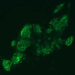
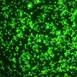
Stem cells. Credit: Julie Mathieu, University of Washington.
The protein HIF1 alpha is beneficial for creating induced pluripotent stem cells (green) from adult human cells. Credit: Julie Mathieu, University of Washington.

Hannele Ruohola-Baker and a team of researchers at the University of Washington recently discovered that two proteins responsible for regulating how cells break down glucose are also essential for stem cell development. The scientists showed that the proteins HIF1 alpha and HIF2 alpha are both required to reprogram adult human cells into pluripotent stem cells, which have the ability to mature into any cell type in the body. Taking a closer look at what each protein does on its own, the researchers found that HIF1 alpha was beneficial for reprogramming throughout the process, whereas HIF2 alpha was required at early stages but was detrimental at later stages of reprogramming. Because the two proteins also play a role in transforming normal cells into cancer cells, the findings could lead to future advances in cancer research.

Learn more:
University of Washington News Release Exit icon
Ruohola-Baker Lab Exit icon
Once Upon a Stem Cell Article from Inside Life Science
Learning About Cancer by Studying Stem Cells Article from Inside Life Science
Sticky Stem Cells Article from Inside Life Science

Basic Research Fuels Medical Advances

0 comments
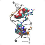
Genetic defect that causes myotonic dystrophy type 2 and used that information to design drug candidates to counteract the disease. Credit: Ilyas Yildirim, Northwestern University.
Scientists revealed a detailed image of the genetic change that causes myotonic dystrophy type 2 and used that information to design drug candidates to counteract the disease. Credit: Ilyas Yildirim, Northwestern University. View larger image

This image may look complicated, but it tells a fairly straightforward tale about basic research: Learning more about basic life processes can pave the way for medical and other advances.

In this example, researchers led by Matthew Disney of the Scripps Research Institute’s Florida campus focused on better understanding the structural underpinnings of myotonic dystrophy type 2, a relatively rare, inherited form of adult-onset muscular dystrophy. While this work is still in the preliminary stages, it may hold potential for someday treating the disorder.

Some 300,000 NIH-funded scientists are working on projects aimed at improving disease diagnosis, treatment and prevention, often through increasing understanding of basic life processes.

Read the complete Inside Life Science article.

Learning More About Our Partners in Digestion

0 comments
Bacteroides ovatus
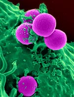
Bacteroides ovatus. Credit: Eric Martens, University of Michigan Medical School.

After eating, we don’t do all the work of digestion on our own. Trillions of gut bacteria help us break food down into the simple building blocks our cells need to function. New research from an international team co-led by Eric Martens of the University of Michigan Medical School has uncovered how a strain of beneficial gut bacteria, Bacteroides ovatus, digests complex carbohydrates called xyloglucans that are found in fruits and vegetables. The researchers traced the microorganism’s digestive ability to a single piece of the genome. They also examined a publicly available set of genomic data, which included information from both humans and their resident bacteria, and found that more than 90 percent of 250 adults harbored at least one Bacteroides strain with xyloglucan-digesting capabilities. These results underscore the importance of the bacteria to human health and nutrition.

This work also was funded by the National Institute of Diabetes and Digestive and Kidney Diseases.

Learn more:
University of Michigan News Release
University of Michigan Host Microbiome Initiative
Gut Reactions and Other Findings About Our Resident Microbes from Inside Life Science
Body Bacteria from Findings Magazine

Resetting Our Clocks: New Details About How the Body Tells Time

0 comments
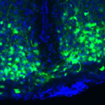
VIP in time-keeping brain cells
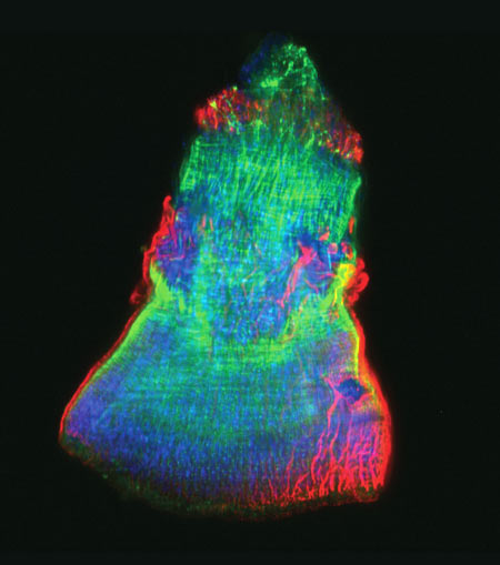
Boosting doses of a molecule called VIP (green) in time-keeping brain cells (blue) helped mice adjust quickly to major shifts in light-dark cycles. Credit: Cristina Mazuski in the lab of Erik Herzog, Washington University in St. Louis.

Springing clocks forward by an hour this Sunday, traveling across time zones, staring at a computer screen late at night or working the third shift are just a few examples of activities that can disrupt our daily, or circadian, rhythms. These roughly 24-hour cycles influence our physiology and behavior, and they’re driven by our body’s network of tiny timekeepers. If our daily routines fall out of sync with our body clocks, sleep, metabolic and other disorders can result.

Researchers funded by the National Institutes of Health have spent decades piecing together the molecular mechanisms of our biological clocks. Now, they’re building on that basic knowledge to better understand the intricate relationship among these clocks, circadian rhythms and physiology—and ultimately, find ways to manipulate the moving parts to improve our modern-day lives.

Continue reading this new Inside Life Science article

Animal Cells ‘Reach Out and Touch’ to Communicate

0 comments
Cytonemes in the fruit fly tracheal system.
Threadlike cytonemes (at right) convey signals between cells in the developing fruit fly tracheal system. Credit: Sougata Roy, University of California, San Francisco. View larger image

Scientists have long known that multicellular organisms use biological molecules produced by one cell and sensed by another to transmit messages that, for instance, guide proper development of organs and tissues. But it’s been a puzzle as to how molecules dumped out into the fluid-filled spaces between cells can precisely home in on their targets.

Using living tissue from fruit flies, a team led by Thomas Kornberg of the University of California, San Francisco, has shown that typical cells in animals can talk to each other via long, thin cell extensions called cytonemes (Latin for “cell threads”) that may span the length of 50 or 100 cells. The point of contact between a cytoneme and its target cell acts as a communications bridge between the two cells.

Until now, only nerve cells (neurons) were known to communicate this way. “This is an exciting finding,” says NIGMS’ Tanya Hoodbhoy. “Neurons are not the only ‘reach out and touch someone’ cells.”

This work also was funded by NIH’s National Heart, Lung, and Blood Institute.

Learn more:

UCSF News Release Exit icon

Meet Jasmine Johnson and Gabe Vela

0 comments
Jasmine Johnson and Gabriel 'Gabe' Vela
Jasmine Johnson and Gabriel “Gabe” Vela
Field: Genetics of sleep and obesity
Worked as researchers at: The Jackson Laboratory, Bar Harbor, Maine
Graduated from high school at: Rockdale Magnet School for Science and Technology in Conyers, Ga.
Now freshman at: Stanford University in Palo Alto, Calif. (Johnson) and Southern Polytechnic State University in Marietta, Ga. (Vela)
Fascinating fact: Johnson presented her research at the 2013 White House Science Fair
Credit: Joe Piergrossi

Jasmine Johnson and Gabe Vela might still be teenagers, but they are also seasoned scientists. It all started 3 years ago, when, as high school juniors, they took the research course Independent Studies in Computational Biology at The Jackson Laboratory in Bar Harbor, Maine. They were hooked. They continued to do research until they graduated, working part-time for 2 academic years and full-time for 2 summers.

They worked with statistical geneticist Gary Churchill, using computational biology to explore the relationship between sleep and obesity. They focused on finding genes that regulate sleep and understanding how sleep affects the body. One goal of the research is to tease out a genetic explanation for why sleep deprivation increases the risk of obesity.

Working in a lab “completely changed what I thought I was going to do with my life,” said Vela. “Now I’m going to focus more on research than anything else.”

For Johnson, the experience provided the opportunity to present her research at the 2013 White House Science Fair, where she hobnobbed with some political hot shots.

“It was an amazing experience,” she said. Having “important White House officials be interested in my project … inspired me.”

Johnson and Vela visited NIH a few months ago and talked with us about their research experiences, their lives and their future goals. Jasmine Johnson & Gabriel Vela on their experience as high school researchers at The Jackson Laboratory in Bar Harbor, Maine.

Learn More
Article Exit icon about Johnson and Vela and other young researchers, from The Jackson Laboratory’s magazine The Search.
Article about the work of Gary Churchill, from NIH’s Findings magazine.

Neanderthal DNA—Still Among Us

0 comments
Map of Europe and Asia showing the presumed range of where Neanderthals lived.
For tens of thousands of years, Neanderthals lived in Europe and Asia (presumed range shown in blue) and interbred with humans, passing on some DNA to present-day people. Credit: Ryulong, Wikimedia Commons.
View larger image

Many of us have some Neanderthal genes. Before Neanderthals went extinct about 30,000 years ago, they interbred with humans living in Europe and Asia. Today’s descendants of those pairings inherited about 2 percent of their genomes from the big-brained hominids.

A research group led by David Reich at Harvard Medical School recently completed an analysis to determine the extent and identity of Neanderthal DNA in modern-day human populations. The group found that many traits in present-day people—including skin characteristics and susceptibility to various diseases—can be traced to Neanderthal DNA.

It also appears that, genetically speaking, Neanderthals and humans weren’t completely compatible. Based on the uneven distribution of Neanderthal DNA in today’s genomes, the scientists concluded that many of the male offspring of Neanderthal-human unions were infertile. In the animal world, this phenomenon is known as hybrid infertility, where the offspring of a male from one subspecies and a female from another have low or no fertility.

Studying human genes passed down through Neanderthals—as well as regions of the human genome notably devoid of Neanderthal DNA—provides an increasingly complete picture of the genetic landscape that contributed to health, disease and diversity among humans today.

Learn more:
Harvard Medical School News Release
Reich Lab Exit icon

Targeting Toxic RNA Molecules in Muscular Dystrophy

0 comments
Genetic defect that causes myotonic dystrophy type 2 and used that information to design drug candidates to counteract the disease. Credit: Ilyas Yildirim, Northwestern University.
Scientists revealed a detailed image of the genetic defect that causes myotonic dystrophy type 2 and used that information to design drug candidates to counteract the disease. Credit: Ilyas Yildirim, Northwestern University. View larger image

Myotonic dystrophy type 2 (DM2) is a relatively rare, inherited form of adult-onset muscular dystrophy that has no cure. It’s caused by a genetic defect in which a short series of nucleotides—the chemical units that spell out our genetic code—is repeated more times than normal. When the defective gene is transcribed, the resulting RNA repeat forms a hairpin-like structure that binds to and disables a protein called MBNL1.

Now, research led by Matthew Disney of The Scripps Research Institute (TSRI), Florida Campus, has revealed the detailed, three-dimensional structure of the RNA defect in DM2 and used this information to design small molecules that bind to the aberrant RNA. These designer molecules, even in small amounts, significantly improved disease-associated defects in a cellular model of DM2, and thus hold potential for reversing the disorder.

Drugs that target toxic RNA molecules associated with diseases such as DM2 are few and far between, as developing such compounds is technically challenging. The “bottom-up” approach that the scientists used to design potent new drug candidates, by first studying in detail how the RNA structure interacts with small molecules, is unconventional, noted Jessica Childs-Disney of TSRI, who was lead author of the paper with Ilyas Yildirim of Northwestern University. But it may serve as an effective strategy for pioneering the use of small molecules to manipulate disease-causing RNAs—a central focus of the Disney lab.

This work also was funded by NIH’s National Cancer Institute.

Learn more:
The Scripps Research Institute News Release Exit icon
Disney Lab