Meet Brad Duerstock

0 comments
Brad Duerstock
Brad Duerstock
Fields: Neuroscience, assistive technology design
Works at: Purdue University, West Lafayette, IN
Hobbies: Gadgetry, architectural design
Bizarre collectible: Ecuadorian shrunken head (not a real one—it’s a replica made from goatskin)
Credit: Andrew Hancock, Purdue University

At the age of 18, Brad Duerstock had a devastating accident. A star member of his high school swim team, Duerstock hit his head during practice in a way that broke his neck and paralyzed all of his limbs. Today, he studies spinal cord injuries much like his own, investigating how the damage occurs and how it could possibly be repaired.

Duerstock has worked to make science accessible to people with disabilities, whether they use wheelchairs, as he does, or have visual or other impairments. For example, he has redesigned laboratory space to make it easier for people with disabilities to navigate and perform tasks.

“I like knowing that what I do can ultimately impact others,” Duerstock says.

Duerstock’s Findings

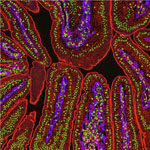
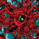
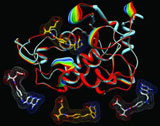
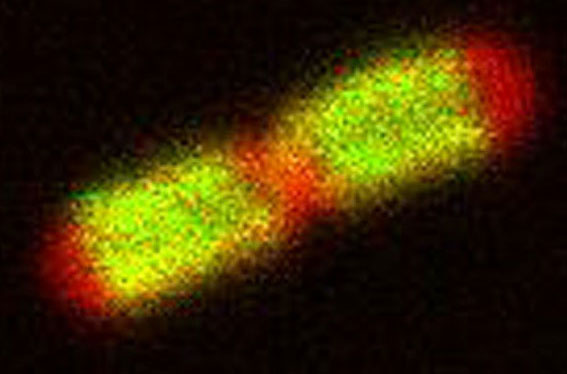
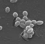
Much of Duerstock’s research deals with what occurs immediately following a nerve injury. In a spinal cord injury, nerve tissue becomes severed or dies. The immune response and bleeding in the injured area can cause extra damage to nerves in the spinal cord. Duerstock and his team have found that a molecule called acrolein is produced in spinal cord injuries and that it kills the nerves it encounters as it spreads around the injury site. They have been investigating a compound called polyethylene glycol (PEG), a polymer that could seal ruptured nerve cell membranes, possibly protecting nerve tissue from further damage immediately following a spinal cord injury.

Duerstock also founded and leads the Institute for Accessible Science (IAS), a community of scientists, students, parents and teachers whose goal is to promote better accommodations for people with disabilities who are studying or working in the sciences. The IAS looks into how to redesign lab spaces and equipment to increase accessibility for people with disabilities, particularly those with limited mobility or vision.

Although Duerstock originally wanted to be a doctor, he believes his true calling is in research. “The sense of discovery and the impact on others are big motivations for me,” says Duerstock. “Being a researcher, you might have a broader impact on society than you would as a practicing physician.”

Content adapted from the NIGMS Findings magazine article Opening Up the Lab.

Cool Video: How Bee Venom Toxin Kills Cells

0 comments
Credit: Huey Huang, Rice University.

Credit: Huey Huang, Rice University.

A new video, starring the toxin in bee venom, might help scientists design new drugs to combat bacterial infections. The video, by Rice University biophysicist Huey Huang Exit icon, condenses 6.5 minutes into less than a minute to show how the toxin, called melittin, destroys an animal or bacterial cell.

What looks like a red balloon is an artificial cell filled with red dye. Melittin molecules are colored green and float on the cell’s surface like twigs on a pond. As melittin accumulates on the cell’s membrane, the membrane expands to accommodate it. In the video, the membrane stretches into a column on the left.

When melittin levels reach a critical threshold, countless pinhole leaks burst open in the membrane. The cell’s vital fluids—red dye in the video—leak out through these pores. Within minutes, the cell collapses.

Many organisms use such a pore-forming technique to kill attacking bacterial cells. This research reveals molecular-level details of the strategy, bringing pharmaceutical scientists a step closer to harnessing it in the design of new antibiotics.

Chemist Phil Baran Joins “Genius” Ranks as MacArthur Fellow

1 comment
Cake decorated with a two-dimensional structure of the molecule, stephacidin B
When Baran’s research team succeeds in synthesizing an important natural product, the group sometimes celebrates with a cake decorated with a two-dimensional structure of the molecule. This molecule, stephacidin B, was isolated from a fungus and has anticancer properties. See images of other Baran lab cakes.

As a newly appointed MacArthur Fellow, Phil Baran is now officially a genius. The MacArthur award recognizes “exceptionally creative” individuals who have made significant contributions to their field and are expected to continue doing so. Baran, a synthetic organic chemist at Scripps Research Institute in La Jolla, Calif., was recognized today for “inventing efficient, scalable, and environmentally sound methods” for building, from scratch, molecules produced in nature. Many of these natural products have medicinal properties. Baran has already concocted a host of natural products, including those with the ability to kill bacteria or cancer cells. In addition to emphasizing the important pharmaceutical applications of his work, Baran embraces its creative aspects: “The area of organic chemistry is such a beautiful one because one can be both an artist and an explorer at the same time,” he said in the MacArthur video interview Exit icon.

Learn more:

NIGMS “Meet a Chemist” Profile of Baran
NIH Director’s Blog Post on Baran’s Recent Work

Protein May Help Reduce Intestinal Injury

5 comments
HB-EGF protein. Credit: National Center for Microscopy and Imaging Research.
HB-EGF has the potential to protect the intestines (magnified here) from different types of injury. Credit: National Center for Microscopy and Imaging Research.

Gail Besner of Nationwide Children’s Hospital and her research team recently found out how the HB-EGF growth factor protein could potentially aid the development of treatments for a number of conditions. Using model systems in two separate studies, the scientists discovered that HB-EGF could protect the intestines from injury by stimulating cell growth and movement and by decreasing substances formed upon intestinal injury that worsen the damage. They also showed that administration of mesenchymal stem cells could further shield the intestines from injury. Future treatments involving a combination of HB-EGF and stem cells could, for example, help cancer patients sustain fewer intestinal injuries resulting from radiation therapy.

This work also was funded by NIH’s National Institute of Diabetes and Digestive and Kidney Diseases.

Learn more:

Nationwide Children’s Hospital News Release Exit icon

How Some Bacteria Colonize the Gut

1 comment
A section of mouse colon with gut bacteria (center, in green). Credit: S. Melanie Lee, Caltech; Zbigniew Mikulski and Klaus Ley, La Jolla Institute for Allergy and Immunology.
A section of mouse colon with gut bacteria (center, in green) residing within a protective pocket. Credit: S. Melanie Lee, Caltech; Zbigniew Mikulski and Klaus Ley, La Jolla Institute for Allergy and Immunology.

Have you ever felt that your gut was trying to tell you something? The guts of germ-free mice have told scientists a few new things about our resident microorganisms. By studying a genus of bacteria called Bacteriodes that live in the gastrointestinal tract, Sarkis Mazmanian of the California Institute of Technology discovered how Bacteriodes species stake their claim in a mouse’s gut. Mazmanian and his colleagues introduced different species of Bacteriodes into germ-free mice to learn how the microbes competed and found that most of them peacefully co-existed. However, when microbes of a species that was already present were introduced, they couldn’t take up residence. Further investigation revealed that Bacteriodes species, due to a set of specific genes, can live in tiny pockets or “crypts” of the colon, where they are sheltered from antibiotics and infectious microbes passing through. Understanding how these microorganisms colonize could help devise ways to correct for abnormal changes in bacterial communities that are associated with disorders like inflammatory bowel disease.

This work also was funded by NIH’s National Institute of Diabetes and Digestive and Kidney Diseases.

Learn more:

California Institute of Technology News Release
Mazmanian Lab
Mazmanian Video Interview Exit icon

Making Strides in Genomic Engineering of Human Stem Cells

1 comment
Genetically engineered human stem cells. Credit: Jeff Miller, University of Wisconsin-Madison.
Genetically engineered human stem cells hold promise for basic biomedical research as well as for regenerative medicine. Credit: Jeff Miller, University of Wisconsin-Madison.

Human pluripotent stem cells (hPSCs) can multiply indefinitely and give rise to virtually all human cell types. Manipulating the genomes of these cells in order to remove, replace or correct specific genes holds promise for basic biomedical research as well as medical applications. But precisely engineering the genomes of hPSCs is a challenge. A research team led by Erik Sontheimer of Northwestern University and James Thomson of the Morgridge Institute for Research at the University of Wisconsin-Madison developed a technique that could be a great improvement over existing, labor-intensive methods. Their approach uses an RNA-guided enzyme from Neisseria meningitidis bacteria—part of a recently discovered bacterial immune system—to efficiently target and modify specific DNA sequences in the genome of hPSCs. The technique could eventually enable the repair or replacement of diseased or injured cells in people with some types of cancer, Parkinson’s disease and other illnesses.

This work also was funded by NIH’s National Center for Advancing Translational Sciences.

Learn more:

Thomson Bio Exit icon

Meet Galina Lepesheva

2 comments
Galina Lepesheva
Galina Lepesheva
Field: Biochemistry
Works at: Vanderbilt University, Nashville, TN
Born, raised and studied in: Belarus
To unwind, she: Reads, travels, spends time with her family

Galina Lepesheva knows that kissing bugs are anything but romantic. When the lights get low, these blood-sucking insects begin feasting—and defecating—on the faces of their sleeping victims. Their feces are often infected with a protozoan (a single-celled, eukaryotic parasite) called Trypanosoma cruzi that causes Chagas disease. Lepesheva has developed a compound that might be an effective treatment for Chagas. She has also tested the substance, called VNI, as a treatment for two related diseases—African sleeping sickness and leishmaniasis.

“This particular research is mainly driven by one notion: Why should people suffer from these terrible illnesses if there could be a relatively easy solution?” she says.

Lepesheva’s Findings

Currently, most cases of Chagas disease occur in rural parts of Mexico, Central America and South America. According to some estimates, up to 1 million people in the U.S. could have Chagas disease, and most of them don’t realize it. If left untreated, the infection is lifelong and can be deadly.

The initial, acute stage of the disease is usually mild and lasts 4 to 8 weeks. Then the disease goes dormant for a decade or two. In about one in three people, Chagas re-emerges in its life-threatening, chronic stage, which can affect the heart, digestive system or both. Once chronic Chagas disease develops, about 60 percent of people die from it within 2 years.

The Centers for Disease Control and Prevention (CDC) has targeted Chagas disease as one of five “neglected parasitic infections,” indicating that it warrants special public health action.

“Chagas disease does not attract much attention from pharmaceutical companies,” Lepesheva says. Right now, there are only two medicines to treat it. They are only available by special request from the CDC, aren’t always effective and can cause severe side effects.

Lepesheva’s research focuses on a particular enzyme, CYP51, that is the target of some anti-fungal medicines. If CYP51 can also act as an effective drug target for the parasites that cause Chagas, her work might help meet an important public health need.

CYP51 is found in all kingdoms of life. It helps produce molecules called sterols, which are essential for the development and viability of eukaryotic cells. Lepesheva and her colleagues are studying VNI and related compounds to examine whether they can block the activity of CYP51 in human pathogens such as protozoa, but do no harm to the enzyme in mammals. In other words, her goal is to cripple disease-causing organisms without creating side effects in infected humans or other mammals.

Lepesheva has tested the effectiveness of VNI on Chagas-infected mice. Remarkably, it has worked 100 percent of the time, curing both the acute and chronic stages of the disease. It acts by preventing the protozoan from establishing itself in the host’s body. If it is similarly effective in humans, VNI could become the first reliable treatment for Chagas disease.

NIH Director Blogs About NIGMS-Funded Research

0 comments
Antifolate drugs (bottom) work by blocking the folate receptor (top). Credit: Charles Dann III, Indiana University.
Caption: Antifolate drugs (bottom) work by blocking the folate receptor (top), starving cancer cells of an essential vitamin. Credit: Charles Dann III, Indiana University.

Within the last few weeks, NIH Director Francis Collins has blogged about several findings that NIGMS helped fund: the identification of a genetic link between hair color and melanoma risk and the solving of human folate receptor structures, which may aid the design of drugs for cancer and inflammatory diseases like rheumatoid arthritis and Crohn’s disease. Both advances are excellent examples of the value and impact of basic research. Want more examples? Check out Curiosity Creates Cures!

Cool Image: Tiny Bacterial Motor

2 comments
Phillip Klebba, Kansas State University.

Credit: Phillip Klebba, Kansas State University.

It looks like a fluorescent pill, but this image of an E. coli cell actually shows a new potential target in the fight against infectious diseases. The green highlights a protein called TonB, which is produced by many gram-negative bacteria, including those that cause typhoid fever, meningitis and dysentery. TonB lets bacteria take up iron from the host’s body, which they need to survive. New research from Phillip Klebba of Kansas State University and his colleagues shows how TonB powers iron uptake. When TonB spins within the cell envelope (the bacteria’s “skin”) like a tiny motor, it produces energy that lets another protein pull iron into the cell. This knowledge may lead to the development of antibiotics that block the motion of TonB, potentially stopping an infection in its tracks.

Learn more:
Kansas State University News Release Exit icon
Klebba Lab Exit icon
Other Cool Images

Gut Microbes Can Inactivate Cardiac Drugs

2 comments
Digoxin bacteria. Credit: CDC.

Bacteria in the gut can inactivate some of the medicines we take. Credit: CDC.

Doctors have known that a medication often prescribed to treat heart failure is inactivated by gut microbes, particularly a bacterial species called E. lenta. Now scientists have a better understanding of why. A Harvard University research team led by Peter Turnbaugh found that the heart drug digoxin turns on two E. lenta genes that help convert the drug into its inactive form, thereby making the medicine less effective. By measuring gene abundance, the scientists could reliably predict whether a microbial community could break down the drug. The researchers also identified a possible way to stop the process: add protein. Their studies using mice showed that a diet high in protein—and the amino acid, arginine, that helps E. lenta grow—increased digoxin absorption. These initial findings suggest that one day it may be possible to tailor digoxin therapy through diet modifications.

Learn more:
Harvard University News Release Exit icon
Turnbaugh lab Exit icon