Animal Cells ‘Reach Out and Touch’ to Communicate

0 comments
Cytonemes in the fruit fly tracheal system.
Threadlike cytonemes (at right) convey signals between cells in the developing fruit fly tracheal system. Credit: Sougata Roy, University of California, San Francisco. View larger image

Scientists have long known that multicellular organisms use biological molecules produced by one cell and sensed by another to transmit messages that, for instance, guide proper development of organs and tissues. But it’s been a puzzle as to how molecules dumped out into the fluid-filled spaces between cells can precisely home in on their targets.

Using living tissue from fruit flies, a team led by Thomas Kornberg of the University of California, San Francisco, has shown that typical cells in animals can talk to each other via long, thin cell extensions called cytonemes (Latin for “cell threads”) that may span the length of 50 or 100 cells. The point of contact between a cytoneme and its target cell acts as a communications bridge between the two cells.

Until now, only nerve cells (neurons) were known to communicate this way. “This is an exciting finding,” says NIGMS’ Tanya Hoodbhoy. “Neurons are not the only ‘reach out and touch someone’ cells.”

This work also was funded by NIH’s National Heart, Lung, and Blood Institute.

Learn more:

UCSF News Release Exit icon

Meet Jasmine Johnson and Gabe Vela

0 comments
Jasmine Johnson and Gabriel 'Gabe' Vela
Jasmine Johnson and Gabriel “Gabe” Vela
Field: Genetics of sleep and obesity
Worked as researchers at: The Jackson Laboratory, Bar Harbor, Maine
Graduated from high school at: Rockdale Magnet School for Science and Technology in Conyers, Ga.
Now freshman at: Stanford University in Palo Alto, Calif. (Johnson) and Southern Polytechnic State University in Marietta, Ga. (Vela)
Fascinating fact: Johnson presented her research at the 2013 White House Science Fair
Credit: Joe Piergrossi

Jasmine Johnson and Gabe Vela might still be teenagers, but they are also seasoned scientists. It all started 3 years ago, when, as high school juniors, they took the research course Independent Studies in Computational Biology at The Jackson Laboratory in Bar Harbor, Maine. They were hooked. They continued to do research until they graduated, working part-time for 2 academic years and full-time for 2 summers.

They worked with statistical geneticist Gary Churchill, using computational biology to explore the relationship between sleep and obesity. They focused on finding genes that regulate sleep and understanding how sleep affects the body. One goal of the research is to tease out a genetic explanation for why sleep deprivation increases the risk of obesity.

Working in a lab “completely changed what I thought I was going to do with my life,” said Vela. “Now I’m going to focus more on research than anything else.”

For Johnson, the experience provided the opportunity to present her research at the 2013 White House Science Fair, where she hobnobbed with some political hot shots.

“It was an amazing experience,” she said. Having “important White House officials be interested in my project … inspired me.”

Johnson and Vela visited NIH a few months ago and talked with us about their research experiences, their lives and their future goals. Jasmine Johnson & Gabriel Vela on their experience as high school researchers at The Jackson Laboratory in Bar Harbor, Maine.

Learn More
Article Exit icon about Johnson and Vela and other young researchers, from The Jackson Laboratory’s magazine The Search.
Article about the work of Gary Churchill, from NIH’s Findings magazine.

Neanderthal DNA—Still Among Us

0 comments
Map of Europe and Asia showing the presumed range of where Neanderthals lived.
For tens of thousands of years, Neanderthals lived in Europe and Asia (presumed range shown in blue) and interbred with humans, passing on some DNA to present-day people. Credit: Ryulong, Wikimedia Commons.
View larger image

Many of us have some Neanderthal genes. Before Neanderthals went extinct about 30,000 years ago, they interbred with humans living in Europe and Asia. Today’s descendants of those pairings inherited about 2 percent of their genomes from the big-brained hominids.

A research group led by David Reich at Harvard Medical School recently completed an analysis to determine the extent and identity of Neanderthal DNA in modern-day human populations. The group found that many traits in present-day people—including skin characteristics and susceptibility to various diseases—can be traced to Neanderthal DNA.

It also appears that, genetically speaking, Neanderthals and humans weren’t completely compatible. Based on the uneven distribution of Neanderthal DNA in today’s genomes, the scientists concluded that many of the male offspring of Neanderthal-human unions were infertile. In the animal world, this phenomenon is known as hybrid infertility, where the offspring of a male from one subspecies and a female from another have low or no fertility.

Studying human genes passed down through Neanderthals—as well as regions of the human genome notably devoid of Neanderthal DNA—provides an increasingly complete picture of the genetic landscape that contributed to health, disease and diversity among humans today.

Learn more:
Harvard Medical School News Release
Reich Lab Exit icon

Cool Image: Visualizing Viral Activity

0 comments
Viral RNA (red) in an RSV-infected cell. Credit: Eric Alonas and Philip Santangelo, Georgia Institute of Technology and Emory University. 

Viral RNA (red) in an RSV-infected cell. Credit: Eric Alonas and Philip Santangelo, Georgia Institute of Technology and Emory University.

What looks like a colorful pattern produced as light enters a kaleidoscope is an image of a cell infected with respiratory syncytial virus (RSV) illuminated by a new imaging technology. Although relatively harmless in most children, RSV can lead to bronchitis and pneumonia in others. Philip Santangelo of the Georgia Institute of Technology and Emory University, along with colleagues nationwide, used multiply-labeled tetravalent RNA imaging probes (MTRIPS) to observe the entry, assembly and replication of RSV inside a living cell. Once introduced into RSV-plagued cells, the MTRIPS latched onto the viral RNA (in the image, red) without altering the level of infectivity. This led to fluorescent RSV viral particles that let the researchers track the viral RNA in host cells and better understand what the virus was doing. The knowledge gained from this new technique might aid in the development of RSV antiviral drugs and possibly a vaccine. Scientists could also one day use the imaging approach to study other RNA viruses, such as the flu and Ebola.

Learn more:
Georgia Tech News Release Exit icon

Transporter Protein Dance Moves

2 comments
Animation depicts the changes that allow a protein transporter to do its job.

In this video, Emad Tajkhorshid of the University of Illinois at Urbana-Champaign explains the molecular dance of transporter proteins, molecules that move substances across the cell membrane.

In this video, Emad Tajkhorshid of the University of Illinois at Urbana-Champaign explains the molecular dance of ABC transporters, a family of molecular machines that utilize ATP to move substances across the cell membrane. Tajkhorshid and his team recently used computational methods to map the movements between two known structural models of MsbA, a bacterial version of a transporter in human cells that helps to export anti-cancer drugs. They then described the individual steps of the molecular motions during the transport cycle. Understanding the process at such a detailed level could suggest new targets for treating a range of diseases, including some drug-resistant cancers that often make more transporter proteins to kick out medications meant to kill them.

Learn more:
University of Illinois at Urbana-Champaign News Release Exit icon
Tajkhorshid Lab

An Experimental Contact Lens to Prevent Glaucoma-Induced Blindness

3 comments
Contact lens. Credit: Peter Mallen, Massachusetts Eye and Ear Laboratory/Kohane Laboratory, Boston Children's Hospital.
An experimental contact lens design releases a glaucoma medicine at a steady rate for up to a month. Credit: Peter Mallen, Massachusetts Eye and Ear Laboratory/Kohane Laboratory, Boston Children’s Hospital.

Like a miniature donut stuffed inside a tiny pita pocket, a common glaucoma medicine held within a biomaterial ring is sandwiched inside this contact lens. In laboratory experiments, the lens, which can also correct vision, releases the eyesight-saving medication at a steady rate for up to a month. Its construction offers numerous potential clinical advantages over the standard glaucoma treatment and may have additional applications, such as delivering anti-inflammatory drugs or antibiotics to the eye. Led by Daniel Kohane and Joseph Ciolino at Harvard Medical School, the researchers who developed the lens are now gearing up to test its effectiveness in additional laboratory studies. They hope a Phase I clinical trial to evaluate the safety and ability of the lens to reduce pressure in the human eye could begin in about a year.

This work also was funded by NIH’s National Eye Institute.

Learn more
:
NEI Glaucoma Awareness Month Resources

Targeting Toxic RNA Molecules in Muscular Dystrophy

0 comments
Genetic defect that causes myotonic dystrophy type 2 and used that information to design drug candidates to counteract the disease. Credit: Ilyas Yildirim, Northwestern University.
Scientists revealed a detailed image of the genetic defect that causes myotonic dystrophy type 2 and used that information to design drug candidates to counteract the disease. Credit: Ilyas Yildirim, Northwestern University. View larger image

Myotonic dystrophy type 2 (DM2) is a relatively rare, inherited form of adult-onset muscular dystrophy that has no cure. It’s caused by a genetic defect in which a short series of nucleotides—the chemical units that spell out our genetic code—is repeated more times than normal. When the defective gene is transcribed, the resulting RNA repeat forms a hairpin-like structure that binds to and disables a protein called MBNL1.

Now, research led by Matthew Disney of The Scripps Research Institute (TSRI), Florida Campus, has revealed the detailed, three-dimensional structure of the RNA defect in DM2 and used this information to design small molecules that bind to the aberrant RNA. These designer molecules, even in small amounts, significantly improved disease-associated defects in a cellular model of DM2, and thus hold potential for reversing the disorder.

Drugs that target toxic RNA molecules associated with diseases such as DM2 are few and far between, as developing such compounds is technically challenging. The “bottom-up” approach that the scientists used to design potent new drug candidates, by first studying in detail how the RNA structure interacts with small molecules, is unconventional, noted Jessica Childs-Disney of TSRI, who was lead author of the paper with Ilyas Yildirim of Northwestern University. But it may serve as an effective strategy for pioneering the use of small molecules to manipulate disease-causing RNAs—a central focus of the Disney lab.

This work also was funded by NIH’s National Cancer Institute.

Learn more:
The Scripps Research Institute News Release Exit icon
Disney Lab

Meet Dave Cummings

0 comments
Dave Cummings. Credit: Marcus Emerson, PLNU.
Dave Cummings
Field: Environmental microbiology
Works at: Point Loma Nazarene University, San Diego, Calif.
Hobbies: Hiking, backpacking, fly-fishing
Dream home: One that doesn’t need a lot of work
Credit: Marcus Emerson, PLNU

In college, as a pre-med student majoring in biology and chemistry, Dave Cummings grew frustrated with the traditional “cookbook” approach to doing labs in his science classes. Turned off by having to follow step-by-step lab procedures that had little to do with scientific discovery, Cummings changed his major to English literature. Studying literature, he says, “helped me find myself” and taught him to think critically.

Ultimately, Cummings says, “I came to realize that it wasn’t the practice of medicine that got me excited, but the science behind it all.” He decided to pursue a graduate degree in biology and—after “knocking on a lot of doors”—was accepted at the University of Idaho, where he earned his master’s and doctoral degrees and discovered his passion for microbiology.

Today, Cummings applies his critical thinking skills to his work as professor of biology back at his alma mater, Point Loma Nazarene University (PLNU), a small university focused on undergraduate education. There he studies the role of urban storm water in spreading genes for antibiotic resistance in natural environments, and pursues his enthusiasm for training the next generation of scientists. He enlists his students in his research, giving them what he calls “real, live, on-the-ground” research experience that relatively few undergraduate students at larger universities receive.

Cummings’ Findings

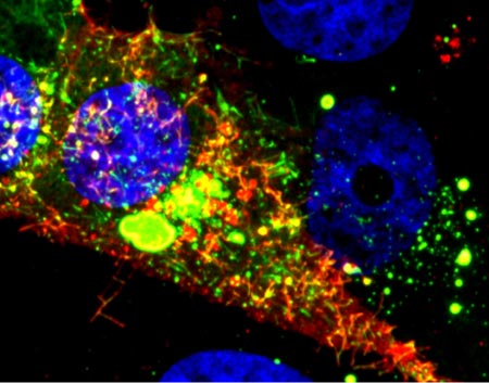
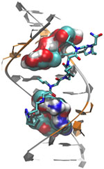
Antibiotic resistance, which can transform once-tractable bacterial infections into diseases that are difficult or impossible to treat, is a major public health challenge of the 21st century. The most common way that bacteria become drug resistant is by acquiring genes that confer antibiotic resistance from other bacteria. Often, such drug resistance genes are found on small, circular pieces of DNA called plasmids that are readily passed from one species of bacteria to another.

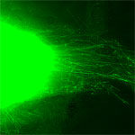
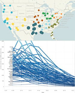
Urban wetlands provide ideal conditions for bacteria to mingle, swap genes and spread antibiotic resistance, notes Cummings. He focuses on the wetlands around San Diego, which act as a giant mixing bowl for storm runoff, human sewage, animal waste, naturally occurring plant and soil microorganisms, and plasmids indigenous to the wetlands.

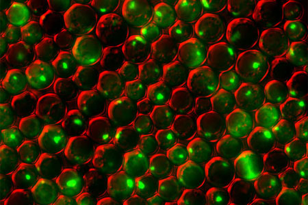
Cummings and his students examine sediment samples from these wetlands in search of plasmids that carry resistance genes. They’ve found that during winter rains, the San Diego wetlands receive runoff containing antibiotic-resistant bacteria and plasmids, which can persist at low, but detectable, levels into the dry summer months.

“We know that urban storm water carries with it a lot of antibiotic-resistant bacteria, and along with that the DNA instructions [or genes] that code for the resistance,” says Cummings. “We have solid evidence of genes encoding resistance to clinically important antibiotics washing … into coastal wetlands in San Diego.”

“We’re trying to understand the scope of the problem, and ultimately what threat that poses to human health,” he says. His concern is that the drug-defying bacterial genes will accumulate in the wetlands, and then “[find] their way back to us, where they will augment and amplify the problem of resistance.”

Precisely how resistance genes might move from the environment into people is not yet known. One way this could occur is through direct contact with contaminated water or sediment by anglers, swimmers, surfers and other recreational users. Fish, birds and insects could also transmit resistance genes from contaminated wetlands to humans.

“There is good evidence elsewhere that birds are important vectors of drug-resistant pathogens, and this is my favorite possibility,” says Cummings. “Hopefully someday we can test that hypothesis.”

Cummings’ studies, done in collaboration with Ryan Botts at PLNU and Eva Top at the University of Idaho, could reveal antibiotic resistance genes with the potential to move into new species of disease-causing bacteria and back into human populations. By identifying these genes and raising awareness of the problem, he hopes to aid future efforts to mitigate the spread of antibiotic resistance.

Local Flu Forecasts Posted on New Web Site

0 comments
Incidence of influenza during the week starting 12/29/2013 (top); influenza incidence forecasts for selected cities (bottom). Credit: Columbia Prediction of Infectious Diseases.

News articles this weekend reported an uptick in flu cases in many parts of the country. When will your area be hardest hit? Infectious disease experts at Columbia University have launched an influenza forecast Web site that gives weekly predictions for rates of flu infection in 94 U.S. cities. The predictions indicate the number of cases in Chicago; Atlanta; Washington, D.C.; and Los Angeles will peak this week, with New York City, Boston, Miami and Providence peaking in following weeks. The forecasts are updated every Friday afternoon, so check back then for any changes.

The forecasting approach, which adapts techniques used in modern weather prediction, relies on real-time observational data of people with influenza-like illness, including those who actually tested positive for flu. The researchers have spent the last couple of years developing the forecasting system and testing it—first retrospectively predicting flu cases from 2003-2008 in New York City and then in real time during the 2012-2013 influenza season in 108 cities.

“People have become acclimated to understanding the capabilities and limitations of weather forecasts,” said Jeffrey Shaman, who’s led the flu forecasting project. “Making our forecasts available on the Web site will help people develop a similar familiarity and comfort.” Shaman and his team are hoping that, just as rainy forecasts prompt more people to carry umbrellas, an outlook for high influenza activity may motivate them to get vaccinated and practice other flu-prevention measures.

This work also was funded by NIH’s National Institute of Environmental Health Sciences.

Learn more:

Columbia University News Release

Cool Images: Holiday Season Cells

0 comments
Yeast cells deficient in zinc and the Tsa1 protein have protein tangles. Credit: Colin MacDiarmid and David Eide, University of Wisconsin-Madison.

Yeast cells deficient in zinc and the Tsa1 protein have protein tangles. Credit: Colin MacDiarmid and David Eide, University of Wisconsin-Madison.

Just in time for the holidays, we’ve wrapped up a few red and green cellular images from basic research studies. In this snapshot, we see a group of yeast cells that are deficient in zinc, a metal that plays a key role in creating and maintaining protein shape. The cells also lack a protein called Tsa1, which normally keeps proteins from sticking together. Green areas highlight protein tangles caused by the double deficiency. Red outlines the cells. Protein clumping plays a role in many human diseases, including Parkinson’s and Alzheimer’s, so knowledge of why it happens—and what prevents it in healthy cells—could aid the development of treatments.

See more festive images!