Tag: Cool Videos

Interview With a Scientist: Thomas O’Halloran, Metal Maestro

2 comments

Inside our bodies is a surprising amount of metal. Not enough to set off the scanners at the airport or make us rich, but enough to fill each of our cells with billions of metal ions, including calcium, iron, copper and zinc. These ions facilitate critical biological functions.

However, too much of any metal can be toxic, while too little can cause disease. Our cells carefully monitor and control their metal content using a whole series of proteins that bind, sense and transport metal ions.

Keeping tabs on why and how metals flow into and out of our cells is a passion of Thomas O’Halloran Exit icon, professor of chemistry and molecular biosciences at Northwestern University in Illinois. For the past three decades, O’Halloran has investigated how fluctuations in the amount of metal ions inside cells influence gene expression, cell growth and other vital functions. Using a variety of approaches, he has uncovered new types of proteins that bind metal ions and investigated the role that imbalances in these ions play in a number of disease-related physiological processes.

One recent focus of his studies has been how zinc regulates oocyte (egg cell) maturation and fertilization. Ultimately, his work could help us better understand infertility, cancer and certain bacterial infections.

Interview With a Scientist: Laura Kiessling, Carbohydrate Scientist

4 comments

The outside of every cell on Earth—from the cells in your body to single-celled microorganisms—is blanketed with a coat of carbohydrates, or sugar molecules, that extend from the cell surface, branching off and bending as they interface with the extra-cellular space. The specific patterns in which these carbohydrates are arranged serve as an ID code that help cells recognize each other. For example, human liver cells have one pattern, while human red blood cells another. Certain diseases can even alter the pattern of surface carbohydrates, which is one way the body can recognize damaged cells. On foreign cells, including invading bacteria such as Streptococcus pneumoniae, the carbohydrate coat is even more distinct.

Laura Kiessling, a professor of chemistry at the University of Wisconsin, Madison, studies how carbohydrate coats are assembled and how cells use these coats to tell friend from foe. The implications of her research suggest strategies for targeting tumors, fighting diseases of inflammation and, as she discusses in this video, developing new classes of antibiotics.

Cool Video: Watching Bacteria Turn Virulent

1 comment

Researchers created an apparatus to study quorum sensing, a communication system that allows some bacteria to cause dangerous infections. Their findings suggest that blocking bacterial communication might lead to a new way to combat such infections. Credit: Minyoung Kevin Kim and Bonnie Bassler, Princeton University.

If you’ve ever felt a slimy coating on your teeth, scrubbed grime from around a sink drain or noticed something growing between the tiles of a shower, you’ve encountered a biofilm. Made up of communities of bacteria and other microorganisms, biofilms thrive where they can remain moist and relatively undisturbed. As they enlarge, biofilms can block narrow passages like medical stents, airways, pipes or intestines. Continue reading “Cool Video: Watching Bacteria Turn Virulent”

The Proteasome: The Cell’s Trash Processor in Action

2 comments

Our cells are constantly removing and recycling molecular waste. On the occasion of Earth Day, we put together this narrated animation to show you one way cells process their trash. The video features the proteasome, a cellular machine that breaks down damaged or unwanted proteins into bits that the cell can re-use to make new proteins. For this reason, the proteasome is as much a recycling plant as it is a garbage disposal.

For more details about the proteasome and other cellular disposal systems, check out our article How Cells Take Out the Trash.

Evolution and Health: A Conversation with Evolutionary Geneticist Dr. Dan Janes on the Occasion of Charles Darwin’s Birthday

5 comments

Today, February 12, is Darwin Day—an occasion to recognize the scientific contributions of 19th-century naturalist Charles Darwin. In this video, our own evolutionary geneticist, Dan Janes, answers questions about Darwin and the role of evolution in health and biomedicine.

For more details about evolution and you, check out our articles Evolution and Health and Everyday Evolution.

How a Cell Knows Friend From Foe

1 comment

We asked the heads of our scientific divisions to tell us about some of the big questions in fundamental biomedical science that researchers are investigating with NIGMS support. This article is the first in an occasional series that will explore these questions and explain how pursuing the answers could advance understanding of important biological processes.

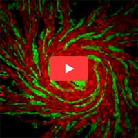
Video screen shot showing different strains of amoeba cells in red and green.
This video shows different strains of amoeba cells in red and green. As cells move toward one another, they use two sets of proteins to recognize others from the same strain. When close relatives meet, their proteins match and the cells join together to form a multicellular structure. When cells from different strains meet, their proteins don’t match, so they can’t aggregate. Credit: Shigenori Hirose, Baylor College of Medicine.

Cells are faced with many decisions: When’s the best time to produce a new protein? To grow and split into two? To treat another cell as an invader? Scientists are working to understand how cells make these and many other decisions, and how these decisions contribute to health and disease.

An active area of research on cell decisions focuses on allorecognition, the ability of an organism to distinguish its own cells from those of another. Immune cells use a system called the major histocompatibility complex (MHC) to identify which cells belong to the body and which are foreign. The particular set of MHC proteins on the outer surface of a cell helps immune cells decide whether it does not belong and should be attacked.

But the system isn’t perfect. Invading pathogens can go undetected, and the body can mistake its own cells for intruders. Continue reading “How a Cell Knows Friend From Foe”

Unprecedented Views of HIV

0 comments

Visualizations can give scientists unprecedented views of complex biological processes. Here’s a look at two new ones that shed light on how HIV enters host cells.

Animation of HIV’s Entry Into Host Cells

Screen shot of the video
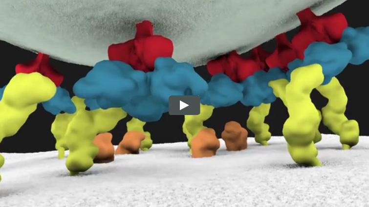
This video animation of HIV’s entry into a human immune cell is the first one released in Janet Iwasa’s current project to visualize the virus’ life cycle. As they’re completed, the animations will be posted at http://scienceofhiv.org Exit icon.

We previously introduced you to Janet Iwasa, a molecular animator who’s visualized complex biological processes such as cells ingesting materials and proteins being transported across a cell membrane. She has now released several animations from her current project of visualizing HIV’s life cycle Exit icon. The one featured here shows the virus’ entry into a human immune cell.

“Janet’s animations add great value by helping us consider how complex interactions between viruses and their host cells actually occur in time and space,” says Wes Sundquist, who directs the Center for the Structural Biology of Cellular Host Elements in Egress, Trafficking, and Assembly of HIV Exit icon at the University of Utah. “By showing us how different steps in viral replication must be linked together, the animations suggest hypotheses that hadn’t yet occurred to us.”

Continue reading “Unprecedented Views of HIV”

Zinc’s Role in Healthy Fertilization

0 comments
Screen shot of the video
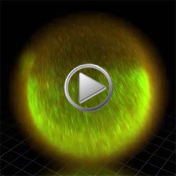
Fluorescent sensors at the cell surface show zinc-rich packages being released from the egg during fertilization. Credit: Northwestern Visualization.

Whether aiding in early growth and development, ensuring a healthy nervous system or guarding the body from illness, zinc plays an important role in the human body.

Husband-and-wife team, Thomas O’Halloran and Teresa Woodruff, plus other researchers at Northwestern University, evaluated the role that zinc plays in healthy fertilization. The study revealed how mouse eggs gather and release billions of zinc atoms at once in events called zinc sparks. These fluxes in zinc concentration are essential in regulating the biochemical processes that facilitate the egg-to-embryo transition.

The scientists developed a series of techniques to determine the amount and location of zinc atoms during an egg cell’s maturation and fertilization as well as in the following two hours. Special imaging methods allowed the researchers to also visualize the movement of zinc sparks in three dimensions.

Continue reading “Zinc’s Role in Healthy Fertilization”

Meet Janet Iwasa

0 comments
Janet Iwasa
Credit: Janet Iwasa
Janet Iwasa
Fields: Cell biology and molecular animation
Works at: University of Utah
Raised in: Indiana and Maryland
Studied at: University of California, San Francisco, and Harvard Medical School
When not in the lab she’s: Keeping up with her two preschool-aged sons
Something she’s proud of that she’ll never try again: Baking a multi-tiered wedding cake, complete with sugar flowers, for a friend’s wedding.

Janet Iwasa wouldn’t have described herself as an artistic child. She didn’t carry around a sketch pad, pencils or paintbrushes. But she remembers accompanying her father, a scientist at the National Institutes of Health, to his lab on the weekends. She’d spend hours doodling in a drawing program on his old Macintosh computer while he worked on experiments.

“I always remember wanting to be a scientist, and that’s probably highly inspired by my dad,” says Iwasa. Her early affinity for art and technology set her on an unusual career path to become a molecular animator. A typical work day now finds her adapting computer programs originally designed to bring characters like Buzz Lightyear to life to help researchers probe complicated, dynamic interactions within cells.

Iwasa’s interest in animation was sparked when she was a graduate student in cell biology, studying a protein called actin, which helps cells to move and change shape. At the time, the only visual representations she had of actin networks were flat, two-dimensional drawings on paper. When she saw an animation of the dynamic movement of a molecule called kinesin, she thought, “Why are we relying on oversimplified, static illustrations [of molecules], when we can be doing something like this video?”

Within a year, she was taking an animation class at a local college. She quickly realized that she would need more intensive instruction to be able to animate complex biological processes. A few summers later, she flew to Hollywood for a 3-month training program in industry-standard animation technology.

The oldest student in that course—and the only woman—Iwasa immediately began thinking about how to adapt a standard animator’s toolkit to illustrate the inner life of cells. A technique used to create the effect of human hair blowing in the wind could also show the movement of an RNA molecule. A chunk of computer code used to make the facets of a soccer ball fall apart and come back together in a different order could be adapted to model virus assembly and disassembly.

Her Findings

Following her training, Iwasa spent 2 years as a National Science Foundation Discovery Corps fellow, producing the Exploring Life’s Origins exhibit with the Boston Museum of Science and the Szostak Lab at Massachusetts General Hospital/Harvard Medical School. As part of the multi-media exhibit, she created animations to illustrate how the simplest living organisms may have evolved on early Earth.

Since then, Iwasa has helped researchers model such complex actions as how cells ingest materials, how proteins are transported across a cell membrane, and how the motor protein dynein helps cells divide.

Screenshot from the video that shows how a protein called clathrin forms a cage-like container that cells use to engulf and ingest materials
Iwasa developed this video to show how a protein called clathrin forms a cage-like container that cells use to engulf and ingest materials.

Iwasa calls her animations “visual hypotheses”: The end results may be beautiful, but the process of animation itself is what encapsulates, clarifies and communicates the science.

“It’s really building the animated model that brings insights,” she says. “When you’re creating an animation, you’re really grappling with a lot of issues that don’t necessarily come up by any other means. In some cases, it might raise more questions, and make people go back and do some more experiments when they realize there might be something missing” in their theory of how a molecular process works.

Now she’s working with an NIH-funded research team at the University of Utah to develop a detailed animation of how HIV enters and exits human immune cells.

Abbreviated CHEETAH, the full name of the group is the Center for the Structural Biology of Cellular Host Elements in Egress, Trafficking, and Assembly of HIV.

“In the HIV life cycle, there are a number of events that aren’t really well understood, and people have different ideas of how things happen,” says Iwasa. She plans to animate the stages of viral infection in ways that reflect different proposals for how the process works, to give researchers a new way to visualize, communicate—and potentially harmonize—their hypotheses.

The full set of Iwasa’s HIV-related animations will be available online as they are completed, at https://scienceofhiv.org, with the first set launching in the fall of 2014.

Learn more:
Janet Iwasa’s TED Talk: How animations can help scientists test a hypothesis
Janet Iwasa’s 3D model of an HIV particle was a winner in the 2014 BioArt contest sponsored by Federation of American Societies for Experimental Biology
NIH Director’s blog post about Iwasa and her HIV video animation

Cool Video: How a Microtubule Builds and Deconstructs

0 comments

A microtubule, part of the cell’s skeleton, builds and deconstructs. Credit: Eva Nogales lab, University of California, Berkeley.

In this animation, tubulin proteins snap into place like Lego blocks to build a microtubule, part of the cell’s skeleton. When construction ends, this long hollow cylinder falls to pieces from its top end. The breakdown is critical for many basic biological processes, including cell division, when rapidly shortening microtubules pull chromosomes into each daughter cell.

Until recently, scientists didn’t know exactly what drove microtubules to fall apart. A research team led by Eva Nogales of the Lawrence Berkeley National Laboratory and the University of California, Berkeley, now has an explanation.

Using high-powered microscopy, the scientists peered into the structure of a microtubule and found how a chemical reaction puts the stacking tubulin proteins under intense strain. The only thing keeping the proteins from springing apart is the pressure from the addition of more tubulin. So when assembly stops, the microtubule deconstructs.

The team also learned that Taxol, a common cancer drug, relieves this tension and allows microtubules to remain intact indefinitely. With microtubules frozen in place, a cancer cell cannot divide and eventually dies.

Because of this research, scientists now better understand both the success behind a common cancer drug and the molecular basis underlying the workings of microtubules.

Learn more:
University of California, Berkeley News Release Exit icon
Nogales Lab