Tag: RNA

Meet Scott Poethig

1 comment
Scott Poethig
Fields: Plant biology, cell and developmental biology, genetics
Works at: University of Pennsylvania
Studied at: College of Wooster, Yale University
Favorite musicians: Nick Drake and Bruce Springsteen
High school job: Radio D.J.
Favorite book: “The Little Prince,” by Antoine de Saint-Exupéry

When Scott Poethig signed up for a developmental biology course in his senior year of college, he expected to learn how organisms transition from single cells to juveniles to adults. He did not expect to learn just how much scientists still didn’t know about this process.

“It was the first course I had taken as an undergraduate where I felt that I could ask a question that there wasn’t an answer to already,” he recalls. “I thought, ‘Wow! This is amazing.’”

Poethig already had an interest in plant biology and an independent research project studying corn viruses. He immediately saw the potential in combining his knowledge of plants with his questions about how organisms grow. “There seemed to be a lot of low hanging fruit in plant development,” he says.

Today, Poethig is the head of a plant development lab at the University of Pennsylvania. His work probes the complex molecular mechanisms that drive the transition from a young seedling to an adult plant that hasn’t yet produced seeds.

“The analogous period in human development is the interval between birth and puberty,” he explains. “People think of puberty as the major developmental transition in postnatal human development, but a lot of change happens before that point.”

His Findings

Poethig discovered that for the mustard plant Arabidopsis, a model organism frequently studied by geneticists, change begins early. Before these plants begin to flower—a sign of reproductive maturity—they undergo a process of vegetative maturation. In Arabidopsis, Poethig found that juvenile plants can be distinguished from adult plants by where hairs are produced on a leaf. Juvenile plants only produce hairs on the upper surface of the leaf, whereas adult plants produce leaves with hairs on both the upper and lower surfaces.

By studying mutant Arabidopsis plants where the adult pattern of hair development is either delayed or advanced, Poethig identified microRNAs as key players in this developmental transition.

MicroRNA molecules commonly block the expression of specific genes. Poethig found that in Arabidopsis, a type of microRNA prevents development. Young plants have high levels of this microRNA and cannot fully mature. When those levels drop, plants progress to adulthood.

MicroRNAs similarly control development in the nematode C. elegans. Scientists study the genetics of this tiny worm to better understand related developmental processes in more complex organisms. Because plants also use microRNAs to regulate development, Poethig’s discoveries may contribute to our understanding of how these molecules govern development in animals, including humans.

Poethig now wants to learn what determines the timing of developmental changes. He asks: “Why do microRNA levels drop? What’s the signal that causes that? What is the plant measuring?” His current hypothesis: sugar.

In a recent study, he found that giving plants additional sugar reduced microRNA levels and sped up development. Meanwhile, mutant plants that couldn’t produce enough sugar on their own through photosynthesis had increased microRNA levels and delayed development compared to normal plants.

This research may one day advance our understanding of how nutrition and genetics interact to affect human development. “In essentially all organisms, aging and the timing of developmental processes are strongly affected by nutrition,” Poethig explains. “In humans, childhood obesity is sometimes associated with early puberty, and it is important to understand the molecular basis for this effect.”

Poethig believes that studying microRNAs in plants may also lead to discoveries in human genetics outside of developmental biology. “MicroRNAs control a wide range of gene activity in plants and animals,” Poethig explains. “In humans, these molecules control the activity of as many as 30 percent of our genes. So understanding how microRNAs work in plants could help us understand their function in humans.”

Besides studying the Arabidopsis plants in his lab, Poethig also studies the plants in his kitchen, and uses his fascination with the history, culture and politics of food to excite others about science. Watch video.

Field Focus: Precision Gene Editing with CRISPR

0 comments
Bacterial cells infected by viruses.
Bacterial cells can be infected by viruses (shown in red and purple) and have evolved ways to defend themselves. Credit: Stock image.

Like humans, bacteria can be infected by viruses and have evolved ways to defend themselves. Researchers are now adapting this bacterial “immune system” to precisely and efficiently edit genes in cells from humans and a wide range of other organisms. Scientists are excited about the tremendous potential of this powerful tool for advancing biomedical research and treating diseases.

The bacterial defense system is called CRISPR, for clustered regularly interspaced short palindromic repeats. A breakthrough in understanding CRISPR came from examining bacteria used by the dairy industry for the production of yogurt and cheese. In a study published in 2007, researchers showed that these bacteria insert viral DNA sequences into their own genomes and use that information to disarm the virus when it attacks again. Subsequent research has shown that the CRISPR system consists of small RNA molecules that target specific viral DNA sequences and proteins that cut the DNA, thus destroying the virus.

Researchers have already adapted CRISPR into a gene-editing tool that’s quicker, cheaper and more precise than existing methods. Researchers can use CRISPR to add, delete, rev up or tone down certain genes as well as create animal models for studying human diseases. The ability to precisely target genes in human cells is expected to speed progress in the development of gene-based therapies.

Although much is known about CRISPR, we still have a lot to learn. For example, how do bacterial cells obtain and insert the viral DNA into their genome? What triggers production of the CRISPR RNA molecules? How are invading viral DNAs targeted for destruction? This last question is answered in part by a pair of findings described in an earlier post, A Crisper View of the CRISPR Gene-Editing Mechanism. We also want to figure out how we can make the CRISPR gene-editing tool even more versatile and precise.

The CRISPR story offers a good example of how studying basic biological processes leads to new—and sometimes unexpected—insights and applications.

Emily Carlson also contributed to this blog post.

Related advances:
CRISPR/Cas9 Protein Complex Can Be Programmed to Recognize and Cleave RNA
CRISPR System Adapted to Reversibly Regulate Gene Expression

Meet Rhiju Das

1 comment
Rhiju Das
Credit: Rhiju Das
Rhiju Das
Fields: Biophysics and biochemistry
Works at: Stanford University
Born and raised in: The greater Midwest (Texas, Indiana and Oklahoma)
Studied at: Harvard University, Stanford University
When he’s not in the lab he’s: Enjoying the California outdoors with his wife and 3-year-old daughter
If he could recommend one book about science to a lay reader, it would be: “The Eighth Day of Creation,” about the revolution in molecular biology in the 1940s and 50s.

At the turn of the 21st century, Rhiju Das saw a beautiful picture that changed his life. Then a student of particle physics with a focus on cosmology, he attended a lecture unveiling an image of the ribosome—the cellular machinery that assembles proteins in every living creature. Ribosomes are enormous, complicated machines made up of many proteins and nucleic acids similar to DNA. Deciphering the structure of a ribosome—the 3-D image Das saw—was such an impressive feat that the scientists who accomplished it won the 2009 Nobel Prize in chemistry.

Das, who had been looking for a way to apply his physics background to a research question he could study in a lab, had found his calling.

“It was an epiphany—it was just flabbergasting to me that a hundred thousand atoms could find their way into such a well-defined structure at atomic resolution. It was like miraculously a bunch of nuts and bolts had self-assembled into a Ferrari,” recounted Das. “That inspired me to drop everything and learn everything I could about nucleic acid structure.”

Das focuses on the nucleic acid known as RNA, which, in addition to forming part of the ribosome, plays many roles in the body. As is the case for most proteins, RNA folds into a 3-D shape that enables it to work properly.

Das is now the head of a lab at Stanford University that unravels how the structure and folding of RNA drives its function. He has taken a unique approach to uncovering the rules behind nucleic acid folding: harnessing the wisdom of the crowd.

Together with his collaborator, Adrien Treuille of Carnegie Mellon University, Das created an online, multiplayer video game called EteRNA Exit icon. More than a mere game, it does far more than entertain. With its tagline “Played by Humans, Scored by Nature,” it’s upending how scientists approach RNA structure discovery and design.

Das’ Findings

Treuille and Das launched EteRNA after working on another computer game called Foldit, which lets participants play with complex protein folding questions. Like Foldit, EteRNA asks players to assemble, twist and revise structures—this time of RNA—onscreen.

But EteRNA takes things a step further. Unlike Foldit, where the rewards are only game points, the winners of each round of EteRNA actually get to have their RNA designs synthesized in a wet lab at Stanford. Das and his colleagues then post the results—which designs resulted in a successful, functional RNAs and which didn’t—back online for the players to learn from.

In a paper published in the Proceedings of the National Academy of Sciences Exit icon, Das and his colleagues showed how effective this approach could be. The collective effort of the EteRNA participants—which now number over 100,000—was better and faster than several established computer programs at solving RNA design problems, and even came up with successful new structural rules never before proposed by scientists or computers.

“What was surprising to me was their speed,” said Das. “I had just assumed that it would take a year or so before players were really able to analyze experimental data, make conclusions and come up with robust rules. But it was one of the really shocking moments of my life when, about 2 months in, we plotted the performance of players against computers and they were out-designing the computers.”

“As far as I can tell, none of the top players are academic scientists,” he added. “But if you talk to them, the first thing they’ll tell you is not how many points they have in the game but how many times they’ve had a design synthesized. They’re just excited about seeing whether or not their hypotheses were correct or falsified. So I think the top players truly are scientists—just not academic ones. They get a huge kick out of the scientific method, and they’re good at it.”

To capture lessons learned through the crowd-sourcing approach, Das and his colleagues incorporated successful rules and features into a new algorithm for RNA structure discovery, called EteRNABot, which has performed better than older computer algorithms.

“We thought that maybe the players would react badly [to EteRNABot], that they would think they were going to be automated out of existence,” said Das. “But, as it turned out, it was exciting for them to have their old ideas put into an algorithm so they could move on to the next problems.”

You can try EteRNA for yourself at http://eternagame.org Exit icon. Das and Treuille are always looking for new players and soliciting feedback.

Learning How Mosquito-Borne Viruses Use Knot-like RNA to Cause Disease

0 comments
A knot-like structure in a section of RNA from a flavivirus
A knot-like structure in RNA enables flaviviruses to cause diseases like yellow fever, West Nile virus and dengue fever, which threaten roughly half the world’s population. Credit: Jeffrey Kieft.

Roughly half the world’s population is now at risk for mosquito-borne diseases other than malaria, such as yellow fever, West Nile virus and dengue fever. These three diseases are caused by flaviviruses, a type of virus that carries its genetic material as a single strand of RNA.

Flaviviruses have found a way not only to thwart our bodies’ normal defenses, but also to harness a human enzyme—paradoxically, one normally used to destroy RNA—to enhance their disease-causing abilities. A team of scientists led by Jeffrey Kieft at the University of Colorado at Denver found that flaviviruses accomplish both feats by bending and twisting a small part of their RNA into a knot-like structure.

The scientists set out to learn more about this unusual ability. First, they determined the detailed, three-dimensional architecture of the convoluted flaviviral RNA. Then, they examined several different variations of the RNA. In doing so, they pinpointed parts that are critical for forming the knot-like shape. If researchers can find a way to prevent the RNA from completing its potentially dangerous twist, they’ll be a step closer to developing a treatment for flaviviral diseases, which affect more than 100 million people worldwide.

This work also was supported by the National Institute of Allergy and Infectious Diseases and the National Cancer Institute.

Learn more:
University of Colorado News Release exit icon
Kieft Lab exit icon

Targeting Toxic RNA Molecules in Muscular Dystrophy

0 comments
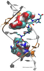
Genetic defect that causes myotonic dystrophy type 2 and used that information to design drug candidates to counteract the disease. Credit: Ilyas Yildirim, Northwestern University.
Scientists revealed a detailed image of the genetic defect that causes myotonic dystrophy type 2 and used that information to design drug candidates to counteract the disease. Credit: Ilyas Yildirim, Northwestern University. View larger image

Myotonic dystrophy type 2 (DM2) is a relatively rare, inherited form of adult-onset muscular dystrophy that has no cure. It’s caused by a genetic defect in which a short series of nucleotides—the chemical units that spell out our genetic code—is repeated more times than normal. When the defective gene is transcribed, the resulting RNA repeat forms a hairpin-like structure that binds to and disables a protein called MBNL1.

Now, research led by Matthew Disney of The Scripps Research Institute (TSRI), Florida Campus, has revealed the detailed, three-dimensional structure of the RNA defect in DM2 and used this information to design small molecules that bind to the aberrant RNA. These designer molecules, even in small amounts, significantly improved disease-associated defects in a cellular model of DM2, and thus hold potential for reversing the disorder.

Drugs that target toxic RNA molecules associated with diseases such as DM2 are few and far between, as developing such compounds is technically challenging. The “bottom-up” approach that the scientists used to design potent new drug candidates, by first studying in detail how the RNA structure interacts with small molecules, is unconventional, noted Jessica Childs-Disney of TSRI, who was lead author of the paper with Ilyas Yildirim of Northwestern University. But it may serve as an effective strategy for pioneering the use of small molecules to manipulate disease-causing RNAs—a central focus of the Disney lab.

This work also was funded by NIH’s National Cancer Institute.

Learn more:
The Scripps Research Institute News Release Exit icon
Disney Lab