Year: 2013

Cool Images: Holiday Season Cells

0 comments
Yeast cells deficient in zinc and the Tsa1 protein have protein tangles. Credit: Colin MacDiarmid and David Eide, University of Wisconsin-Madison.

Yeast cells deficient in zinc and the Tsa1 protein have protein tangles. Credit: Colin MacDiarmid and David Eide, University of Wisconsin-Madison.

Just in time for the holidays, we’ve wrapped up a few red and green cellular images from basic research studies. In this snapshot, we see a group of yeast cells that are deficient in zinc, a metal that plays a key role in creating and maintaining protein shape. The cells also lack a protein called Tsa1, which normally keeps proteins from sticking together. Green areas highlight protein tangles caused by the double deficiency. Red outlines the cells. Protein clumping plays a role in many human diseases, including Parkinson’s and Alzheimer’s, so knowledge of why it happens—and what prevents it in healthy cells—could aid the development of treatments.

See more festive images!

One Mutation Leads to Another—At Least in Yeast

0 comments
DNA mutation. Credit: Stock image.
Newly discovered genetic effect in yeast could shed light on carcinogenesis. Credit: Stock image.

Cancer cells typically include many gene mutations, extra or missing genes, or even the wrong number of chromosomes. Scientists know that certain genetic changes lead to ones elsewhere. But they’ve had a chicken-and-egg problem trying to figure out which changes trigger which others—or whether mutations accumulate randomly in tumors.

New research led by J. Marie Hardwick Exit icon of Johns Hopkins University sheds light on the issue. She found that incapacitating a single gene in yeast cells—regardless of which gene it was—spurred mutations in one or two other genes. The process was anything but random: If, say, gene X was knocked out, yeast cells almost always developed a secondary mutation in gene Y. It’s as if knocking out one gene disrupts the genomic balance enough that the cell must alter a different gene to compensate.

Significantly, the secondary mutations—but not the original ones—caused altered yeast cell characteristics, including traits linked to cancer. Also, many of the secondary mutations occurred in genes associated with cancer in humans, further suggesting that these secondary changes might play a role in carcinogenesis.

This new information will help researchers better understand the chain of genetic events that lead to cancer. It might also prompt scientists to reevaluate years of research that attributed changes in cell behavior or appearance to a given gene knockout.

This work also was funded by NIH’s National Institute of Neurological Disorders and Stroke.

Learn more:
Johns Hopkins University News Release Exit icon

Abnormal Mitochondria Might Cause Resistance to Radiation Therapy

2 comments
Mitochondria. Credit: Judith Stoffer.
Bean-shaped mitochondria are cells’ power plants. The highly folded inner membranes are the site of energy generation. Credit: Judith Stoffer. View larger image

Why some cancers are resistant to radiation therapy has baffled scientists, but research on abnormalities in mitochondria, often described as cells’ power plants, could offer new details. A research team led by Maxim Frolov Exit icon of the University of Illinois at Chicago learned that the E2F gene, which plays a role in the natural process of cell death, contributes to the function of mitochondria. Fruit flies with a mutant version of the E2F gene had misshapen mitochondria that produced less energy than normal ones. Flies with severely damaged mitochondria were more resistant to radiation-induced cell death. Studies using human cells revealed similar effects. The work could help explain why people with cancer respond differently to radiation therapy and might aid the development of drugs that enhance mitochondrial function, thereby improving the effectiveness of radiation therapy.

This work also was funded by NIH’s National Cancer Institute.

Learn more:
University of Illinois at Chicago News Release Exit icon

New Models Predict Where E. coli Strains Will Thrive

0 comments
Illustration of E. coli. Credit: Janet Iwasa, University of Utah.
Illustration of E. coli. Credit: Janet Iwasa, University of Utah (image available under a Creative Commons Attribution-NonCommercial-ShareAlike license Exit icon). View larger image

Like plants and animals, different types of E. coli thrive in different environments. Now, scientists can even predict which environments—such as the bladder, stomach or blood—are most amenable to the growth of various strains, including pathogenic ones. A research team led by Bernhard Palsson Exit icon of the University of California, San Diego, accomplished this by using genome data to reconstruct the metabolic networks of 55 E. coli strains. The metabolic models, which identify differences in the ability to manufacture certain compounds and break down various nutrients, shed light on how certain E. coli strains become pathogenic and how to potentially control them. One approach could be depriving the deadly strains of the nutrients they need to survive in their niches. The researchers plan to use their new method to study other bacteria, such as those that cause staph infections.

This work also was funded by NIH’s National Cancer Institute.

Learn more:
University of California, San Diego News Release

Stop the (Biological) Clock

0 comments
Molecular structure of the three proteins in blue-green algae’s circadian clock.  Credit: Johnson Lab, Vanderbilt University.
Molecular structure of the three proteins in blue-green algae’s circadian clock. Credit: Johnson Lab, Vanderbilt University.

Many microorganisms can sense whether it’s day or night and adjust their activity accordingly. In tiny blue-green algae, the “quartz-crystal” of the time-keeping circadian clock consists of only three proteins, making it the simplest clock found in nature. Researchers led by Carl Johnson of Vanderbilt University recently found that, by manipulating these clock proteins, they could lock the algae into continuously expressing its daytime genes, even during the nighttime.

Why would one want algae to act like it’s always daytime? The kind used in Johnson’s study is widely harnessed to produce commercial products, from drugs to biofuels. But even when grown in constant light, algae with a normal circadian clock typically decrease production of biomolecules when nighttime genes are expressed. When the researchers grew the algae with the daytime genes locked “on” in constant light, the microorganism’s output increased by as much as 700 percent. This proof of concept experiment may be applicable to improving the commercial production of compounds such as insulin and some anti-cancer drugs.

Learn more:

Vanderbilt University Press Release Exit icon
Johnson Laboratory Exit icon
Circadian Rhythms Fact Sheet

Meet Shanta Dhar

1 comment
Shanta Dhar
Shanta Dhar
Fields: Chemistry and cancer immunotherapy
Works at: University of Georgia, Athens
Born and raised in: Northern India
Studied at: Indian Institute of Science, Bangalore; Johns Hopkins University, Baltimore, Md.; and Massachusetts Institute of Technology, Cambridge, Mass.
To unwind: She hits the gym
Credit: Frankie Wylie, Stylized Portraiture

The human body is, at its most basic level, a giant collection of chemicals. Finding ways to direct the actions of those chemicals can lead to new treatments for human diseases.

Shanta Dhar, an assistant professor of chemistry at the University of Georgia, Athens (UGA), saw this potential when she was exposed to the field of cancer immunotherapy as a postdoctoral researcher at the Massachusetts Institute of Technology. (Broadly, cancer immunotherapy aims to direct the body’s natural immune response to kill cancer cells.) Dhar was fascinated by the idea and has pursued research in this area ever since. “I always wanted to use my chemistry for something that could be useful [in the clinic] down the line,” she said.

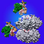
A major challenge in the field has been training the body’s immune system—specifically the T cells—to recognize and attack cancer cells. The process of training T cells to go after cancer is rather like training a rescue dog to find a lost person: First, you present the scent, then you command pursuit.

The type of immune cell chiefly responsible for training T cells to search for and destroy cancer is a called a dendritic cell. First, dendritic cells present T cells with the “scent” of cancer (proteins from a cancer cell). Then they activate the T cells using signaling molecules.

Dhar’s Findings

Dhar’s work focuses on creating the perfect trigger for cancer immunotherapy—one that would provide both the scent of cancer for T cells to recognize and a burst of immune signaling to activate the cells.

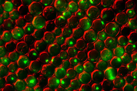
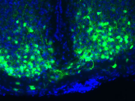
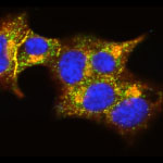
Using cells grown in the lab, Dhar’s team recently showed that they could kill most breast cancer cells using a new nanotechnology technique, then train T cells to eradicate the remaining cancer cells.

For the initial attack, the researchers used light-activated nanoparticles that target mitochondria in cancer cells. Mitochondria are the organelles that provide cellular energy. Their destruction sets off a signaling cascade that triggers dendritic cells to produce one of the proteins needed to activate T cells.

Because the strategy worked in laboratory cells, Dhar and her colleague Donald Harn of the UGA infectious diseases department are now testing it in a mouse model of breast cancer to see if it is similarly effective in a living organism.

For some reason, the approach works against breast cancer cells but not against cervical cancer cells. So the team is examining the nanoparticle technique to see if they can make it broadly applicable against other cancer types.

Someday, Dhar hopes to translate this work into a personalized cancer vaccine. To create such a vaccine, scientists would remove cancer cells from a patient’s body during surgery. Next, in a laboratory dish, they would train immune cells from the patient to kill the cancer cells, then inject the trained immune cells back into the patient’s body. If the strategy worked, the trained cells would alert and activate T cells to eliminate the cancer.

Detailing Key Structures of HIV

1 comment
Structure of Env. Credit: The Scripps Research Institute.
This model shows a birds-eye view of the structure of Env, a protein on the HIV surface that enables it to infect cells, bound to neutralizing antibodies. Credit: The Scripps Research Institute.

In a statement on World AIDS Day 2013, NIH leaders wrote, “In the 25 years that have passed since the first annual commemoration of World AIDS Day, extraordinary scientific progress has been made in the fight against HIV/AIDS. That progress has turned an HIV diagnosis from an almost-certain death sentence to what is now for many, a manageable medical condition and nearly normal lifespan. We have come far, yet not far enough.”

One area of progress is in understanding the structural biology of HIV, the virus that causes AIDS. Capturing details about the virus’ shape has helped scientists better understand how HIV operates and pinpoint its Achilles’ heels. Recently, scientists got a closer look at two key pieces of the virus.

In one study, researchers developed the most detailed picture yet of Env, a three-segment protein on the HIV surface that allows the virus to infect cells. The work illuminated the complex process by which the protein assembles, undergoes radical shape changes during infection and interacts with neutralizing antibodies, which can block many strains of HIV from infecting human cells. The findings also may guide the development of HIV vaccines.

In another study, researchers created the best image yet of the cocoon-like container, or capsid, that carries HIV’s genome. Capsids have been difficult to study because individual imaging techniques had not produced high enough detail. By combining several cutting-edge imaging methods, the scientists pieced together the individual polygonal units of the capsid like a jigsaw puzzle to determine its structure in detail. Now that they know how HIV’s inner vessel looks, the research team is searching for its cracks—potential targets for drug development.

Learn more:
Imaging HIV’S Inner Shell Article from Inside Life Science
Key HIV Protein Structure Revealed Article from NIH Research Matters
Interactive HIV Structural Model

Cool Image: Tick Tock, Master Clock

0 comments
Master clock in mouse brain with the nuclei of the clock cells shown in blue and the VIP molecule shown in green. Credit: Cristina Mazuski in the lab of Erik Herzog, Washington University in St. Louis.

Master clock in mouse brain with the nuclei of the clock cells shown in blue and the VIP molecule shown in green. Credit: Cristina Mazuski in the lab of Erik Herzog, Washington University in St. Louis.

Our biological clocks play a large part in influencing our sleep patterns, hormone levels, body temperature and appetite. A small molecule called VIP, shown in green, enables time-keeping neurons in the brain’s central clock to coordinate daily rhythms. New research shows that, at least in mice, higher doses of the molecule can cause neurons to get out of synch. By desynchronizing mouse neurons with an extra burst of VIP, Erik Herzog of Washington University in St. Louis found that the cells could better adapt to abrupt changes in light (day)-dark (night) cycles. The finding could one day lead to a method to reduce jet lag recovery times and help shift workers better adjust to schedule changes.

Learn more:

Washington University in St. Louis News Release Exit icon
Circadian Rhythms Fact Sheet
Tick Tock: New Clues About Biological Clocks and Health Article from Inside Life Science
A Light on Life’s Rhythms Article from Findings Magazine

Protein Triggers Inflammatory Responses in Hemorrhage and Sepsis

0 comments
Doctors helping a patient. Credit: U.S. Navy.
Inflammation is part of the body’s natural response to trauma, but when it becomes widespread, it can lead to sepsis. Credit: U.S. Navy.

Inflammation is part of the body’s natural response to trauma, playing a vital role in wound healing and prevention of infection. However, when inflammation becomes widespread, or systemic, it can lead to sepsis, a condition that can damage organs and cause death. Scientists led by Ping Wang of the Feinstein Institute for Medical Research have found a way to potentially target harmful systemic inflammation. They identified a protein–cold-inducible RNA-binding protein (CIRP)–that triggers inflammatory responses during hemorrhagic shock and sepsis. Wang then hypothesized that blocking CIRP activity might mitigate the body’s overall inflammatory response and improve patient survival. In a preclinical study using mice, an antibody against CIRP decreased mortality after hemorrhage and sepsis. The molecule could lead to the development of an anti-CIRP drug.

This work also was funded by the NIH Office of the Director and NIH’s National Heart, Lung, and Blood Institute.

Learn more:

Fact Sheets on Physical Trauma and Sepsis
The Body’s Response to Traumatic Injury Video

Mapping Approach Yields Insulin Secretion Pathway Insights

0 comments
TMEM24 protein (green) and insulin (red) in pancreatic beta cells (yellow).  Credit: Balch Lab, the Scripps Research Institute.
The interactions of TMEM24 protein (green) and insulin (red) in pancreatic beta cells are shown in yellow. Credit: Balch Lab, the Scripps Research Institute.
View larger image

The identities of the proteins that drive insulin production and release from pancreatic beta cells have largely been a mystery. In new work from the lab of William Balch of the Scripps Research Institute, researchers isolated and then identified all the insulin-bound proteins from mouse beta cells. The results provided a roadmap of the protein interactions that lead to insulin production, storage and secretion. The researchers used the roadmap to identify a protein called TMEM24, which was abundant in beta cells and binds readily to insulin. Balch and his team uncovered that TMEM24, whose involvement in insulin secretion was previously unknown, effectively regulates slower insulin release and could have a key role in maintaining control of glucose levels in the blood. The scientists hope that this roadmap of insulin-interacting proteins will lead to the development of new, targeted approaches to treating type 2 diabetes and a similar insulin-related condition called metabolic syndrome.

Learn more:

The Scripps Research Institute News Release Exit icon
Balch Lab Exit icon