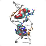
Scientists revealed a detailed image of the genetic change that causes myotonic dystrophy type 2 and used that information to design drug candidates to counteract the disease. Credit: Ilyas Yildirim, Northwestern University. View larger image
This image may look complicated, but it tells a fairly straightforward tale about basic research: Learning more about basic life processes can pave the way for medical and other advances.
In this example, researchers led by Matthew Disney of the Scripps Research Institute’s Florida campus focused on better understanding the structural underpinnings of myotonic dystrophy type 2, a relatively rare, inherited form of adult-onset muscular dystrophy. While this work is still in the preliminary stages, it may hold potential for someday treating the disorder.
Some 300,000 NIH-funded scientists are working on projects aimed at improving disease diagnosis, treatment and prevention, often through increasing understanding of basic life processes.
Researchers found that the antibiotic trovafloxacin cuts off a channel for communication between cells and interferes with a cell-death process. Credit: Stock image.
Many compounds that show promise as new antibiotics for treating bacterial infections never make it to the clinic because they turn out to be toxic to humans as well as to bacteria. A research team led by Kodi Ravichandran of the University of Virginia recently gained insights into why one such antibiotic, trovafloxacin, harms human cells. They found that the compound cuts off a channel for communication between cells, which in turn interferes with how dying cells are broken down and recycled by the body. Roughly 200 billion cells in the human body die and are replaced every day as part of a routine cleanup process, and interference in this process by trovafloxacin may have contributed to the serious liver damage seen in some patients in clinical trials of the drug. Understanding how trovafloxacin causes toxicity in people may help researchers re-engineer this and related compounds to make them safe and effective for use in fighting bacterial infections.
Jeff Shaman Field: Climatology Works at: Columbia University’s Mailman School of Public Health, N.Y. Favorite high school subject: Biology First job: Guide at the Franklin Institute in Philadelphia, Pa. Alternative career: Opera singer Credit: Anne Foulke
Before he wrote any scientific papers, Jeff Shaman wrote operas. At the premiere of one of his operas, an 80-minute story about psychoanalysis, reviewers said the work “crackle[d] with invention.”
After 4 years of training to become an opera singer, Shaman realized that the work wouldn’t offer him career stability. He started thinking about his other interests. After college, where he majored in biology with a focus on ecology, he had volunteered to help with HIV clinical trials and developed a fascination with understanding infectious diseases. He wondered if the quantitative tools and methods used to study the physical sciences—another interest area—could inform how contagions spread and possibly even lead to systems for monitoring or predicting their transmission.
So Shaman returned to school—this time, for advanced degrees in earth and environmental sciences. He now studies the relationship between soil wetness and mosquito-borne diseases such as malaria in Africa and West Nile in Florida.
“I love science—probing questions, thinking about problems, finding solutions, pursuing my ideas,” says Shaman.
His Findings
A few years ago, Shaman took some of his scientific compositions in another direction by focusing on seasonal flu outbreaks. For more than 60 years, researchers have linked seasonal flu outbreaks with environmental data like humidity and temperature. Shaman analyzed this work and figured out that absolute humidity, rather than relative humidity, was the best predictor of outbreaks. Now he’s applied state-of-the-art mathematical modeling and real-time observational estimates of influenza incidence to predict when outbreaks will likely occur.
His forecasting technique mimics that used by meteorologists to predict weather conditions like temperatures, precipitation and even hurricane landfall. Shaman’s version incorporates variables like how transmissible a virus is, the number of days people are contagious and sick, and how much humidity is in the air.
The flu forecasts build on a series of studies in which Shaman and his colleagues used data from previous influenza seasons to test their predictions and improve reliability of their model. The work culminated with real-time predictions for 108 cities during the 2012-2013 influenza season. The forecasts could reliably estimate the peaks of flu outbreaks up to 9 weeks before they occurred.
For the 2013-2014 flu season, the researchers continued to make weekly predictions. But instead of first publishing the results in a scientific journal, they posted them on a newly launched influenza forecasts Web site where the public could view the projections.
“People understand the limitations and capabilities of weather forecasts,” says Shaman. “Our hope is that people will develop a similar familiarity with the flu forecasts and use that information to make sensible decisions.” For instance, the prediction of high influenza activity may motivate them to get vaccinated and practice other flu-prevention measures.
As he waits for the start of the next flu season, Shaman continues to tweak his forecast system to improve its reliability. He’s also beginning to address other questions, such as how to predict multiple outbreaks of different influenza strains and how to predict the spread of other respiratory illnesses.
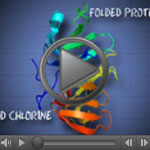
Exposure to hypochlorous acid causes bacterial proteins to unfold and stick to one another, leading to cell death. Credit: Video segment courtesy of the American Chemistry Council. View video
Spring cleaning often involves chlorine bleach, which has been used as a disinfectant for hundreds of years. But our bodies have been using bleach’s active component, hypochlorous acid, to help clean house for millennia. As part of our natural response to infection, certain types of immune cells produce hypochlorous acid to help kill invading microbes, including bacteria.
Researchers funded by the National Institutes of Health have made strides in understanding exactly how bleach kills bacteria—and how bacteria’s own defenses can protect against the cellular stress caused by bleach. The insights gained may lead to the development of new drugs to breach these microbial defenses, helping our bodies fight disease.
During a live online chat dubbed “Cell Day,” scientists at NIGMS recently fielded questions about the cell and careers in research from middle and high school students across the country. Here’s a sampling of the questions and answers, some of which have been edited for clarity or length.
What color are cells?
While cells with lots of iron, like red blood cells, may be red, usually cells are colorless.
How many different types of cells can be found inside the human body?
There are about 200 cell types and a few trillion total cells in the human body. That does not include bacteria, fungi and mites that live on the body.
Is it possible to have too many or not enough cells?
The answer depends on cell type. For example, within the immune system, there are many examples of diseases that are caused by too many or not enough cells. When too many immune cells accumulate, patients get very large spleens and lymph nodes. When too few immune cells develop, patients have difficulty fighting infections.
How fast does it take for a cell to produce two daughter cells?
Some cells, for example bacterial ones, can produce daughter cells very fast when nutrients are available. The doubling time for E. coli bacteria is 20 minutes. Other cells in the human body take hours or days or even years to divide.
Do skin cells stretch or multiply when you gain weight?
The size of cells is tightly regulated and maintained so they do not stretch much. As the surface area of the body increases with weight gain, the number of skin cells increases.
Why do cells self-destruct?
The term for cellular self-destruction is “apoptosis” or “programmed cell death.” Apoptosis is very important for normal development of humans and other animals as it ensures that we do not have too many cells and that “unhealthy” cells can be eliminated without causing harm to the surrounding cells. For instance, did you know that human embryos have webbing between their fingers and toes (just like ducks!)? Apoptosis eliminates the cells that form the web so that you are born with toes and fingers.
In what field is there a need for new scientists?
I would say that there is a need for scientists who can work at the interface between the biological and biomedical sciences and the data sciences. Knowing sophisticated mathematics and having computer skills to address questions like ‘what does this biomedical data tell us about particular diseases’ is still a challenge.
What is a scientist’s daily work day like? Is all of your time spent in a lab testing or like in an office throwing ideas around?
There are lots of different kinds of jobs a scientist can have. Many work in labs where they get to do experiments AND throw ideas around. Working in a lab is a lot of fun—you learn things about the world that no one has known before (how cool is that?). Other important jobs that scientists can do include writing about science as a journalist, helping other scientists patent new technologies they invent as a patent agent or lawyer, or working on important scientific policy issues for the government or other organizations.
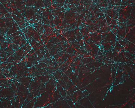
Neurons activated with red or blue light using algae-derived opsins. Credit: Yasunobu Murata/McGovern Institute for Brain Research at MIT.
The nerve cells, or neurons, lit up in blue and red in this image of mouse brain tissue are expressing algae-derived, light-sensitive proteins called opsins. To control neurons with light, scientists engineer the cells to produce particular opsins, most of which respond to light in the blue-green range. Then they shine light on the cell to activate it. Now, a team of researchers led by Ed Boyden of the Massachusetts Institute of Technology and Gane Ka-Shu Wong of the University of Alberta has discovered an opsin that responds to red light preferentially, enabling them to manipulate two groups of neurons simultaneously with different colors of light and get a more comprehensive look at how those two sets of brain cells interact. Other opsins have shown potential for vision restoration in animal studies, and, because red light causes less damage to tissue than blue-green light, this new opsin might eventually be used for such treatments in humans.
A 5-year, randomized clinical trial helped resolve a long-standing debate about how best to manage sepsis patients.
For years, doctors have debated the best ways to identify, predict and treat sepsis. The condition, which is usually triggered by infection, is marked by body-wide inflammation and can lead to a dangerous drop in blood pressure known as septic shock. Sepsis affects more than 800,000 people each year and kills about 20 to 30 percent of them. It’s the most expensive condition treated in U.S. hospitals, costing more than $20 billion a year.
Now, a nationwide, 5-year clinical trial that set out to compare three different treatment approaches has shown that survival of patients with septic shock was the same regardless of whether they received treatment based on structured, standardized medical plans (protocols) or the usual high-level standard of care. If patients were diagnosed shortly after the onset of sepsis and treated promptly with fluids and antibiotics, they did equally well whether they received treatment based on either of two specific protocols—one less invasive than the other—or got the usual, high-level care provided by the academic hospitals where the study was conducted.
According to the study’s leaders, the trial “helps resolve a long-standing clinical debate about how best to manage sepsis patients, particularly during the critical first few hours of treatment,” and shows that “there is not a mandated need for more invasive care in all patients.”
Fat cells such as these listen for incoming signals like FGF21, which tells them to burn more fat. Credit: David Gregory and Debbie Marshall. All rights reserved by Wellcome Images.
Living things are chatty creatures. Even when they’re not making actual sounds, organisms constantly communicate using chemical signals that course through their systems. In multicellular organisms like people, brain cells might call, “I’m in trouble!” signaling others to help mount a protective response. Single-celled organisms like bacteria may broadcast, “We have to stick together to survive!” so they can coordinate certain activities that they can’t carry out solo. In addition to sending out signals, cells have to receive information. To help them do this, they use molecular “ears” called receptors on their surfaces. When a chemical messenger attaches to a receptor, it tells the cell what’s going on and causes a response.
Scientists are following the dialogue, learning how cell signals affect health and disease. They’re also starting to take part in the cellular conversations, inserting their own comments with the goal of developing therapies that set a diseased system right.
Continue reading this new Inside Life Science article
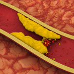
Artery with fat deposits and a formed clot. Credit: Stock image. View larger image
Heart disease is the leading cause of death for both men and women in the United States, according to the Centers for Disease Control and Prevention. One treatment challenge is developing non-invasive ways to direct medication to damaged or clogged arteries, which can block blood flow and increase the risk for heart attack and stroke. A team led by Naren Vyavahare at Clemson University has engineered extremely tiny particles—nanoparticles—that offer a promising step forward.
Healthy arteries have elastic fibers that make the arteries flexible. But, in most cardiovascular diseases, the fibers get damaged. The new nanoparticles, which can deliver drugs, attach only to damaged fibers and could enable site-specific drug delivery to minimize off-target side effects. The nanoparticles also allow drugs to be released over longer periods of time, potentially increasing the drugs’ effectiveness. The new biomaterial was tested in rodent models for studying vascular disease, so it is still in the early stages of development.
This work also was funded by NIH’s National Heart, Lung, and Blood Institute.
Bacteroides ovatus. Credit: Eric Martens, University of Michigan Medical School.
After eating, we don’t do all the work of digestion on our own. Trillions of gut bacteria help us break food down into the simple building blocks our cells need to function. New research from an international team co-led by Eric Martens of the University of Michigan Medical School has uncovered how a strain of beneficial gut bacteria, Bacteroides ovatus, digests complex carbohydrates called xyloglucans that are found in fruits and vegetables. The researchers traced the microorganism’s digestive ability to a single piece of the genome. They also examined a publicly available set of genomic data, which included information from both humans and their resident bacteria, and found that more than 90 percent of 250 adults harbored at least one Bacteroides strain with xyloglucan-digesting capabilities. These results underscore the importance of the bacteria to human health and nutrition.
This work also was funded by the National Institute of Diabetes and Digestive and Kidney Diseases.
Learn more:
University of Michigan News Release
University of Michigan Host Microbiome Initiative
Gut Reactions and Other Findings About Our Resident Microbes from Inside Life Science
Body Bacteria from Findings Magazine


 Exposure to hypochlorous acid causes bacterial proteins to unfold and stick to one another, leading to cell death. Credit: Video segment courtesy of the American Chemistry Council. View video