
Wildlife photos can be truly stunning, and cute cat pictures are a cornerstone of the internet. But zooming in on the early lives of fish, insects, and worms can have equally wonderful results. Using powerful microscopes, researchers are revealing the complexity and beauty of animal development.
 Credit: James E. Hayden, The Wistar Institute, Philadelphia, PA.
This image captures the spiral-shaped ovary of an anglerfish in cross section. Once matured, these eggs will be released in a gelatinous, floating mass. For some species of anglerfish, this egg mass can be up to 3 feet long and include nearly 200,000 eggs.
 Credit: Philipp Keller, Bill Lemon, Yinan Wan, and Kristin Branson, Janelia Farm Research Campus, Howard Hughes Medical Institute, Ashburn, VA.
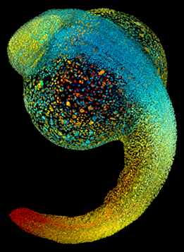
Just 22 hours after fertilization, this zebrafish embryo is already taking shape. The portion that will become the head is at the top of the image, above the bulge of the yolk sac, and the portion that will become the tail is at the bottom. By 36 hours, all of the major organs will have started to form. The zebrafish’s rapid growth and see-through embryo make it ideal for scientists studying how organs develop.
 Credit: Bo Wang and Phillip A. Newmark, University of Illinois at Urbana-Champaign.
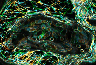
The parasitic worm that causes schistosomiasis—an infection that’s rare in the United States but common in other parts of the world—hatches in water and grows up in a freshwater snail. Here, the worms are larvae inside a snail’s body. Once mature, the worms swim back into the water, where they can infect people through skin contact. Initially, an infected person might have a rash, itchy skin, or flu-like symptoms, but the real damage is done over time to internal organs.
 Credit: Kevin Edwards, Johny Shajahan, and Doug Whitman, Illinois State University.
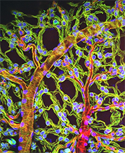
The lubber grasshopper, found throughout the southern United States, is frequently used in biology classes to teach students about the respiratory system of insects. Unlike mammals, which have red blood cells that carry oxygen throughout the body, insects have breathing tubes that carry air through their exoskeleton directly to where it’s needed. This image shows the breathing tubes embedded in the weblike sheath cells that cover developing egg chambers.
Visit our Image and Video Gallery for more cool images. And send us yours for possible inclusion!