It’s the spookiest time of the year! To celebrate Halloween, we’re showcasing scientific images that capture the spirit of the holiday, from a brain shaped like a bat to protein “cobwebs” in a quail embryo. Check out our image and video gallery for even more scientific photos, illustrations, and videos.


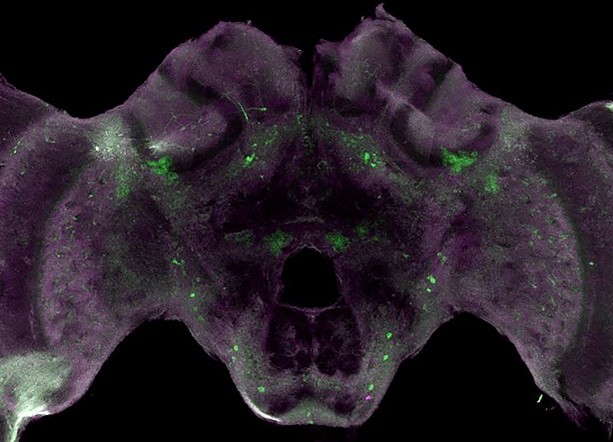
This image may bring to mind a bat spreading its wings, or you may feel like a skull is peering at you, but what’s actually shown is a honeybee brain. The bright-green spots are tyrosine hydroxylase, an enzyme that allows the brain to produce dopamine. Dopamine is involved in many important functions—such as the ability to experience pleasure—in both bees and humans.
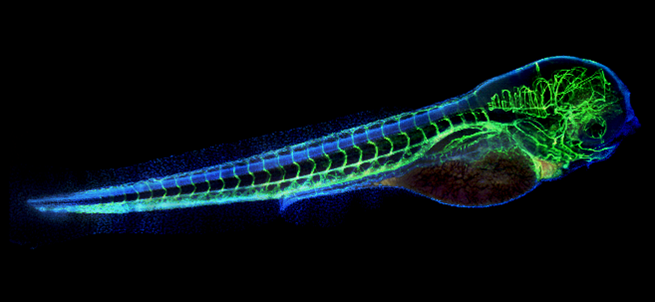
The eerie green “skeleton” in this photo is really the vasculature (blood vessels) of a zebrafish embryo. The blue areas are cell bodies. Zebrafish are useful research organisms for studying development because their eggs and embryos are see-through, making it easy for scientists to watch changes take place.
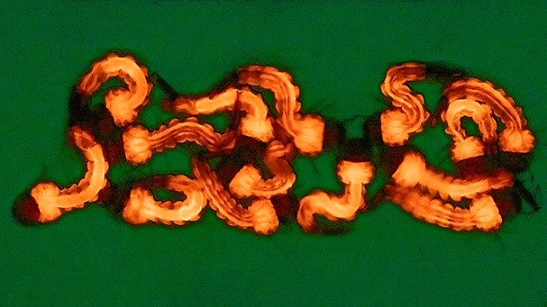
Researchers edited the genes of these creepy-crawly mosquito larvae using a technique called CRISPR (clustered regularly interspaced short palindromic repeats). This species of mosquito, Culex quinquefasciatus, can transmit diseases including West Nile virus, Japanese encephalitis virus, and avian malaria. The gene-editing toolkit used on these larvae could ultimately help stop Culex quinquefasciatus from spreading pathogens.
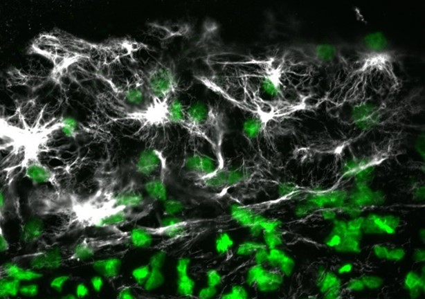
The “cobwebs” in this image are actually a protein called vimentin in a quail embryo. The green spots are cell nuclei. Vimentin is part of the cytoskeleton and helps cells maintain their structure and resist mechanical stress. The protein is found in many animals and in humans.

