Author: Alisa Zapp Machalek

Alisa Machalek Headshot.

Posts by Alisa Zapp Machalek

Scientists Shine Light on What Triggers REM Sleep

1 comment
Illustration of a brain.
While studying how the brain controls REM sleep, researchers focused on areas abbreviated LDT and PPT in the mouse brainstem. This illustration shows where these two areas are located in the human brain. Credit: Wikimedia Commons. View larger image

Has the “spring forward” time change left you feeling drowsy? While researchers can’t give you back your lost ZZZs, they are unraveling a long-standing mystery about sleep. Their work will advance the scientific understanding of the process and could improve ways to foster natural sleep patterns in people with sleep disorders.

Working at Massachusetts General Hospital and MIT, Christa Van Dort, Matthew Wilson, and Emery Brown focused on the stage of sleep known as REM. Our most vivid dreams occur during this period, as do rapid eye movements, for which the state is named. Many scientists also believe REM is crucial for learning and memory.

REM occurs several times throughout the night, interspersed with other sleep states collectively called non-REM sleep. Although REM is clearly necessary—it occurs in all land mammals and birds—researchers don’t really know why. They also don’t understand how the brain turns REM on and off.

Continue reading “Scientists Shine Light on What Triggers REM Sleep”

E. Coli Bacteria as Medical Sensors and Hard Drives?

1 comment
E.Coli
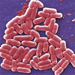
Modified E. coli bacteria can serve as sensors and data storage devices for environmental and medical monitoring. Credit: Centers for Disease Control and Prevention. View larger image

E. coli bacteria help us digest our food, produce vitamin K and have served as a model organism in research for decades. Now, they might one day be harnessed as environmental or medical sensors and long-term data storage devices Exit icon.

MIT researchers Timothy Lu and Fahim Farzadfard modified the DNA of E. coli cells so that the cells could be deployed to detect a signal (for example, a small molecule, a drug or the presence of light) in their surroundings. To create the modified E. coli, the scientists inserted into the bacteria a custom-designed genetic tool.

When exposed to the specified signal, the tool triggers a series of biochemical processes that work together to introduce a single mutation at a specific site in the E. coli’s DNA. This genetic change serves to record exposure to the signal, and it’s passed on to subsequent generations of bacteria, providing a continued record of exposure to the signal. In essence, the modified bacteria act like a hard drive, storing biochemical memory for long periods of time. The memory can be retrieved by sequencing the bacteria or through a number of other laboratory techniques.

Continue reading “E. Coli Bacteria as Medical Sensors and Hard Drives?”

Meet Alfred Atanda Jr.

1 comment
Alfred Atanda Jr.
Credit: Cynthia Brodoway, Nemours/Alfred I. duPont
Hospital for Children
Alfred Atanda Jr.
Fields: Pediatric orthopedic surgery, sports medicine
Works at: Nemours/Alfred I. duPont Hospital for Children
Blogs: as Philly.com’s Sports Doc at http://bit.ly/sportsdoc Exit icon
Family fact: Youngest of seven children
Musical skills: Piano and trumpet
Kitchen talent: Baking chocolate desserts for his pediatrician wife and their two young children

As a kid, Alfred Atanda loved science, sports and tinkering. He dreamed of being a construction worker or an engineer. Today, he works on one of the most complex construction projects of all: the human body.

As a pediatric orthopedic surgeon, Atanda focuses on sports medicine and injuries to children. He has a special passion for young baseball pitchers who have torn the ulnar collateral ligament (UCL) in the elbow of their throwing arm.

This sort of injury is most often caused by overuse. Many small tears accumulate over a long period, resulting in pain and slower, less accurate pitches. Decades ago, this sort of damage occurred almost exclusively in elite athletes. Now, Atanda sees it in children as young as 12 years old. He aims not only to study and treat these injuries, but also to find ways to prevent them.

His Findings

Atanda was first introduced to research on UCL injuries while working alongside team physicians for the Phillies, the professional baseball team in Philadelphia. The physicians wanted to determine whether ultrasound imaging could detect early warning signs—slight anatomical changes in the ligament—before the damage became severe enough to warrant an operation known as Tommy John surgery.

The research focused on Phillies pitchers who had no pain or other symptoms of injury. The multi-year project showed that the UCL in the throwing elbows of these players got progressively thicker and weaker compared to the same ligament in the players’ nonthrowing elbows. The scientists concluded that these physical changes are caused by prolonged exposure to professional-level pitching.

Alfred Atanda Jr. with Joe Piergrossi
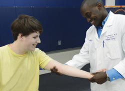
Atanda examines the elbow of a young patient. Courtesy: Cynthia Brodoway, Nemours/Alfred I. duPont Hospital for Children

Atanda wondered whether ultrasound imaging could also detect early signs of UCL damage in young pitchers—those in Little League through high school. There has been a dramatic rise in the number of young pitchers who are experiencing the same injuries and undergoing the same surgery as the pros.

Atanda secured funding for this project from an Institutional Development Award (IDeA). The IDeA program builds research capacities in states like Delaware, where Atanda works, that historically have received low levels of funding from the National Institutes of Health.

Atanda’s project began about a year ago, and has examined 55 young athletes so far.

“We found similar results to what we found with the Phillies,” Atanda says, indicating that the UCL in the throwing elbows of young athletes was noticeably thicker than the UCL in the nonthrowing elbows. And the damage seems progressive, he says: “We saw that these ligaments got thicker as the pitchers got older and had more pitching experience.”

The immediate goal of this project, which he hopes to continue for another 3 years, is prophylaxis. “We’re trying to prevent any kind of overuse elbow injuries and the need for Tommy John surgeries later on,” Atanda says. He also hopes to establish quantitative correlations between pitching behavior and anatomical changes.

Atanda also has longer-term aspirations. “My goal is to change the culture in sports for young athletes in general,” he says. “I want to show there are downsides to pitching so much.”

In addition to championing pitch count limits recommended by the American Sports Medicine Institute, Atanda advises a focus away from excess competition and toward getting exercise, enjoying social inter­action, building self-confidence and having fun.

Atanda’s research is funded by the National Institutes of Health through grant P20GM103464

Content adapted from the NIGMS Findings magazine article Game Changer

Molecules Known to Damage Cells May Also Have Healing Power

0 comments
Free radicals in an ying-yang symbol
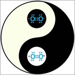
Biology in balance: Molecules called free radicals—like the peroxide molecules illustrated here—have a reputation for being dangerous. Now, they’ve revealed healing powers. In worms, at least. Credit: Stock image

When our health is concerned, some molecules are widely labeled “good,” while others are considered “bad.” Often, the truth is more complicated.

Consider free radical molecules. These highly reactive, oxygen-containing molecules are well known for damaging DNA, proteins and other molecules in our bodies. They are suspected of contributing to premature aging and cancer. But now, new research shows they might also have healing powers.

Using the oft-studied laboratory roundworm known as C. elegans, a research group led by Andrew Chisholm at the University of California, San Diego, made a surprising discovery. Free radicals, specifically those made in cell structures called mitochondria, appear necessary for skin wounds to heal. In fact, higher (but not dangerously high) levels of the molecules can actually speed wound closure.

If further research shows the same holds true in humans, the work could point to new ways to treat hard-to-heal wounds, like diabetic foot ulcers.

This work was funded in part by NIH under grants R01GM054657 and P40OD010440.

Nobel Prize for Powerful Microscopy Technology

0 comments
Fibroblasts
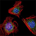
The cells shown here are fibroblasts, one of the most common cells in mammalian connective tissue. These particular cells were taken from a mouse. Scientists used them to test the power of a new microscopy technique that offers vivid views of the inside of a cell. The DNA within the nucleus (blue), mitochondria (green) and cellular skeleton (red) is clearly visible. Credit: Dylan Burnette and Jennifer Lippincott-Schwartz, Eunice Kennedy Shriver National Institute of Child Health and Human Development, National Institutes of Health.

William E. Moerner was at a conference in Brazil when he learned he’d be getting a Nobel Prize in chemistry. “I was incredibly excited and thrilled,” he said of his initial reaction.

An NIGMS grantee at Stanford University, Moerner received the honor for his role in achieving what was once thought impossible—developing super-resolution fluorescence microscopy, which is so powerful it allows researchers to see and track individual molecules in living organisms in real time.

Nobel recipients usually learn of the prize via a phone call from Stockholm, Sweden, in early October. For those in the United States, the call typically comes between 2:30 a.m. and 5:45 a.m.

Every year, the NIGMS communications office prepares for the Nobel Prize announcements in physiology or medicine and chemistry, the categories in which our grantees are most likely to be recognized. If the Institute played a significant role in funding the prize-winning research, we work quickly to provide information and context to reporters covering the story on tight deadlines. We issue a statement, identify an in-house expert on the research and arrange interviews with reporters. It’s all to help get the word out about the research and the taxpayers’ role in supporting it.

This year’s in-house expert, Cathy Lewis, shared her thoughts on the prize to Moerner in an NIGMS Feedback Loop post. You can also read this year’s statement and see a full list of NIGMS-supported Nobel laureates.

Cool Image: Training Cells to Devour Dying Neighbors

2 comments
A healthy cell that has ingested dying cells.

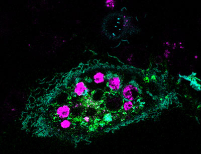
A healthy cell (green) that has ingested dying cells (purple). Credit: Toru Komatsu, University of Tokyo.

In this image, a healthy cell (green) has engulfed a number of dying cells (purple) just as a predator might ingest wounded or dying prey. A team of researchers is hoping to use this same strategy at the cellular level to help treat infection, neurodegenerative diseases or cancer.

Our bodies routinely use a process called phagocytosis to rid themselves of unhealthy cells. Takanari Inoue at Johns Hopkins University and collaborators at the University of Tokyo set out to investigate the molecular underpinnings of phagocytosis. Their goal was to endow laboratory-grown human cells with phagocytotic skills, namely the ability to recognize, swallow and digest dying cells. The scientists tried to do this by inserting into the cells two molecules known to play a role in phagocytosis.

The engineered cells accomplished the first two tasks—they recognized and surrounded dying cells. But they didn’t digest what they’d engulfed. Now the researchers are looking for a molecular trigger to get the engineered cells to complete this last task.

Eventually, the scientific team aims to build artificial cells that are programmed to target and destroy abnormal cells, such as those ravaged by bacteria, cancer or other diseases.

Learn more:
Johns Hopkins News Release Exit icon
How Cells Take Out the Trash Article from Inside Life Science
Cellular Suicide: An Essential Part of Life Article from Inside Life Science

An Insider’s Look at Life: Magnified, an Airport Exhibit of Stunning Microscopy Images

0 comments
Bubonic plague bacteria on part of the digestive system in a rat flea
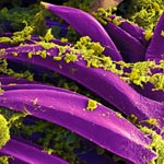
What looks like pollen on petals is actually bubonic plague bacteria on the digestive spines of a flea, viewed through a powerful microscope. Credit: B. Joseph Hinnebusch, Elizabeth Fischer and Austin Athman, NIH’s National Institute of Allergy and Infectious Diseases.

Science. Art. Airports. I’ve never used those three words together before. But I’ve been doing it a lot lately while working on Life: Magnified, an exhibit of 46 striking scientific images created by scientists around the country using state-of-the-art microscopes.

The show is at Washington Dulles International Airport, where more than a million travelers will see it over its 6-month run. As our director said in a recent post on another NIGMS blog, “What a great way to share the complexity and beauty of biomedical science with such a large public audience!”

We’ve also set up an online gallery, where the colorful images can be viewed and freely downloaded for research, educational and news media purposes.

The project itself was quite an adventure. When we asked scientists to send us images, our fears of not getting enough attractive, high-resolution options were drowned by a deluge of more than 600 submissions. Then we worried how to sort through them all and make final selections. Doing so required several rounds of online viewing, various ranking systems and a panel of experts. Then the images were printed as large, digital negatives on transparency film.

Artist's rendition of a network diagram. Credit: Allison Kudla, Institute for Systems Biology.
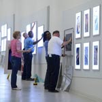
The midnight installation of Life: Magnified involved five people (that’s me in pink), a ladder and lots of rags and glass cleaner. Credit: Woody Machalek.

Dulles is a pretty busy place, so we set up the exhibit when it was quietest—the middle of the night (10 p.m. to 1:30 a.m., to be exact). We swapped out images from the previous photography exhibit and installed the Life: Magnified ones in LED lightboxes mounted in the airport’s Gateway Gallery. It was an eerie and exhilarating feeling to be in a noiseless, nearly empty airport without heavy bags and a long walk to a departure gate.

Less than 12 hours later, I was surprised to receive an e-mail from someone who had passed through the exhibit and sent a few photos. He called the images “stunning.” Similar sentiments were expressed by Science, NBC News online Exit icon, The Atlantic Exit icon, The Washington Post Exit icon, National Geographic Exit icon and other publications.

Now I’m being asked about next steps, including whether the exhibit will travel. We’re investigating a variety of options. For now, I hope you’re able to see the exhibit in person. If not, take a look at the images online and see which ones you enjoy most.

Learning How Mosquito-Borne Viruses Use Knot-like RNA to Cause Disease

0 comments
A knot-like structure in a section of RNA from a flavivirus
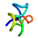
A knot-like structure in RNA enables flaviviruses to cause diseases like yellow fever, West Nile virus and dengue fever, which threaten roughly half the world’s population. Credit: Jeffrey Kieft.

Roughly half the world’s population is now at risk for mosquito-borne diseases other than malaria, such as yellow fever, West Nile virus and dengue fever. These three diseases are caused by flaviviruses, a type of virus that carries its genetic material as a single strand of RNA.

Flaviviruses have found a way not only to thwart our bodies’ normal defenses, but also to harness a human enzyme—paradoxically, one normally used to destroy RNA—to enhance their disease-causing abilities. A team of scientists led by Jeffrey Kieft at the University of Colorado at Denver found that flaviviruses accomplish both feats by bending and twisting a small part of their RNA into a knot-like structure.

The scientists set out to learn more about this unusual ability. First, they determined the detailed, three-dimensional architecture of the convoluted flaviviral RNA. Then, they examined several different variations of the RNA. In doing so, they pinpointed parts that are critical for forming the knot-like shape. If researchers can find a way to prevent the RNA from completing its potentially dangerous twist, they’ll be a step closer to developing a treatment for flaviviral diseases, which affect more than 100 million people worldwide.

This work also was supported by the National Institute of Allergy and Infectious Diseases and the National Cancer Institute.

Learn more:
University of Colorado News Release exit icon
Kieft Lab exit icon

Study Comparing Sepsis Treatment Methods Shows Equivalent Survival Rates

0 comments
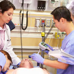
Doctors and a patient in a hospital
A 5-year, randomized clinical trial helped resolve a long-standing debate about how best to manage sepsis patients.

For years, doctors have debated the best ways to identify, predict and treat sepsis. The condition, which is usually triggered by infection, is marked by body-wide inflammation and can lead to a dangerous drop in blood pressure known as septic shock. Sepsis affects more than 800,000 people each year and kills about 20 to 30 percent of them. It’s the most expensive condition treated in U.S. hospitals, costing more than $20 billion a year.

Now, a nationwide, 5-year clinical trial that set out to compare three different treatment approaches has shown that survival of patients with septic shock was the same regardless of whether they received treatment based on structured, standardized medical plans (protocols) or the usual high-level standard of care. If patients were diagnosed shortly after the onset of sepsis and treated promptly with fluids and antibiotics, they did equally well whether they received treatment based on either of two specific protocols—one less invasive than the other—or got the usual, high-level care provided by the academic hospitals where the study was conducted.

According to the study’s leaders, the trial “helps resolve a long-standing clinical debate about how best to manage sepsis patients, particularly during the critical first few hours of treatment,” and shows that “there is not a mandated need for more invasive care in all patients.”

Learn more:
NIGMS News Release
University of Pittsburg News Release Exit icon
New England Journal of Medicine Article
Sepsis Fact Sheet

Visualizing Vessels

0 comments
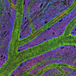
Blood vessels in a mouse retina
Blood vessels in a mouse retina visualized using cutting-edge imaging technology. Credit: Tom Deerinck and Mark Ellisman, NCMIR.

For poets and lovers, the eyes are the windows of the soul. For scientists and doctors, blood vessels at the back of the eye are windows into many diseases.

Blood vessel abnormalities can indicate a variety of serious conditions such as atherosclerosis (hardening of the arteries), heart attacks and strokes. But most vessels are buried beneath skin and other tissues, making them difficult to examine without surgery.

There’s one exception—in the eye. Unlike anywhere else in the body, larger vessels on the retina at the back of the eye are directly visible through the pupil, requiring essentially only light and magnifying lenses to view.

These vessels are used to diagnose glaucoma and diabetic eye disease. Because they display characteristic changes in people with high blood pressure, some researchers hope retinal vessels might one day help predict an impending stroke, congestive heart failure or other diseases stemming from dangerously high blood pressure.

The medical importance of retinal vessels piqued the interest of scientists funded by the National Institutes of Health at the National Center for Microscopy and Imaging Research (NCMIR) at the University of California, San Diego, who captured this micrograph image of mouse retinal vessels.

Continue reading this new Inside Life Science article