Author: Alisa Zapp Machalek

Alisa Machalek Headshot.

Posts by Alisa Zapp Machalek

New Technology May Help Reduce Serious and Costly Post-Surgical Infections—Using Nothing but Air

7 comments

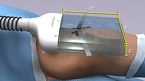
According to a recent estimate, implant infections following hip and knee replacement surgeries in the U.S. may number 65,000 by 2020, with the associated healthcare costs exceeding $1 billion. A new small, high-tech device could have a significant impact on improving health outcomes and reducing cost for these types of surgeries. The device, Air Barrier System (ABS), attaches on top of the surgical drape and gently emits HEPA-filtered air over the incision site. By creating a “cocoon” of clean air, the device prevents airborne particles—including the bacteria that can cause healthcare-associated infections—from entering the wound.

Air Barrier System
The Air Barrier System creates a “cocoon” of clean air (gray area with size indicated) over a surgical site to remove airborne contaminants and reduce the risk of infection in patients who are receiving an artificial hip, a blood vessel graft, a titanium plate in the spine or other implants.

Scientists recently analyzed the effectiveness of the ABS device in a clinical study—funded by NIGMS—involving nearly 300 patients. Each patient needed an implant, such as an artificial hip, a blood vessel graft in the leg or a titanium plate in the spine. Because implant operations involve inserting foreign materials permanently into the body, they present an even higher risk of infection than many other surgeries, and implant infections can cause life-long problems.

The researchers focused on one of the most common causes of implant infections—the air in the operating room. Although operating rooms are much cleaner than almost any other non-hospital setting, it’s nearly impossible to sterilize the entire room. Instead, the scientists focused on reducing contaminants directly over the surgical site. They theorized that if the air around the wound was cleaner, the number of implant infections might go down. Continue reading “New Technology May Help Reduce Serious and Costly Post-Surgical Infections—Using Nothing but Air”

Cool Tools: Pushing the Limits of High-Resolution Microscopy

1 comment

Cell biologists would love to shrink themselves down and actually see, touch and hear the inner workings of cells. Because that’s impossible, they have developed an ever-growing collection of microscopes to study cellular innards from the outside. Using these powerful tools, researchers can exhaustively inventory the molecular bits and pieces that make up cells, eavesdrop on cellular communication and spy on cells as they adapt to changing environments.

In recent years, scientists have developed new cellular imaging techniques that allow them to visualize samples in ways and at levels of detail never before possible. Many of these techniques build upon the power of electron microscopy (EM) to see ever smaller details.

Unlike traditional light microscopy, EM uses electrons, not light, to create an image. To do so, EM accelerates electrons in a vacuum, shoots them out of an electron gun and focuses them with doughnut-shaped magnets onto a sample. When electrons bombard the sample, some pass though without being absorbed while others are scattered. The transmitted electrons land on a detector and produce an image, just as light strikes a detector (or film) in a camera to create a photograph.

This image, showing a single protein molecule, is a montage. It was created to demonstrate how dramatically cryo-EM has improved in recent years. In the past, cryo-EM was only able to obtain a blobby approximation of a molecule’s shape, like that shown on the far left. Now, the technique yields exquisitely detailed images in which individual atoms are nearly visible (far right). Color is artificially applied. Credit: Veronica Falconieri, Subramaniam Lab, National Cancer Institute.

Transmission electron microscopes can magnify objects more than 10 million times, enabling scientists to see the outline and some details of cells, viruses and even some large molecules. A relatively new form of transmission electron microscopy called cryo-EM enables scientists to view specimens in their natural or near-natural state without the need for dyes or stains.

In cryo-EM—the prefix cry- means “cold” or “freezing”—scientists freeze a biological sample so rapidly that water molecules do not have time to form ice crystals, which could shove cellular materials out of their normal place. Cold samples are more stable and can be imaged many times over, allowing researchers to iteratively refine the image, remove artifacts and produce even sharper images than ever before. Continue reading “Cool Tools: Pushing the Limits of High-Resolution Microscopy”

The Irresistible Resistome: How Infant Diapers Might Help Combat Antibiotic Resistance (sort of)

5 comments
Gautam Dantas
Credit: Pablo Tsukayama, Ph.D.,
Washington University School of Medicine
Gautam Dantas
Born: Mumbai, India
Most proud of: His family, which brings him joy and pride
Favorite lab tradition: OOFF! Official Optional Formal Fridays, when members of his lab can dress up, eat bread—made in the lab’s own bread machine—and drink beer and wine together at the end of the day
When not in the lab, he: Enjoys home brewing, pickling and canning, and spending time with his wife and children. He also attends musical performances, including those of his wife, who sings in the St. Louis Symphony Chorus
Advice to aspiring scientists: Pursue hobbies, take risks, explore beyond your comfort zone. “You can do a Ph.D., but also have other experiences.” He says his own outside activities refine his focus in the lab, keep him grounded and help him be an empathetic mentor to his students. Plus, he met his wife while singing in the chorus of Macalester College in St. Paul, Minnesota

When I Grow Up…

Gautam Dantas remembers the day in 10th grade when he first wanted to be a scientist. It was the day he had a new biology teacher, a visiting researcher from the U.S. The teacher passionately described his own biochemical studies of how organisms live together in communities. By the end of the class, Dantas had resolved to earn a Ph.D. in biochemistry.

He ended up doing much more—gaining expertise in computational biology, protein design and synthetic biology. He now combines his skills and knowledge in multifaceted research that spans four departments at Washington University in St. Louis. His goal: to better understand and help combat a vital public health threat—drug-resistant bacteria.

“Our motivation is that we are living in the antibiotic era, and antibiotic resistance is getting out of control,” Dantas says. “We have very few new antibiotics we can use, so we’re kind of scrambling [to find new ways to treat bacterial diseases].”

His research focuses on one of the groups most vulnerable to bacterial infections—newborn babies.

According to his lab’s website Exit icon, the research is “at the interface of microbial genomics, ecology, synthetic biology, and systems biology,” and it aims “to understand, harness, and engineer the biochemical processing potential of microbial communities.” They do it by scrounging around in infant diapers.

Antibiotic Angst

Since their introduction in the 1940s, antibiotic drugs have saved countless lives. Simultaneously, they weeded out strains of bacteria easily killed by the drugs, allowing drug-resistant strains to thrive. Every year, at least 2 million people in the U.S. become infected and at least 23,000 die from drug-resistant bacteria, according to the Center for Disease Control and Prevention. Continue reading “The Irresistible Resistome: How Infant Diapers Might Help Combat Antibiotic Resistance (sort of)”

NIGMS Is on Instagram!

0 comments

Science is beautiful.

For several years, we’ve used this blog to highlight pictures we think are cool, scientifically relevant and visually striking. The images were created by NIGMS-funded researchers in the process of doing their research. Many come from our Life: Magnified collection, which features dozens of stunning photos of life, close-up. We’ll continue to bring you interesting images and information here on Biomedical Beat, but if you can’t get enough of them, we have a new way to share our visual content with you: Instagram.

We’re pleased to announce the launch of our NIGMS Instagram account. We’ll highlight gorgeous images, and bring you the science behind them—straight from our mobile device to yours. Instagram lets us label our images with subject-specific hashtags. You can find our pictures by going on Instagram and searching for #NIGMS. If you’re already an Instagram user, you can follow us @NIGMS_NIH. You can even see our page using a web browser at https://www.instagram.com/nigms_nih/ Exit icon. Let us know what you think!

Also, if you have any stunning images or videos that relate to scientific areas supported by NIGMS, please send them to us. They might end up on our Instagram feed!

See those finger-like projections? They are called villi. This image shows the small intestine, where most of the nutrients from the food we eat are absorbed into the bloodstream. The villi increase the organ’s surface area, making nutrient absorption more efficient. Credit: National Center for Microscopy and Imaging Research.
#science Exit icon #biology Exit icon #research Exit icon #cells Exit icon #cellbiology Exit icon #microscopy Exit icon #nigms Exit icon #scienceisbeautiful Exit icon #sciart Exit icon #nigms Exit icon #nih Exit icon #NCMIR Exit icon #UCSD Exit icon #smallbowel Exit icon  #nutrients Exit icon #nutrient Exit icon #anatomyphysiology Exit icon

We need zinc. It’s an essential nutrient for growth and development, fending off invading microbes, healing injuries, and all sorts of cellular processes. We get the mineral through our diet, but people in certain parts of the world don’t get enough. Researchers study how plants acquire and process zinc, hoping to find ways to increase the nutrient in food crops. Using synchrotron X-ray fluorescence technology, scientists created this heat map of zinc in a leaf from a plant called Arabidopsis thaliana (zinc levels from lowest to highest: blue, green, red, white). Credit: Suzana Car and Mary Lou Guerinot, Dartmouth College.
#science Exit icon #biology Exit icon #research Exit icon #botany Exit icon #plant Exit icon #arabidopsis Exit icon #zinc Exit icon #microscopy Exit icon #synchrotron Exit icon #x-ray Exit icon #modelorganism Exit icon #leaf Exit icon #heatmap Exit icon #scienceisbeautiful Exit icon #nigms Exit icon #nih Exit icon

Beautiful brain! This image shows the cerebellum, which is the brain’s locomotion control center. Every time you shoot a basketball, tie your shoe or chop an onion, your cerebellum fires into action. Found at the base of your brain, the cerebellum is a single layer of tissue with deep folds like an accordion. People with damage to this region of the brain often have difficulty with balance, coordination and fine motor skills. Credit: Tom Deerinck and Mark Ellisman, NCMIR
#science Exit icon #biology Exit icon #research Exit icon #cerebellum Exit icon #neuroscience Exit icon #neurobiology Exit icon #brain Exit icon #brains Exit icon #brainimages Exit icon #neuroanatomy Exit icon #microscopy Exit icon #scienceisbeautiful Exit icon #sciart Exit icon #nigms Exit icon #nih Exit icon #NCMIR Exit icon #UCSD Exit icon

Scientists can learn a lot by studying pigment cells, which give animals their colorful skins, eyes, hair and scales. We can even gain insight into skin cancers, like melanoma, that originate from pigment cells. Pigment cells can form all sorts of patterns, like these stripes on the fin of a pearl danio, a type of tropical minnow. Credit: David Parichy, University of Washington.
#biology Exit icon #marinebiology Exit icon #marinescience Exit icon #cell Exit icon #pigmentcell Exit icon #xanthophore Exit icon #science Exit icon #research Exit icon #cellbiology Exit icon #microscopy Exit icon #scienceisbeautiful Exit icon #scienceiscool Exit icon #zebrafish Exit icon #universityofwashington Exit icon

Lighting Up the Promise of Gene Therapy for Glaucoma

0 comments
Retinal ganglion cells in the mouse.

Retinal ganglion cells in the mouse retina that do (bright, yellow spots throughout) and do not (blue streaks, mostly along the edges) contain a specific gene that scientists introduced with a virus. Credit: Kenyoung (“Christine”) Kim, Wonkyu Ju and Mark Ellisman, National Center for Microscopy and Imaging Research, University of California, San Diego.

What looks like the gossamer wings of a butterfly is actually the retina of a mouse, delicately snipped to lay flat and sparkling with fluorescent molecules. Researchers captured this image while investigating the promise of gene therapy for glaucoma, a progressive eye disease. It all happened at the National Center for Microscopy and Imaging Research Exit icon (NCMIR) at the University of California, San Diego.

Glaucoma is the leading cause of irreversible blindness. It is characterized by the slow, steady death of certain nerve cells in the retina. If scientists can prevent the death of these cells, which are called retinal ganglion cells, it might be possible to slow the progression of glaucoma. Some researchers are examining the possibility of using gene therapy to do just that.

A major challenge of gene therapy is finding a way to get therapeutic genes into the right cells without damaging the cells in any way. Scientists have had success using a non-disease-causing virus (adeno-associated serotype 2) for this task. Continue reading “Lighting Up the Promise of Gene Therapy for Glaucoma”

Newly Identified Cell Wall Construction Workers: A Novel Antibiotic Target?

0 comments
SEDS

A family of proteins abbreviated SEDS (bright, pink) help build bacterial cell walls, so they are a potential target for new antibiotic drugs. Credit: Rudner lab, Harvard Medical School.

Scientists have identified a new family of proteins that, like the targets of penicillin, help bacteria build their cell walls. The finding might reveal a new strategy for treating a range of bacterial diseases.

The protein family is nicknamed SEDS, because its members help control the shape, elongation, division and spore formation of bacterial cells. Now researchers have proof that SEDS proteins also play a role in constructing cell walls. This image shows the movement of a molecular machine that contains a SEDS protein as it constructs hoops of bacterial cell wall material.

Any molecule involved in building or maintaining cell walls is of immediate interest as a possible target for antibiotic drugs. That’s because animals, including humans, don’t have cell walls—we have cell membranes instead. So disabling cell walls, which bacteria need to survive, is a good way to kill bacteria without harming patients.

This strategy has worked for the first antibiotic drug, penicillin (and its many derivatives), for some 75 years. Now, many strains of bacteria have evolved to resist penicillins—and other antibiotics—making the drugs less effective.

According to the Centers for Disease Control and Prevention, drug-resistant strains of bacteria Exit icon infect at least 2 million people, killing more than 20,000 of them in the U.S. every year. Identifying potential new drug targets, like SEDS proteins, is part of a multi-faceted approach to combating drug-resistant bacteria.

The Extracellular Matrix, a Multitasking Marvel

1 comment

In part II of this series, we reveal how the ECM helps body cells move around, a process vital for wounds to heal and a fetus to grow. Here we introduce the extracellular matrix (ECM) and discuss how it makes our tissues stiff or squishy, solid or see-through.

When we think about how our bodies are made and what they do, we usually focus on organs, tissues and cells. These structures have well-known roles. But around, within and between them is a less understood material that also plays an essential part in making us what we are.

This gelatinous filler material is known as the extracellular matrix (ECM). Once thought to be the biological equivalent of bubble wrap, we now know that the ECM is a dynamic, physiologically active component of all our tissues. It guides cell shape, orientation and function.

The ECM is found in all of our body parts. In some tissues, it’s a thin layer separating cells, like mortar between bricks. In other tissues, it’s the major constituent.

The ECM is most prevalent in connective tissue, the material that forms our skeletons, cushions our internal organs and winds between blood vessels and around nerves. In connective tissue, the ECM is more abundant than the cells suspended within it.

The extracellular matrix meets the needs of each body part. In teeth and bones, it’s rock-hard. In corneas, it’s a transparent gel that acts like a camera lens. In tendons, it forms strong fibers that bind muscle to bone. Credit: Stock image.

What makes the ECM truly unique is its variability: Its texture, composition and functions vary by body part. That’s because the ECM’s deceptively simple recipe of water, fibrous proteins and carbohydrates has virtually endless variations.

In general, the fibrous proteins give the ECM its texture and help cells adhere properly. Carbohydrates in the ECM absorb water and swell to form a gel that acts as an excellent shock absorber. Continue reading “The Extracellular Matrix, a Multitasking Marvel”

Cool Video: Watching Bacteria Turn Virulent

1 comment

Researchers created an apparatus to study quorum sensing, a communication system that allows some bacteria to cause dangerous infections. Their findings suggest that blocking bacterial communication might lead to a new way to combat such infections. Credit: Minyoung Kevin Kim and Bonnie Bassler, Princeton University.

If you’ve ever felt a slimy coating on your teeth, scrubbed grime from around a sink drain or noticed something growing between the tiles of a shower, you’ve encountered a biofilm. Made up of communities of bacteria and other microorganisms, biofilms thrive where they can remain moist and relatively undisturbed. As they enlarge, biofilms can block narrow passages like medical stents, airways, pipes or intestines. Continue reading “Cool Video: Watching Bacteria Turn Virulent”

Ticks, Mice and Microbes—Studying Disease Spread

0 comments
Maria Diuk-Wasser
Credit: Oscar Gonzalez (Diuk-Wasser’s husband)
Maria Diuk-Wasser
Hometown: Buenos Aires, Argentina
Childhood dream job: Veterinarian
Hobbies: Hiking and gardening with her son (age 10) and daughter (age 7)
Favorite music: Salsa
Worksite: Lab at Columbia University and forests in coastal New England

Maria Diuk-Wasser grew up on the 10th floor of an apartment building in the middle of a bustling city. With no forests or meadows nearby, she read book after book about the natural world and surrounded herself with houseplants.

“I yearned for nature,” Diuk-Wasser says. “But my parents couldn’t provide it. They’re city people. They didn’t know anything about hiking or camping.”

These days, Diuk-Wasser still spends a lot of time in a city—she’s a professor at Columbia University in New York, the most populous city in the U.S.—but she also gets plenty of time in the woods. She hikes for hours through coastal New England forests, some of the loveliest in the country, searching for what many consider less-than-lovely inhabitants: mice and ticks. Continue reading “Ticks, Mice and Microbes—Studying Disease Spread”

Designing Drugs That Kill Invasive Fungi Without Harming Humans

0 comments
Top to bottom: Cryptococcus, Candida, Aspergillus, Pneumocystis
Invasive fungal infections kill more than 1 million people worldwide every year. Almost all of these deaths are due to fungi in one of these four groups. Credit: Centers for Disease Control and Prevention.

Invasive fungal infections—the kind that infect the bloodstream, lung and brain—are inordinately deadly. A big part of the problem is the lack of drugs that are both effective against the fungi and nontoxic to humans.

The situation might change in the future though, thanks to the work of a multidisciplinary research team led by chemist Martin Burke at the University of Illinois. For years, the team has focused on an antifungal agent called amphotericin B (AmB for short). Although impressively lethal to fungi, AmB is also notoriously toxic to human cells.

Most recently, the research team chemically modified the drug to create compounds that kill fungi, but don’t disrupt human cells. The scientists explain it all in the latest issue of Nature Chemical Biology.

Invasive fungal infections are so intractable because most antifungal drugs aren’t completely effective. Plus, fungi have a tendency to develop resistance to them. AmB is a notable exception. Isolated 50 years ago from Venezuelan dirt, AmB has evaded resistance and remains highly effective. Unfortunately, it causes side effects so debilitating that some doctors call it “ampho-terrible.” At high doses, it is fatal.

For decades, scientists believed that AmB molecules kill fungal cells by forming membrane-piercing pores, or ion channels, through which the cells’ innards leak out. Last year, Burke’s group overturned this well-established concept using evidence from nuclear magnetic resonance, chemistry and cell-based experiments. The researchers showed that AmB molecules assemble outside cells into lattice-like structures. These structures act as powerful sponges, sucking vital lipid molecules, called ergosterol, right out of the fungal cell membrane, destroying the cell. Continue reading “Designing Drugs That Kill Invasive Fungi Without Harming Humans”