
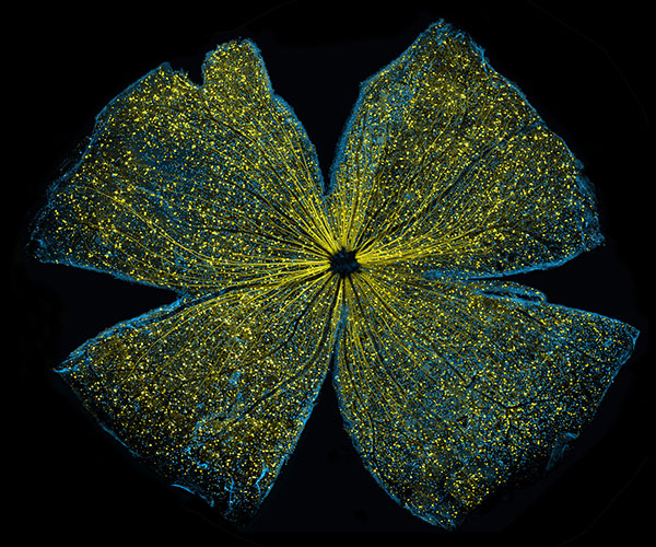
What looks like the gossamer wings of a butterfly is actually the retina of a mouse, delicately snipped to lay flat and sparkling with fluorescent molecules. Researchers captured this image while investigating the promise of gene therapy for glaucoma, a progressive eye disease. It all happened at the National Center for Microscopy and Imaging Research ![]() (NCMIR) at the University of California, San Diego.
(NCMIR) at the University of California, San Diego.
Glaucoma is the leading cause of irreversible blindness. It is characterized by the slow, steady death of certain nerve cells in the retina. If scientists can prevent the death of these cells, which are called retinal ganglion cells, it might be possible to slow the progression of glaucoma. Some researchers are examining the possibility of using gene therapy to do just that.
A major challenge of gene therapy is finding a way to get therapeutic genes into the right cells without damaging the cells in any way. Scientists have had success using a non-disease-causing virus (adeno-associated serotype 2) for this task.
— Kenyoung (“Christine”) Kim, lead researcher”
Here’s how it works: Researchers insert the desired gene into the virus, then let the virus do what it does best—enter cells. Once inside, the virus splits apart to release its genetic material, which gets incorporated into the genome of the host (mice, in this case). Then the inserted genes function just like other genes normally found in the host genome.
This image shows that the process worked—at least for some of the cells. Retinal ganglion cells that contain a functional copy of the test gene are yellow (bright spots throughout). Retinal ganglion cells that don’t are blue (streaks, mostly along the edges). These results bring the concept of gene therapy for glaucoma one step closer.
But the research was about more than delivering genes or treating eye diseases. It also showcases a powerful technique pioneered by NCMIR for obtaining high-resolution images of large samples, such as an entire mouse retina. The technique is similar to Google Earth in that it computationally stitches together many small, high-resolution images.
The directors of NIH’s two dozen or so institutes were so impressed with this detailed image of a whole mouse retina that they awarded it first place in a recent art contest held in conjunction with the 2016 Combined Federal Campaign, an annual, government-wide charity fundraiser.
For more incredible microscopy images from this facility, visit the NIGMS Image and Video Gallery and search for NCMIR.